




 IJCSMB Home



Shh-Gli1 Pathway in Alzheimer’s Disease: Molecular Dynamics and Clinical Significance
Nabeela Khanam1*, Lama Misba2 and Shagufta Moin3
1TB & Respiratory Diseases, J.N. Medical College, Aligarh Muslim University, Aligarh, India
2Interdisciplinary Biotechnology Unit, Aligarh Muslim University, Aligarh, India
3Department of Biochemistry, J. N. Medical College, Aligarh Muslim University, Aligarh, India
Submission: May 21,2026; Published:July 07, 2026
*Corresponding author: Nabeela Khanam, TB & Respiratory Diseases, J.N. Medical College, Aligarh Muslim University, Aligarh, India
How to cite this article: Nabeela K, Lama M, Shagufta M. Shh-Gli1 Pathway in Alzheimer’s Disease: Molecular Dynamics and Clinical Significance. Int J Cell Sci & Mol Biol. 2026; 8(2): 555734.DOI: 10.19080/IJCSMB.2026.08.555734
Abstract
Alzheimer’s disease (AD), a complex, heterogenous, and progressive form of neurodegenerative dementia. It is anticipated that the prevalence of AD is rising in the aging population. Therefore, we need to identify new potential biomarkers, that will help diagnose AD in its early stages. However, in adult neural tissues Sonic hedgehog (Shh) signaling pathway has emerged as an important modulator through different processes. Hence, the clinical application of Shh in various neurodegenerative diseases including AD remains unclear. Thus, we aim to know how Shh-Gli1 cell signaling pathway plays a potential role in AD rats and also determines potential interactions with p53 for AD development. Our results showed that rats from AlCl3 and STZ-induced AD models showed similar anxiogenic behavior. Also, rats from AlCl3 and STZ-induced AD models showed cognitive impairment and altered memory and learning patterns as compared to the control group. Thereafter, gene expression assay showed significant up-regulation of Shh and Gli1, whereas no significant changes were found in p53 expression in AD rat brain tissue samples as compared to control. Further, histology staining showed morphologically damaged nuclei in the AlCl3 and STZ-induced AD rat groups whereas in the control group, arranged round nuclei were seen. IHC showed high expression of Shh and Gli1, however, low expression of p53 was observed in the induced AD rat’s groups as compared to the control rat group. Hence, we concluded that the dysregulated Shh, Gli1 and P53 may act as the predicted potential biomarkers in the diagnosis and treatment of AD.
Keywords: Alzheimer’s disease; hedgehog; Shh; Gli1; p53
Abbreviations: AD: Alzheimer’s Disease , Shh: Sonic hedgehog ; NFT: Neurofibrillary Tangles ; APP: Amyloid Precursor Protein; ApoE: Apolipoprotein E; PSEN-1: Presenilin-1; PSEN-2: Presenilin-2; AlCl3: Aluminum Chloride; BBB: Blood-Brain Barrier; SW: Southwest; NW: Northwest ; NE: northeast; SE: Southeast; MWM: Morris Water Maze; OFT: Open Field Test; PFA: Paraformaldehyde; CNS: Central Nervous System
Introduction
Alzheimer’s disease (AD) is the most common neurodegenerative disorder in people above the age of 65 years. It is a prevalent form of dementia often characterized by progressive loss of cognitive abilities [1,2]. Every year, around 4.6 million new cases are reported worldwide [3] and present cerebral atrophy, especially in the hippocampus, parietal and temporal lobes, as well as by the presence of neurofibrillary tangles (NFT) and senile plaques. The accumulation of β-amyloid (Aß) induces neurotoxicity and oxidative stress associated with the disease progression. Both genetic and environmental risk factors play a role in AD manifestation. The risk factors include decreased reserve brain capacity, gross brain shrinkage, and low mental achievement in early life, but age remains the greatest risk factor. Mutations in the dominant genes such as Amyloid precursor protein (APP), Presenilin-1 (PSEN-1), Presenilin-2 (PSEN-2) and apolipoprotein E (ApoE) are also associated with AD [4,5]. Some other risk factors including air pollution, diet, metals, infections, and many others may induce oxidative stress and inflammation, hence increasing the risk of developing AD. However, the brain is a potential target for aluminum toxicity [6]. In the brain, aluminum accumulates mainly in the frontal cortex and hippocampus [7,8]. Numerous studies reported that aluminum is one of the major heavy metals that participated in the initiation and progression of neurodegenerative ailments, as it directly affects the numerous metabolic cascades in the nervous system, and may cause specific encephalopathy with dementia [9,10].
The utilization of aluminum chloride (AlCl3) is highly compounded because it is found in various commercially manufactured products like toothpaste, foods, medicines, and packaged drinking water [11]. AlCl3 is then absorbed into the blood-brain barrier (BBB), accumulates in the brain, and plays the role of a causative agent of AD [12]. Epidemiological and animal studies showed that the incorporation of aluminum in the hippocampus leads to anomalous Aß accumulation, neuroinflammation and thus neuronal necrosis, resulting in cognitive dysfunction, impaired cholinergic projections, apoptotic neuronal death and phosphorylated tau overexpression [13-18]. Sporadic dementia induced by intra-ventricular injection of streptozotocin (STZ) is a well-defined non-transgenic animal experimental model that mimics late-onset AD. STZ injections decrease cerebral glucose uptake and produce multiple other effects that resemble molecular, pathological, and behavioral changes associated with sporadic form of Alzheimer's disease such as spatial memory impairment and cholinergic cell death in the hippocampus [19,20]. Various cellular pathways might be involved in the regulation of Aβ metabolism. These pathways including the Shh-Gli1 cell signaling pathway could not only be linked with altered amyloidogenic or non-amyloidogenic processing of amyloid precursor protein (APP) but also might prevent clearance of extracellular or intracellular Aβ [21- 24]. In the Shh-Gli1 cell signaling pathway, Sonic hedgehog (Shh) sets a chain of events in target cell, leading to the activation and repression of target genes by Gli gene family of transcription factors.
The activated Gli1 regulates the expression of many target genes that control cell growth, survival, and differentiation in a wide range of cells, including neurons. Gli1 is a critical effector of the Shh pathway; indeed, Gli1 is also a transcriptional downstream target of Shh cell signaling pathway [25]. Microglial-mediated neuronal apoptosis is also mediated by p53 in AD. This modified p53 is considered either mutated or misfolded in AD and is suggested to be one of the potential biomarkers for AD [26]. Henceforth, we also attempted to check the expression of p53 in the AD group. Although AD was identified many years ago, there is no effective treatment and early-stage biomarker for AD yet. Current treatments only provide temporary and modest improvement in cognitive impairment and are considered to be symptomatic treatments. Hence, there is a need to identify novel and effective biomarkers as well as therapeutic targets for AD. As a result, this research was conducted to identify the dysregulation of Shh, Gli1, and P53 which may act as potential biomarkers or therapeutic targets for AD.
Material and Methods
Animal model
200-250g weight male Wistar rats were obtained from Central Animal House Facility, Jawaharlal Nehru Medical College, Faculty of Medicine, Aligarh Muslim University (A.M.U) one week prior to the experiments, housed individually with temperature (25±1°C), humidity (60±10%) approved by the Institutional Animal Ethics Committee and carried out as per CPCSEA guidelines India (Reg no. 401/GO/Re/S/2001/CPCSEA). They were divided into Control group (n=6), AlCl3 group (n=6) and streptozotocin (STZ (#14653 Sigma Aldrich) group (n=6). The control group received saline solutions intraperitoneally while AD groups were treated with AlCl3 and STZ for 28 days at doses of 0.71 mg/kg/day and 33 mg/kg/day respectively.
Morris Water Maze (MWM)
The MWM tank was 132 cm in diameter and 60 cm in height, filled with water (25±2°C) to a depth of 45 cm. The dye was added to make the water opaque. The tank was divided into four equal quadrants by two virtual perpendicular lines crossing at the center. The four quadrants were named southwest (SW), northwest (NW), northeast (NE), and southeast (SE). A platform of 10 cm diameter was placed in the middle of the target quadrant (SW) of the tank, and the platform was submerged 2.5 cm below the surface of the water for the rat to climb on it, to escape from the water. The test was completed in six days. On the first day the rats were trained to remember the visible platform and from the second day, the platform was hidden to record escape latency and three trials each day by placing the rat in different quadrants (NW, NE and SE) up to five days. The duration of each trial was 60 s only because more than 60 s rats were aggressive. If the rat failed to locate the platform within 60 seconds, the experimenter would gently guide it onto the platform to reinforce its memory of the platform's position for 30 seconds. Thereafter, on the sixth day, the probe trial was carried out in which the platform was removed, and the rat was allowed to search the platform for 60 s, during which the time spent in the target quadrant was recorded by the ANY-Maze system, V4.3 (Steolting, IL, USA). After the end of each trial, the rat was dried with a towel and placed in their home cages.
Open field test (OFT)
The OFT arena consisted of a 60×60cm black wooden apparatus, surrounded by 30 cm high walls and equally divided into 15×15cm squares on the floor by white colored lines. It was a two-day procedure. The test was started by placing the rat at the center of the arena and allowing it to explore freely for 5 min, the behavior was recorded by the overhead camera attached to ANY-Maze system V4.3 (Stoelting, IL, USA).
Tissue sampling
After the behavioral experiments, rats were anesthetized by ketamine (75mg/kg; i.p.) and euthanized by cervical dislocation. Rat brains were quickly isolated and placed in ice-cold phosphate buffer saline. The isolated tissue samples were kept in RNAlater (#76104, Qiagen, Germany) and stored at -200C for molecular analysis and in paraformaldehyde (PFA) for histopathological experiments.
RNA isolation
RNA from rat brain tissue samples was isolated and purified by the TRI reagent (#T9424, Sigma-Aldrich). After homogenization, 200 µl of chloroform per ml of TRI reagent was added and shaken vigorously for 15 seconds. Samples were then centrifuged at 12,000×g for 15minutes at 2–8°C. Aqueous phase containing RNA was carefully collected in a fresh tube, without disturbing the lower layer. Add 500µl of isopropanol per ml of TRI reagent, followed by centrifugation at 12,000×g for 15minutes at 4°C, which leads to precipitation of RNA in small translucent to opaque white pellet at the bottom of the tube. The supernatant of the tube was discarded and 1 ml 75% ice-cold ethanol per ml of TRI used was added to wash the pellet. The pellet was dissolved in nuclease-free water. RNA concentration was determined spectroscopically (Shimadzu, UV-1800, Germany) by measuring A260/A280 absorbance ratios. RNA integrity was assessed by agarose gel electrophoresis. 1μg of total RNA was used to prepare cDNA.
Reverse-transcriptase quantitative PCR
The cDNA (#4368814, MERCK) was synthesized, and real-time PCR (Applied Biosystems) was done. Real-time quantitative PCR was performed by Step-One (Applied Biosystems) with SYBR green PCR master mix (#04710924001, Thermo-Scientific). The reaction mixture contained 150 ng of a cDNA sample and appropriate PCR primers. The cycle profile included an initial denaturation at 95°C for 10 min, followed by 40 cycles of amplification consisting of denaturation at 95°C for 15 s, annealing at 60°C for 30s, and extension at 72°C for 30 s. Each sample was run in triplicate and the means and standard deviations were determined Table 1.
Histopathological analysis of rat brain tissue by H&E
Paraformaldehyde (PFA) fixed hindbrain tissues were separated and dehydrated in a series of alcohol gradients and xylene. Paraffin-embedded samples were further cut into thin layers of 4μm thickness by cryostat microtome and picked on gelatine-coated slides and dried in an incubator. Slides were dewaxed in xylene for 20minutes and rehydrated in a series of alcohol gradients and dipped in distilled water and stained with hematoxylin. After cleaning the overstrained sections with acid alcohol, they were stained with Eosin. Slides were again dipped in a series of alcohol gradients in descending order. Finally, slides were placed in xylene for 20minutes, then cleaned and mounted with DPX mounting.
Immunohistochemical analysis of rat brain tissue
Rat brain samples were fixed in 4% PFA. Fixed tissues were dehydrated (in series of 50% to 100% ethanol for 30 min), xylene and embedded in paraffin wax. The paraffin-sectioned samples were dewaxed, rehydrated, and incubated with primary antibodies (0.1µg/100µL) against Shh (#NBP2-22139, Thermo Fisher Scientific) and Gli-1(#PA5-32206, Thermo Fisher Scientific) for 30 mins. Non-covalently bound antibodies were washed out with PBS‐T buffer four times (10 min). Then, incubation with 0.05µg/100µl anti‐mouse secondary antibody‐fluorescein isothiocyanate (#NBP2- 30348H, Novus Biological) tagged per sample was performed for 30 min, followed by the washing steps. The slides were cleaned and mounted with the Ibidi mounting medium (#50305707), Thermo Fisher Scientific), and images were acquired by a fluorescent microscope (Nikon, Tokyo, Japan).
Statistical analysis:
All results were analyzed by one-way ANOVA using Graph pad Prism 7. The results are expressed as mean ± SE.
Results
Assessment of anxiogenic behavior
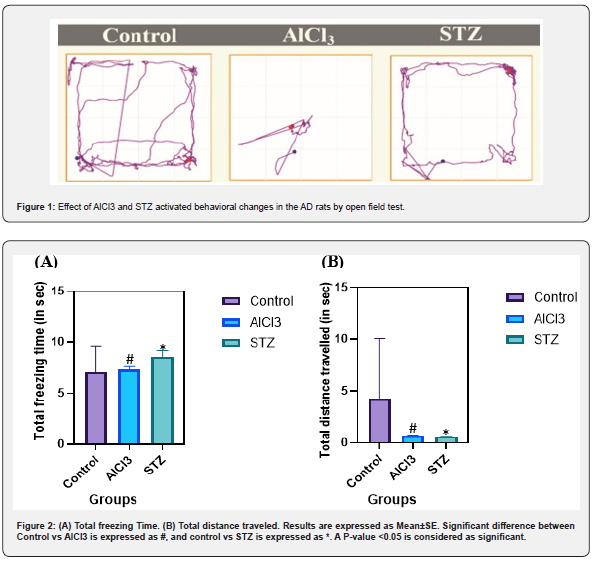
To assess the effect of AlCl3 and STZ on rats, the Open Field Test was carried out (Figure 1). Rats from AlCl3 and STZ-induced AD models showed similar anxiogenic behavior. Total distance travel and freezing time were found to be statistically significant when AD groups were compared to the control group (Figure 2A and B).
6 Assessment of cognitive impairment
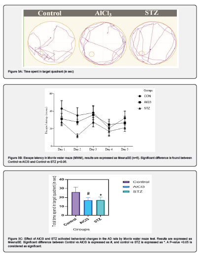
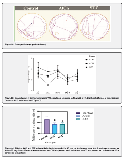
Moreover, in Morri’s water maze, first time spent by rats in each quadrant (Figure 3A) was seen and also significant decrease was observed in the escape latency (Figure 3B). Rats from AlCl3 and STZ-induced AD models showed cognitive impairment as compared to the control. AlCl3 and STZ rats groups presented altered memory and learning patterns (Figure 3C).
Reverse transcriptase PCR and Real-Time PCR for analysis of gene expression
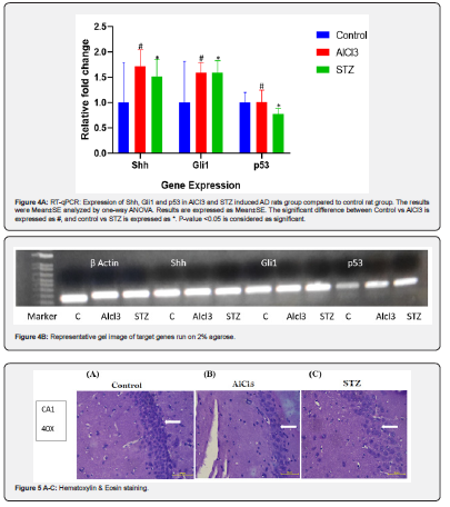
RT-PCR showed high expression of Shh and Gli1 transcript in AD groups of rat brain tissue compared to control group. Thereafter, we checked these genes' expression by quantitative PCR. RT-qPCR expression showed a significant up-regulation of Shh and Gli1 as compared to control. Moreover, we did not find any significant changes in p53 expression in induced AD rat tissue samples compared to control (Figure 4A & B).
Histopathological analysis to check pyknotic damage
Paraffin-sectioned samples were stained with H&E to determine the morphological changes in rat tissue samples, the control group showing arranged round nuclei and morphologically damaged nuclei in the AlCl3 and STZ-induced AD rats groups (Figure 5A-C).
DAPI Staining
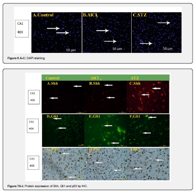
DAPI staining of 4μm paraffin sections of hippocampus of AlCl3 and STZ induced AD rats, compared with control rats. In DAPI staining we have found damaged and shrinkaged nuclei in AlCl3 and STZ-induced AD rats groups compared to control group (Figure 6A-C).
Immunohistochemical analysis of AD rat brain for differential expression of Shh, Gli1 and p53
IHC showed high expression of Shh and Gli1 in induced AD rat’s groups as compared to control group (Figure 7A, B, C, D, E & F). Although, low expression of p53 was observed in the induced AD rat’s groups as compared to the control rat group (Figure 7A-I).
Discussion
Few studies explored the role of the Shh-Gli1 cell signaling pathway in neurodegenerative diseases but to the best of our knowledge, none of them reported the role of the Shh-Gli1 cell signaling pathway in AD. In this study, we explored the role of Shh-Gli1 cell signaling pathway in an AD-induced rat model. AD rat models were developed by AlCl3 and STZ and thereafter we explored the potential role of the Shh-Gli1 cell signaling pathway in these AD-induced rat models. Moreover, AlCl3 model is widely used to induce AD in rats but compared to AlCl3, STZ is less invasive and induces a similar sporadic AD pathology as AlCl3. According to Campbell et al, AlCl3 is a possible contributing factor in Alzheimer’s disease [27-29]. AlCl3 has been implicated in Alzheimer's disease, Parkinsonism, and Dementia complex and causes extensive damage to the nervous system, to date the mechanism of AlCl3 neurotoxicity has not been fully elucidated [30]. Evidence for the contribution of AlCl3 to AD remains contradictory [31,32]. However, epidemiological studies have indicated a link between AlCl3 in drinking water and AD and a variety of human and animal studies have implicated learning and memory deficits after AlCl3 exposure [33-35].
We also used STZ as AD inducer in one group of rats. IICV injections of STZ in rats impair brain biochemistry, cerebral glucose and energy metabolism, cholinergic transmission, and increase the generation of free radicals, ultimately leading to cognitive deficits [36,37]. Collectively, these effects are similar to sporadic dementia of Alzheimer's type in humans [38]. In our study, we observed that the level of Shh-Gli1 is up-regulated in AlCl3 and STZ induced AD rats group compared to control group. This pathway is very crucial for the normal embryonic and adult brain development. When the pathway is downregulated due to multiple factors, brain tumors and damage to the hippocampus leading to AD may be caused [39]. According to previous studies, Shh signaling pathway is activated after injury and helps in tissue repair mechanisms in the adult organism [40, 41]. In the early patterning of the embryonic brain, Shh-Gli1 signaling pathway plays a crucial role in regulating the polarity of the central nervous system (CNS) as well as guiding the ventral patterning in the spinal cord [42]. Some previous studies suggested that Shh plays an important role in establishing the ventral spinal cord, inducing the basal lamina, and forming motor neurons among several other functions during the early stage of embryonic development [43]. According to Li et al, blockage of the Shh pathway may reduce apoptosis of hippocampal neurons to improve spatial learning and memory capacity in AD mice [44]. Shh affects the proliferation, differentiation, survival, and apoptosis of neural precursors [45]. Reilly et al. found that Shh exerts an important function on cholinergic neuron development, and its receptor PTCH1 is specifically expressed in cholinergic neurons of the adult rat basal forebrain, suggesting a therapeutic value of Shh in AD [46, 47]. Furthermore, we investigated the expression of p53. Although p53 showed a significant contribution to neurodegeneration and synaptic plasticity in previous studies [48] we did not find any significant difference in p53 expression in AD group compared to control.
Some previous studies suggested that p53 is a biomarker for AD and expression and activity of p53 are highly regulated [49]. p53 plays an important role in many areas of cellular physiology and biology, ranging from cellular development and differentiation to cell cycle arrest and apoptosis [50]. p53 expression and activity are tightly regulated, such that p53 protein product is either rapidly degraded or exists in a latent form in unstressed cells. Moreover, p53 activation depends upon complex post-translational modifications [51]. According to C Lanni et al, p53 is conformationally altered and plays a role as a novel candidate biomarker for AD. According to them, an anomalous and detectable conformational state of p53 is expressed in peripheral blood cells from sporadic AD patients and not in age-matched non-AD subjects, thus supporting the existence of a new putative marker for AD [52]. Recently, Uberti et al. described a correlation between p53 and AD. In AD fibroblasts they showed an impairment of the p53 signaling pathway.
Conclusion
As far as various studies on AD have been done but till now, many problems have not been solved. Presently, the treatments for AD provide only temporary improvement. The development of new technical tools, animal experimental models, and gene transcription, and protein detection technologies may provide the circumstances necessary for humans to defeat neurodegenerative illnesses. Therefore, it is crucial to carefully examine how the Shh-Gli1 signaling pathway contributes to the development of the central nervous system and neurodegenerative disorders. Finding novel treatment targets and researching precise diagnostic and prognostic biomarkers will also raise hopes for curing neurodegenerative illnesses, including AD. Henceforth, our study suggests that Shh and Gli1 may act as potential biomarkers in the diagnosis of AD. In aged neural tissues, the role of Shh-Gli1 may also provide a therapeutic possibility, mainly in devastating neurodegenerative disorders including AD.
References
- Villemagne VL, Burnham S, Bourgeat P, Belinda B, Kathryn AE, et al. (2013) Amyloid β deposition, neurodegeneration, and cognitive decline in sporadic Alzheimer’s disease: a prospective cohort study. Lancet Neurol 12(4): 357-367.
- Alzheimer’s Disease International. World Alzheimer Report (2015) London: Alzheimer’s Disease International; 2015.
- Alzheimer’s in the Middle East Population, 6 2015.
- Caroline VC, Christine VB, Kristel S (2016) The genetic landscape of Alzheimer disease: Clinical implications and perspectives. Genet Med 18(5): 421-430.
- Khanahmadi M, Farhud DD, Maryam M (2015) Genetic of Alzheimer’s disease: A narrative review article. Iran J Public Health 44(7): 892-901.
- Laraib L, Sadia S, Zehra B, Saiqa T, Sidrah S, et al. (2019) Acute aluminum chloride toxicity revisited: Study on DNA damage and Histopathological, biochemical and neurochemical alterations in rat brain. Life Sci 217:202-211.
- Cheng XJ, Jing XG, Yi-Peng P, Jiao L, Ting X, et al. (2019) Tacrine-hydrogen sulfide donor hybrid ameliorates cognitive impairment in the aluminum chloride mouse model of Alzheimer’s disease ACS Chemical Neuroscience 10(8): 3500-3509.
- Raozhou L, Xiaohong C, Wenming L, Yifan H, Peiqing L, et al. (2008) Exposure to metal ions regulates mRNA levels of APP and BACE1 in PC12 cells: blockage by curcumin. Neuroscience Letters 440(3): 344-347.
- Exley C (2014) What is the risk of aluminum as a neurotoxin?. Expert Rev Neurother 14(6): 589-591.
- Cao Z, Fan W, Chunyu X, Jian Z, Yanfei L (2017) Hypericum perforatum extract attenuates behavioral, biochemical, and neurochemical abnormalities in Aluminum chloride-induced Alzheimer’s disease rats. Biomed Pharmacother 91: 931-937.
- Mirza A, King A, Troakes C, Exley C (2017) Aluminum in brain tissue in familial Alzheimer’s J Trace Elem Med Biol 40: 30-36.
- Willhite CC, Nataliya AK, Robert AY, Nagaraj KY, Thomas MW, et al. (2014) Systematic review of potential health risks posed by pharmaceutical, occupational and consumer exposures to metallic and nanoscale aluminum, aluminum oxides, aluminum hydroxide and its soluble salts Critical Reviews in Toxicology 4 (Suppl 4): 1-80.
- Chiroma SM, Mohamad THB, Che NMT, Zuhair A, Saravanan J, et al. (2019) Protective effect of Centella asiaticaagainst D-galactose and aluminum chloride induced rats: Behavioral and ultrastructural approaches. Biomed Pharmacother 109: 853-864.
- Walton JR, Wang MX (2009) APP expression, distribution and accumulation are altered by aluminum in a rodent model for Alzheimer’s disease. Journal of Inorganic Biochemistry 103(11): 1548-1554.
- Campbell A, Kumar AA, La Rosa FG, Prasad KN, Bondy SC (2000) Aluminum increases levels of beta-amyloid and ubiquitin in neuroblastoma but not in glioma cells. Proceedings of the Society for Experimental Biology and Medicine 223(4): 397-402.
- Kawahara M, Kato M, Kuroda Y (2001) Effects of aluminum on the neurotoxicity of primary cultured neurons and on the aggregation of 𝛽-amyloid protein. Brain Res Bull 55(2): 211-217.
- Gulya K, Rakonczay Z, Kasa P (1990) Cholinotoxic effects of aluminum in rat brain. J Neurochem 54(3): 1020-1026.
- El-Sebae AH, Abdel GME, Shalloway D, Abou ZMM, Blancato J, et al. (1993) Aluminum interaction with human brain tau protein phosphorylation by various kinases. J Environ Sci Health B 28(6): 763-777.
- Correia SC, Renato XS, George P, Xiongwei Z, Paula IM, et al. (2011) Insulin-resistant brain state: the culprit in sporadic Alzheimer's disease?. Ageing Res Rev 10(2): 264-273.
- Kamat PK (2015) Streptozotocin induced Alzheimer's disease like changes and the underlying neural degeneration and regeneration mechanism. Neural Regen Res 10(7): 1050-1052.
- Scheuner D, Eckman C, Jensen M, Song X, Citron M, et al. (1996) Secreted amyloid 𝛽 -protein similar to that in the senile plaques of Alzheimer’s disease is increased in vivo by the presenilin 1 and 2 and APP mutations linked to familial Alzheimer’s Nat Med 2(8): 864-870.
- Ertekin TN, Younkin LH, Yager DM, Parfitt F, Baker MC, et al. (2008) Plasma amyloid beta protein is elevated in late-onset Alzheimer disease families. Neurology 70: 596-606.
- Villemagne VL, Samantha B, Pierrick B, Belinda B, Kathryn AE, et al. (2013) Amyloid β deposition, neurodegeneration, and cognitive decline in sporadic Alzheimer's disease: a prospective cohort study. Lancet Neurol 12(4): 357-367.
- Kosaka T, Imagawa M, Seki K, Arai H, Sasaki H, et al. (1997) The beta APP717 Alzheimer mutation increases the percentage of plasma amyloid-beta protein ending at Abeta-42(43). Neurology 48(3): 741-745.
- Levine AJ (1997) p53, the cellular gatekeeper for growth and division. Cell 88(3): 323-331.
- Xavier GM, Maisa S, William B, Anahid AB, Finn G, et al. (2016) Hedgehog receptor function during craniofacial development. Dev Biol 415(2): 198-215.
- Tayyab M, Farheen S, Mariyath MPM, Nabeela K, Mobarak HM, et al. (2019) Antidepressant and Neuroprotective Effects of Naringenin via Sonic Hedgehog‑GLI1 Cell Signaling Pathway in a Rat Model of Chronic Unpredictable Mild Stress. NeuroMolecular Med 21(3): 250-261.
- Liu D, Zhen W, Ze G, Kai X, Qingrui Z, et al. (2014) Effects of curcumin on learning and memory deficits, BDNF, and ERK protein expression in rats exposed to chronic unpredictable stress. Behav Brain Res 271: 116-121.
- Campbell A (2002) The potential role of aluminum in Alzheimer's Nephrol Dial Transplant 17 Suppl 2: 17-20.
- Niu Q, Yang Y, Zhang Q, Niu P, He S, et al. (2007) The relationship between Bcl-gene expression and learning & memory impairment in chronic aluminum-exposed rats. Neurotox Res 12(3): 163-169.
- Flaten TP (2001) Aluminum as a risk factor in Alzheimer's disease, with emphasis on drinking water. Brain Res Bull 55(2): 187-196.
- Gupta VB, Anitha S, Hegde ML, Zecca L, Garruto RM, et al. (2005) Aluminum in Alzheimer’s disease: are we still at a crossroad ?. Cell Mol Life Sc 62(2): 143-158.
- Buraimoh AA, Ojo SA, Hambolu JO, Adebisi SS, et al. (2011) Effects of Oral Administration of Aluminum Chloride on the Histology of the Hippocampus of Wistar Rats. Curr Res J Biol Sci 3(5): 509-515.
- Exley et al (2005) The aluminum-amyloid cascade hypothesis and Alzheimer disease. Subcell Biochem 38: 225-234.
- Yokel RA (2000) The toxicology of Aluminum in brain: review. Neurotoxicology 21(5): 813-828.
- Hoyer S, Lannert H (1999) Inhibition of the neuronal insulin receptor causes Alzheimer-like disturbances in oxidative/energy brain metabolism and in behavior in adult rats. Ann N Y Acad Sci 893(1): 301-303.
- Ishrat T, Nasrul HMD, Badruzzaman KM, Seema Y, Muzamil A, et al. (2009) Amelioration of cognitive deficits and neurodegeneration by curcumin in rat model of sporadic dementia of Alzheimer's type (SDAT). Eur Neuropsychopharmacol 19(9): 636-647.
- Hoyer S, Nitsch R, Oesterreich K (1991) Predominant abnormality in cerebral glucose utilization in late-onset dementia of the Alzheimer type: a cross-sectional comparison against advanced late-onset and incipient early-onset cases. J Neural Transm Park Dis. Dement Sect 3(1): 1-14.
- Ahlgren SC, Bronner FM (1999) Inhibition of sonic hedgehog signaling in vivo results in craniofacial neural crest cell death. Curr Biol 9(22): 1304-1314.
- Yin VT, Esmaeli B (2017) Targeting the Hedgehog pathway for locally advanced and metastatic basal cell carcinoma. Curr Pharm Des 23(4): 655-659.
- Eli EB, Aneeka C, Mohamed HF, Charles GE (2007) Hedgehog Signaling Promotes Medulloblastoma Survival via BclII. Am J Pathol 170(1): 347-355.
- Shahi MH, Lorente A, Castresana JS (2008) Hedgehog signaling in medulloblastoma, glioblastoma and neuroblastoma. Oncol Rep 19(3): 681-688.
- Kvon EZ, Olga KK, Uirá SM, Iros B, Marco O, et al. (2016) Progressive Loss of Function in a Limb Enhancer during Snake Evolution. Cell 167 (3): 633-642.
- Patel SS, Sunil T, Diksha S, Neeraj M, Malairaman U (2017) Targeting Sonic Hedgehog Signaling in Neurological Disorders. Neurosci Biobehavioral Rev 74 (Pt A): 76-97.
- Li J, Jifan F, Yang L, Thach VH, Weston G, et al. (2015) BMP-SHH Signaling Network Controls Epithelial Stem Cell Fate via Regulation of its Niche in the Developing Tooth. Developmental Cel 33(2): 125-135.
- Chen SD, Jenq LY, Yi-Chun L, Ching C A, Ding IY (2020) Emerging Roles of Inhibitor of Differentiation-1 in Alzheimer’s Disease: Cell Cycle Reentry and beyond. Cells 9 (7): 1746.
- Lee SH, Young CB, Jin HK, Ra KA, Min S, et al. (2017) Late Treatment with Choline alfoscerate (L-alpha Glycerylphosphorylcholine, α-GPC) Increases Hippocampal Neurogenesis and Provides protection against Seizure-Induced Neuronal Death and Cognitive Impairment. Brain Res 1654(Pt A): 66-76.
- Reilly JO, Irina DK, Kevin PW, Nagesh KM, Karen LA (2002) Cooperative Effects of Sonic Hedgehog and NGF on Basal Forebrain Cholinergic Neurons. Mol Cell Neurosci 19(1): 88-96.
- Vogelstein B, Lane D, Levine AJ (2000) Surfing the p53 network. Nature 408(6810): 307-310.
- Ji H, Jiangyong M, Xiangjian Z, Yuanyuan D, Haichao L, et al. (2012) Inhibition of sonic hedgehog signaling aggravates brain damage associated with the down-regulation of Gli1, Ptch1 and SOD1 expression in acute ischemic stroke. Neusrosci Lett 506(1): 1-6.
- Lanni C, Racchi M, Mazzini G, Ranzenigo A, Polotti R, et al. (2008) Conformationally altered p53: a novel Alzheimer’s disease marker?. Mol Psychiatry 13(6): 641-647.
- Lee SJ, Dong CK, Bo-Hwa C, Hyunjung H, Kyong TK (2006) Regulation of p53 by activated protein kinase C-delta during nitric oxide-induced dopaminergic cell death. J Biol Chem 281(4): 2215-2224.

























