


The Prevalence of Bovine Trypanosomiasis in JabiTehnan District of Amhara Regional State, Ethiopia
Melak Wondie1* and Tewodros Alemneh2*
1Woreta Town Office of Agriculture and Environmental Protection, Ethiopia
2University of Gondar, Ethiopia
Submission: July 03, 2018;Published: July 16, 2018
*Corresponding author: Tewodros Alemneh: Faculty of Veterinary Medicine, University of Gondar, Ethiopia, Tel: 251-920-499-820; Email: tedyshow@gmail.com
How to cite this article: Melak W, Tewodros A. The Prevalence of Bovine Trypanosomiasis in JabiTehnan District of Amhara Regional State, Ethiopia. Int J cell Sci & mol biol. 2018; 4(5): 555649. DOI: 10.19080/IJCSMB.2018.04.555649.
Abstract
Cross sectional study was conducted in Jabi Tehnan District of West Gojjam Administrative Zone of Amhara Regional State, Ethiopia to determine the prevalence of bovine trypanosomiasis. In the parasitological survey, blood samples of 164 cattle were examined using a buffy coat technique. The Packed Cell Volume (PVC) value of each animal was also measured using hematocrit reader. The overall prevalence of trypanosomiasis was found to be 15.24% and it consists of 9.76% and 20.73% in Adankegne and Ergib peasants’ association, respectively (X2=5.783, p=0.056). The most positive cases were due to Trypanosoma congolense (T. congolense ) (80%) followed by Trypanosoma vivax (T. vivax)(20%). The mean(PCV) values of parasitaemic and aparasitaemic animals during the study period were 20.75% and 25.07%, respectively. The variation in mean PCV values were statistically significant (p=0.01). The study also demonstrated statistically significant (X2=13.886, p=0.001) variations in prevalence between sexes of cattle, which were 10.67% and 19.1% in female and male animals, respectively. The present prevalent study generated valuable information on the epidemiology of bovine trypanosomosis in the study area and revealed that trypanosomosis was an important disease affecting the livestock production
Keywords: PCV; Prevalence; Trypanosoma congolense; Trypanosoma vivax; Bovine
Introduction
Livestock is backbone of the socio-economic system of most of the rural communities of Africa [1]. Ethiopia is known for its large and diverse livestock resource endowments. Livestock is primarily kept on small holdings where it provides drought power for crop production, manure for soil fertility and fuels, serves as a sources of family diet and sources of cash income (from sale of livestock and livestock products). Despite large livestock population, Ethiopia fails to optimally utilize this resource due to different constrains facing the livestock subsector. Shortage of nutrition, reproductive insufficiency, management constraints and animal disease are the major constraints [2]. One of the diseases hampering the livestock subsector is trypanosomosis [3]. Trypanosomosis is a complex disease of protozoa that is caused by different species of unicellular parasites (trypanosome) found in the blood and other tissues of vertebrates, including livestock, wild life and people [4]. Trypanosomosis limited to the extension of natural herds particularly in Africa were the presence of the tsetse fly density access to woodland and savanna areas with good grazing potential [3]. It is a serious constraint to agricultural production in extensive areas of the tsetse infested regions which accounts over 10 million squares of the tropical Africa [5].Ethiopia is one of the countries suffering from the impact of trypanosomosis. In Ethiopia, it is estimated that some 10 to 14 million heads of cattle and an equivalent number of small ruminants together with a significant number of equines and camels, are exposed to the risk of trypanosomosis [6]. Six species of trypanosomes are recorded in Ethiopia and the most important trypanosomes in terms of economic loss in domestic livestock are the tsetse transmitted species T. congolense, T. vivax and T. brucei [3].
Tsetse flies in Ethiopia are confined to western and south-western parts of the country between 33°C and 38°C E longitude and 5°C and 12°C N latitude. It is estimated to cover an area of 140, 000, 220, 000 km2[7]. Tsetse infested areas follow the major river systems; namely, Abay (Blue Nile), Baro, Akobo, Didessa, Ghibe and Omo river systems [8]. Five species of Glossina (Glossina morsitans submorsitans, G. pallidipes, G. tachinozdes, G.f. fuscipes and G. longipennis) have been recorded in Ethiopia [3]. According to National Tsetse and Trypanosomosis Investigation and Control Center [7], tsetse transmitted animal trypanosomosis still remains as one of the largest causes of livestock production losses in Ethiopia. The effects of trypanosomosis is not only the direct losses resulting from mortality, morbidity, infertility of the infested animals and costs of controlling the disease, but also due to indirect losses, which include exclusion of livestock and animal power-based crop production from the huge fertile tsetse infected areas. Annual estimated losses for Ethiopia as a result of trypanosomosis is roughly $200 million, in terms of mortality and morbidity losses in livestock (excluding utilization of fertile land for crop and livestock production) and the costs included in controlling the disease [9].
The most prevalent trypanosome species in tsetse infested areas of Ethiopia are T. congolense and T. vivax. Rowlands et al. [10] reported a prevalence of 37% for T. congolense in Southeastern Ethiopia. Abebe and Jobre [11] reported an infection rate of 58% for T. congolense , 31.2% for T. vivaxand 3.5 % for T. bruceiin Southern Ethiopia. In the same report it is also indicated that 8.71% infection rate was recorded in the highlands (tsetse free areas) of which 99% is due to T. vivax. Different workers [12- 14] indicated a prevalence of 17.2%, 21% and 12 % in Metekel district, in upper Didesa Valley and Southern Rift valley areas of tsetse transmitted regions, respectively, and the dominant species was T. congolense .
In the western part of Amhara Regional State bordering the Abay river basin, one of the north western tsetse belt areas of Ethiopia, tsetse transmitted trypanosomes are becoming a serious threat for livestock production and agricultural activity in particular. Reports made by the Regional Veterinary Laboratory in 1999 indicated the presence of tsetse fly transmitted trypanosomosis in three districts of the region (Bure, Jabi Tehnan, and Ankesha) bordering the Abay valley areas. A preliminary survey conducted in Dembecha district by the Ethiopian Science and Technology Commission and West Gojjam Veterinary Office in 2001 indicated a trypanosome infection rate of 23% with a dominant species of T. congolense and tsetse fly identified was G. morsitans. Therefore, this study was undertaken to determine the prevalence of bovine trypanosomosis, to identify the dominant species of trypanosomes involved, and to assess the PCV values of cattle in relation to the risk factors associated with the disease.
Materials and Methods
Study area
The study was conducted in Jabi Tehnan district of west Gojjam Administrative Zone of Amhara Regional State. The district covers an area of 112,772.1 ha and bordered by Quarit and DegaDamot in East, Burie in West, Sekela in North, and Dembecha and Abay River in the South. The annual mean temperature for most part of the district is 14-32°C and the elevation varies from 1500-2300 mm above sea level (m a. s. 1) with mean annual rain fall of 1250mm. The livestock populations that are found in Jabi Tehnan district include cattle, sheep, goats, horses, mule, donkey and poultry. Among these animals, cattle are the dominant species raised in the area. The cattle population in the district is estimated to be about 187,481[15] (Figure 1).

Study animals
The study was conducted on local Zebu cattle. These animals were raised in different villages of Adankegne and Ergib of Jabi Tehnan district. The animals examined in this particular study were representing different Kebeles. Sex and body conditions of cattle were also being recorded accordingly.
Study design
The retrospective data of cross sectional survey was conducted to determine the prevalence of bovine trypanosomosis. The two sites were selected based on their higher prevalence of trypanosomosis than any other Kebeles of Jabi Tehnan district.
Sample size and sampling methods
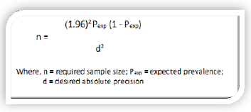
The sample size was calculated using previous prevalence of 11.7% by [17] and desired absolute precision of 5% as per the standard procedure described by Thrusfield [18] shown below. An estimated minimum sample size of 159 cattle was obtained; however, we were able to examine 164 cattle for our study.
Study Method and Procedure
Buffy coat technique
Blood was collected from an ear vein using heparinized microhematocrit capillary tube and the tube was sealed. A heparinized capillary tube containing blood was centrifuged for 5 minutes at 12,000rpm. After centrifugation, trypanosomes were usually found in or just above the buffy coat layer. The capillary tube was out using a diamond tipped pen 1mm below the buffy coat to include the upper most layers of the red blood cells and 3mm above to include the plasma. The content of the capillary tube was expressed on to slide, homogenized on to a clean glass slide and covered with cover slip. The slide was examined under x40 objectives and x10 eye piece for the movement of parasite [19].
Measuring of packed cell volume (PCP)
Blood samples were obtained by puncturing the marginal ear vein with a lancet and collected directly into a capillary tube. The capillary tubes were placed in micro-hematocrit centrifuge with sealed end outer most. The tube was loaded symmetrically to ensure good balance. After screwing the rotary cover and closing the centrifuge lid, the specimens were allowed to revolve at 12,00rpm for 5 minutes [4,20]. Tubes were then placed in hematocrit and the readings were expressed as a percentage of packed red cells to the total volume of whole blood. Animals with PCV ≤ 24% were considered to be anemic [21].
Data analysis
Row data on individual animals and parasitological examination results were inserted into MS Excel spread sheets to create a data-base. Students t-test were employed to compare between the two-independent mean PCV values of animals from an individual site (peasant’s association). Chi-square test was also employed to assess the association between the risk factors and the disease. While analyzing data, p-values (p)<0.05 were registered as statistically significant. Otherwise, recorded as insignificant.
Result
Prevalence


Out of the total 164 (75 females and 89 males) cattle examined, 25 (15.24%) were found positive to trypanosomosis. The prevalence varied between different study areas, in which 9.76% (n = 8) and 20.73% (n = 17) were recorded at Adankegne and Ergib peasant’s association, respectively. The variation in the prevalence of bovine trypanosomosis between the study sites were not statistically significant (X2= 5.783; p = 0.056) (Table 1 and Figure 2). The most prevalent trypanosome species in the study area was T. congolense (80%) followed by T. vivax(20%) (Table l and Figure 2). The prevalence of bovine trypanosomosis showed statistically significance difference between sexes of cattle, in which, higher in male animals (19.1%) as compared to females (10.67%) (X2= 13.886; p = 0.001) (Table 2 and Figure 3).


Hematological findings

Discussion
The study revealed that the prevalence of bovine trypanosomosis in the area was 15.24% (25/164) which was higher compared with the previous findings of Bitew et al. [17] in the same area (11.7%). The difference in prevalence might be due the site from which the blood samples were collected. However, there were tsetse control intervention, and continuous treatment of sick animals as well as deforestation for the cultivation of land. These activities could have led to the reduction of tsetse fly population along with the decline of tsetse borne trypanosomosis in the study area. But the continuous and longtime utilization of trypanocidal drugs particularly Diminazin aceturate in the study area contribute for the development of drug resistance, so that the prevalence of trypanosomosis was higher than the previous finding due to the above reasons.
In this study, two species of trypanosomes; namely, T. congolense and T. vivax were retrieved from inspected cattle. Majority of infections were also due to T. congolense. The higher proportion of T. congolense infection in the study area was in agreement with trypanosome species prevalence data from other tsetse infested region of Ethiopia where T. congolense is the most prevalent species in cattle [11]. In the same report it was also indicated that in tsetse free area of highlands, 99% of prevalence was due to T. vivax [12-14]. But in this study area, the prevalence of T. vivaxwas less than T. congolense in both peasant associations because the two sites are located adjacent to tsetse infested belts. Leak [22] and Degneh et al. [23] also indicated that T. vivax was highly susceptible to treatment while the problems of drug resistance were higher in T. congolenseM.
In the current study, higher infection rate of trypanosomosis was detected in males (19.1%) as compared to in female cattle (10.67%) with statistically significant difference (X2= 13.886; p = 0.001). Different researchers work supported this finding [22- 25]. Although the variation was not statistically significant, Yalew and Fantahun [26], and Teferi and Biniam [27] had also reported higher prevalence of bovine trypanosomes in males than in females (X2 = 0.85, p=0.35 and X2= 0.10, p>0.05, respectively). According to Gemtessa and Dera [28], the higher prevalence of trypanosomes in males rather than in females might be related to the hardworking of male animals. Similarly, the variation in the prevalence between the two sexes might also be associated with that male animals travel longer distances to tsetse abundant areas for draught and ploughing purposes, and the journey creates stress leading to susceptibility to the infection [23,)].In contrast to this study,Kitila et al. [30] at Yayo District Illuababora Zone of Western Oromia and Tamirat et al. [31] at Enemorena Ener Woreda of Gurage Zone were found higher prevalence of bovine trypanosomosis in female cattle than males.
Comparing the mean PCV values of cattle, significantly (p=0.01) low PCV was recorded in parasitaemic animals (25.07%) (SD = 0.989; df = 6; t-value = 8.069) than in aparasitaemic animals (20.75%) (SD = 1.601; df = 152; t-value = 40.316). This finding was in line with previous works conducted at different regions of Ethiopia by many authors [22,25]. In the absence of other diseases causing anemia, a low PCV value of individual animals is a good indicator of trypanosome infection [23,32]. Trypanosomosis might adversely lower the PCV values of infected animals [33]. A survey conducted in cattle in Hawagelan District of West Wellega Zone [34] revealed that the mean PCV of trypanosome infected animals was significantly lower (20.8±3.2 %) compared to non-infected animals (24.9±3.8 %). A later study in Northwest Ethiopia [35] in cattle experimentally infected with T. vivaxi solates also showed that the mean PCV, Hb and total RBC count were lower (p < 0.001) in all infected groups than in noninfected control animals. In Nigeria, domestic ruminants that were naturally infected with trypanosomes had significantly lower (p<0.05) PCV and RBC counts compared to uninfected animals [36]. Lower herd average PCVs for trypanosomepositive cattle compared to trypanosome-negative cattle have also been reported from Ghana [37], Zambia [32], Cameroon [38] and Gabon [39].
In spite of the fact that trypanosome infection has significant association with risk factors such as age and body condition scoring, as reported by many scholars, this study had not demonstrated and regarded as limitations.
Conclusion
From this study it is possible to conclude that trypanosomosis is an important disease and a potential threat affecting the health and productivity of cattle. The major species of trypanosomes in the study area were T. congolense and T. vivax. To sum up, infection with trypanosomosis negatively affects PCV and body condition of animals. This indicated that trypanosome infection of cattle causes loss of body weight and production. Trypanosomosis control measures should be targeted on tsetse fly destruction and control methods such as pour-on and effective trypanocidal drug applications. Similarly, rearing or raising of trypanosomosis resistance cattle breeds is now a day in practical. Otherwise, the problems will increase through the aide of global warming. In conclusion, further study on the occurrence of tsetse and trypanosomosis at different season of the year at different altitudes and species of animals should be conducted.
Acknowledgement
The Authors would like to thank Bahir Dar Regional Veterinary Diagnostic and Investigation Center for logistic and material support in the realization of this study. And we would also to adders our thanks to Dr. HabtamuTamrat for his constructive comments and editing the entire work.
Conflict of Interest
Authors declare that no competing of interests in the publication of this work.
References
- Elnasri H (2005) Prevalence and Ranking of Bovine trypanosomiasis in Unity State, Sudan, MSc Thesis, University of Khartoum, Faculty of Veterinary Medicine, Unity State, Sudan, pp.1-76.
- Bekele J, Asmare K, Abebe G, Ayelet G, Esayas G (2010) Evaluation of Deltamethrin applications in the control of tsetse and trypanosomosis in the Southern Rift Valley areas of Ethiopia. Vet Parasitol168: 177-184.
- Getechew A (2005) Review Article trypanasomiasis in Ethiopia. Ethiopia J Biol Sci27(1): 1-8.
- Uilenberg G (1998) A field guide for the diagnosis, treatment and prevention of African animal Trypanosomosis. FAO of the United Nations, Rome, Italy, pp.11-41.
- Slingenbergh J (1992) Tsetse control and Agricultural Development in Ethiopia. World Anim70-71: 30-36.
- Stein J (2011) Trypanotolerance and Phenotypic Characteristics of Four Ethiopian Cattle Breeds Doctoral Thesis, Swedish University of Agricultural Sciences, Faculty of Vet Med AnimSci, Uppsala, Sweden, pp. l-63.
- NTTICC (2004) National Tsetse and Trypanosomosis Investigation and Control Center report for the period 7th July 2001 - 6 July 2002 Bedelle Ethiopia, p. 3.
- Langridge WP (1976) Tsetse and Trypanosomosis. Survey of Ethiopia Ministry Overseas Department UK, England, pp. 1-40.
- IAEA (1996) Government of Federal Democratic Republic of Ethiopia. Draft Projects Document Integrating the Sterile Insect Technique to Eradicate tsetse from the Southern Rift Valley of Ethiopia.
- Rowlands GJ, Mulatu W, Authie, leak SGA, Peregrine AS (1993) Epidemiology of Bovine Trypanosomosis in the Ghibe Valley, Southwest Ethiopia. Acta Trop53: 135-150.
- Abebe G, Jobre Y (1996) Trypanosomosis: a threat to cattle production in Ethiopia. Revue de MédecineVétérinaire 147: 897-902.
- Afewerk Y (1998) Field Investigation on the Application of drug Resistant population of Trypanosomes in Metekel district, Faculty of veterinary Medicine, Addis Ababa university and Frei University Berlin,Northwest Ethiopia.
- Banchat B (2001) Integration of Tsetse Survey Data and Agroecological characteristics from Remotely sensed and field observations in a Geographic information system Southern Rift valley of Ethiopia. Faculty of Veterinary Medicine, MSc Thesis, Addis Ababa University, Ethiopia.
- Tewelde N (2001) Study on the Occurrence of Drug Resistant Trypanosome in cattle in the Farming in Tsetse Control Areas (FITCA) project in Western Ethiopia. Faculty of Veterinary Medicine, MSc Thesis, Addis Ababa University, Ethiopia.
- CSA, Federal democratic republic of Ethiopia (2009) Agricultural sample survey. Statistical Bull 446: 85-87.
- Adugna A (2018) Land forms, Climate and Economy. Retrieved June 28, 2018, from Ethiopian Demography and Health:
- Bitew M, Amedie Y, Abebe A, Tolosa T (2011) Prevalence of bovine trypanosomosis in selected areas of JabiTehenan district, West Gojam of Amhara regional state, Northwestern Ethiopia. Afr J Agric Res6(1): 140-144.
- Thrusfield M (2005) Veterinary Epidemiology, (3rdedn), Blackwell Science, Ltd., London, UK, England, pp. 228-246.
- Paris J, Murray M, Mcodim BF (1982) A comparative evaluation of the Parasitiological techniques currently available for the diagnosis of African Trypanosomiasis in cattle. Acta Trop 39: 307-316.
- Kebede B, Hailu S, Terefe G (2016) Prevalence and Pathogenic Significance of Trypanosomosis on Sheep and Goats of Mareka District, Dawro Zone, Southwestern Ethiopia. J Anim Sci Livest Prod1:1.
- OIE (2008) Standardized techniques for the diagnosis of tsetse transmitted trypanosomiasis. OIE Terrestrial Manual: 49.
- Leak SGA (1999) Tsetse Biology and Ecology: Their Role in the Epidemiology and control of Trypanosomosis. CABI publishing in association with the ILRI: 152-210.
- Degneh E, Ashenafi H, Terefe G, Kassa T, Kebede N, et al. (2018) A Crosssectional Study of Bovine Trypanosomosis in Sayo District, Oromia Regional State, Western Ethiopia. International Journal of Nutrition and Food Sciences7: 56-64.
- Mulugeta D, Menkir S, Kebede A (2013) Prevalence and seasonal incidence of bovine trypanosomosis in Birbir valley, Baro-Akobo river system, Western Ethiopia, J Vet Med Anim Health5(5): 138-143.
- Gona Z, Teshale A, Tilahun A (2016) Study on prevalence of bovine trypanosomosis and density of its vectors in three selected districts of Wolaita Zone, Southern Ethiopia. Journal of Veterinary Medicine and Animal Health8(9): 128-135.
- Yalew ST, Fantahun B (2017) Prevalence of Bovine Trypanosomosis and its Associated Risk Factors in Bambasi Woreda, Western Ethiopia. J Dairy Vet Anim Res5(2): 00132.
- Teferi B, Biniam T (2018) Study on Bovine Trypanosomosis and Tse Tse Fly Challenge in Darimu District of Birbir Valley, South Western Ethiopia. Vet Sci Res3(1): 000153.
- Gemtessa T, Dera KL (2017) Study on Prevalence of Bovine Trypanosomosis in Dale Wabera District, Kellam Wollega Zone, Western Ethiopia. Int J Anim Sci1(1): 1002.
- Alemu G, Alemneh T (2017) The Prevalence of Bovine Trypanosomiasis in Quara Woreda, North Western of Ethiopia. J Vet Med Res 4(1): 1067.
- Kitila G, Kebede B, Guta D, Bekele F, Wagari M, Tilahun B and Jaleta D (2016) Epidemiological Investigation of Bovine Trypanosomosis and its Vector Apparent Densities in Yayo District Illuababora Zone, Western Oromia, Ethiopia. Research & Reviews: Journal of Veterinary Sciences3(1): 4-9.
- Tamirat TG, Tsegaye L (2018) Prevalence of Bovine Trypanosomosis in Gurage Zone Enemorena Ener Woreda, Ethiopia. Austin J Nutri Food Sci6(2): 1104.
- Marcotty T, Simukoko H, Berkvens D, Vercruysse J, Praet N et al. (2008) Evaluating the use of packed cell volume as indicator of trypanosomal infections in cattle in eastern Zambia. Prevent Vet Med 87: 288-300.
- Girma K, Meseret T, Tilahun Z, Haimanot D, Firew L, et al. (2014) Prevalence of Bovine Trypanosomosis, its Vector Density and Distribution in and Around Arbaminch, Gamogofa Zone, Ethiopia. Acta Parasitologica Globalis5 (3): 169-176.
- Bekele M, Nasir M (2011) Prevalence and host related risk factors of bovine trypanosomosis in Hawagelan District, West Wellega Zone, Western Ethiopia. Afr J Agric Res6(22): 5055-5060.
- Dagnachew S, Bezie M, Terefe G (2015) Comparative clinicohaematological analysis in young Zebu cattle experimentally infected with Trypanosoma vivax isolates from tsetse infested and non-tsetse infested areas of Northwest Ethiopia. Acta Vet Scand57: 24.
- Ohaeri CC, Eluwa MC (2011) Abnormal biochemical and hematological indices in trypanosomiasis as a threat to herd production. Vet Parasitol177(3-4): 199-202.
- Ganyo EY, Boampong JN, Masiga DK (2018) Haematology of N’Dama and West African Short Horn cattle herds under natural challenge in Ghana [version 1; referees: 1 approved, 1 approved with reservations, 1 not approved] Trypanosoma vivax.:314
- Achukwi MD, Musongong GA (2009) Trypanosomosis in the Doayo/ Namchi (Bos taurus and zebu White Fulani (Bos indicus) cattle in Faro Division, North Cameroon. J Appl Biosci. 15: 807-814.
- Cossic BGA, Adjahoutonon B, Gloaguen P (2017) Trypanosomiasis challenge estimation using the diminazene aceturate (Berenil) index in Zebu in Gabon. Trop Anim Health Prod 49(3): 619-624


