Studies Oniridovirus Infection in Grouper Fish (EpinephelusSp.) In Seribu Islands, Indonesia
NurulSulfi Andini1*, Kurniasih2 and Tri Untari3
1Gadjah Mada University, Indonesia
2Pathology Department, Gadjah Mada University, Indonesia
3Microbiology Department, Gadjah Mada University, Indonesia
Submission: May 28, 2018;Published: July 05, 2018
*Corresponding author: NurulSulfiAndini,Post graduate Student, Veterinary Science, Faculty of Veterinary Medicine Gadjah Mada University, Indonesia, Email: nsandini242@gmail.com
How to cite this article: NurulSulfi A, Kurniasih, Tri Untari. Studies Oniridovirus Infection in Grouper Fish (EpinephelusSp.) In Seribu Islands, Indonesia. Int J cell Sci & mol biol. 2018; 4(5): 555647. DOI: 10.19080/IJCSMB.2018.04.555647.
Abstract
Grouper fish has a high interest aquaculture in Indonesia, Singapore, Taiwan and China. Iridovirus infection can cause mass mortality in groupers. The outbreak of Iridovirus disease was often found in several marine aquacultures in islands of Indonesia, including Seribu Islands.The aim of research is to diagnose Iridovirus infection in grouper fish in Seribu Islands based on Polymerase Chain Reaction and immunehistochemistry methods. The number of 18 grouper fish was collected from the location of outbreak. Spleen was treated with Polymerase Chain Reaction method. Spleen, kidney, liver, gonad and gill were processed for all results were analyzed descriptively. Results of this research show 3 samples from Tidung Island was infected by Iridovirus and 15 samples showed negative result with PCR test. Result of immunohistochemistry tests of three positive samples were the presence of antigen-antibody reaction in several organ such as spleen, liver, gill, gonade, and eye. The inclution body was brown color within the cells.
Abbreviation: Iridovirus; Grouper; Immunohistochemistry; PCR
Introduction
Grouper fish is one of the fish with high economic value, not only for import also for export. Indonesia is the main producer of groupers; the production of grouper fish cultivation in 1999 was 759 tons and in 2005 increased to 6,493 tons with total value around Rp. 116.891.489.000 [1]. Seribu islands are one of the areas producing Grouper fish farming in Indonesia. Viral infections that often cause mass mortality in grouper were viral nervous necrosis (VNN) and Iridovirus [2]. Iridovirus caused high mortality in more than 30 species of marine fish. One of the most common diagnostic techniques for gold standard testing of viral disease was using Polymerase Chain Reaction [3].The purpose of research is to diagnose Iridovirus infection in Grouper fish (Epinephelus sp.) in KepulauanSeribubased on PCR and immunohistochemistry methods.
Materials and Methods
Number of 18 grouperfish measuring 10-15cm with clinical signs were collected from two location Seribu islands. Fish were autopsied, and then internal organs such as gills, spleen, liver, gonade and kidney were fixed with phosphate buffered formalin 10% for immunohistochemistry and with absolute ethanol of solution for PCR analysis.
MolecularTest
Tissue samples wereextracted using Gene aid (gSYNC DNA Extraction KIT). The extraction product was then amplified. Primers used for PCR amplification were 1-F primers (5 ‘CTCAAACACTCTGGCTCATC 3’) and 1-R (5 ‘GCACCAACACATCTCCTATC 3’) (Kurita et al., 1998).Thermal cycler was prepared with the following programs: pre denaturation (temperature of 94°C for 60 seconds); denaturation (temperature of 94°C for 30 seconds), annealing (temperature of 57°C for 60 seconds); extension (temperature of 72°C for 60 seconds); Final heat (Temperature 72°C for 5 minutes). Reactions carried out as many as 30 cycles, final extension 72°C for 5 minutes [3]. Theamplification product was the electrophoresize using 2% of agarose gel.
MonoclonalAntibody
Antibody was made from Rabbit serum. Three Rabbits were maintained and injected intraperitoneally with Iridoviruses vaccine weekly with a dosage of 0,5mL, 1mL, 2mL, and 3mL for 4 weeks in a row. Blood was taken 10mL for each rabbit at the fourth week. Serum was precipitated with ammonium sulfate until 3 times and filtered withdialysis membrane inPBS pH 7,2 for 12 hours and was repeated until 3 times. The final product obtained was immunoglobulin G which was used for antibody Iridovirus[4].
Immunohistochemistry
Tissue samples were processed in several stages such as fixation, dehydration, clearing, embedding, paraffin blocking, sectioning, coloring and mounting[5]. Staining for immunohistochemistry use DAB chromogen was added NovolinkTM 37 Polymer and incubate for 5 minutes. Immunohistochemistry procedure was using Novocastra Polymer Detection Systems [6].
Result
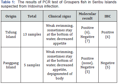
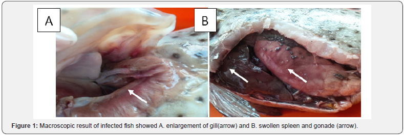
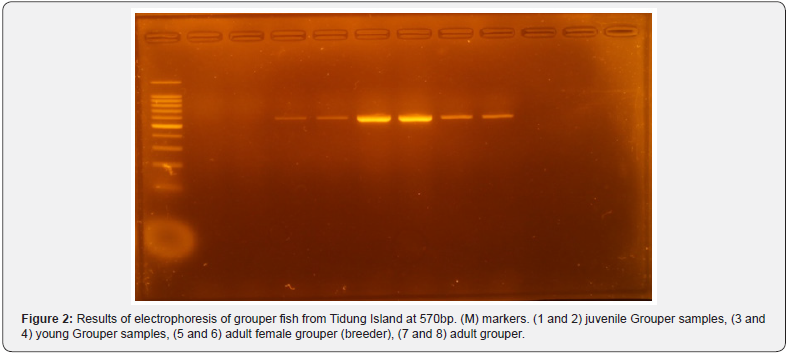
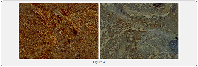
The female adult of grouper fish with positive infection of Iridovirus from Tidung island showed the enlargement of several organs such as gills, spleen, liver, and gonade(Figure 1A &1B).The result of molecular test using PCR method with a total of 18 samples consisted of 13 samples from Tidung Island and 5 samples from Panggang Island (Table 1).Positive result PCR was only 6 from adult grouper fish originated from TidungIsland from total samples of 18 fish. Electrophoresis result of positive Iridovirus had a bright band at 570bp(Figure 2). Result of PCR of samples from PanggangIsland (5 samples) were all negative. Result of immunohistochemistry showed the presence of inclusion body, brown color in parenchyma cell of spleen, eye, gonade, and kidney (Figure 3).


Iridovirus infection in grouper fish usually had clinical symptoms such as dwelling in the bottom of water, weak swimming, and decreasedappetite; there is a blackened part of the fish body [7]. However, fish samples with similar clinical symptoms showed negative result of iridovirus infection. Iridoviruses are relatively stable in aquatic environments, but it is unclear whether subsequent die-offs reflect the reintroduction of the virus, the activation of latent or subclinical infections in the surviving host population [8]. The negative result also probably caused by improper environmental condition like inappropriate season. Mortality was observed in experimental infections of ISKNV between 25 and 34°C [9]. Outbreaks of Iridovirus have been seen mostly in the summer season at water temperatures of 25°C and above, andsource of grouper larvae that will be distributed came from infected area of iridovirus[3].In addition, it is likely due to the spreading time of grouper fish into the floating net cage and the replication process of the virus to the newly entered fish. According to the cultivators in the Seribu Islands the Grouper fish is just distributed in that month so the size is still within the range of 9-10cm [10]. The other reason that probably cause the negative result is the relation between spreading time of grouper fish into the floating net cage and the replication process of the virus to the newly entered fish. In trial test for Red Sea Bream Iridoviral Disease for non vaccinated group, viral antigenwas detected in all the 10 fish examined at 6 week [11]. while according to the cultivation the fish in Panggang Island just spread in 2 weeks.
Conclusion
Iridovirus infection in grouper fish was only found in Tidung island, Seribu islands, based on the molecular and immunohistochemistry. Iridovirus was firstly found in the gonade, so that, the disease could be transmitted vertically by infected spermatozoa.
Acknowledgement
The research was supported by project of PT UPT-UGM, Department of culture and Education, Indonesia in2018.
References
- Hudaya, Masri (2015) Analysis of Economic Economy of Cultivation Farms In Palau Tidung Kepulauan Seribu DKI Jakarta. An Sosial Universitas Indraprasta PGRI.
- Koesharyani I, Mahardika K, Sugama, Yuasa K (2001) Detection using polymerase chain reaction (PCR) Iridovirus causing death of municipalities and municipalities, Epinepheluscoioides: A collection of makalahteknologibudidayalaut and a sea farming in Indonesia. Dep. KelautandanPerikanan RI works with JICA: 28-234.
- OIE (2009) Chapter 10.8 Red Sea bream iridoviral disease in Aquatic animal health code.
- Amanu S, Sulistiyono D, Suardana N (2016) Detection of Fish Disease Caused by Iridovirus on Grouper (Epinephellus sp.) And Pomfret Star (Trachinotus blochii) with Co-agglutination Method in Tanjungpinang, Indonesia. J Agricultural Science and Technology 21: 121-128.
- Slaoui M, Fiette L (2011) Histopathology procedures: From Tissue Sampling to Histopathological Evaluation. Methods Mol Bio 691: 69- 82.
- Anonymous DAB Chromogen Manual 2015 Dako.
- Mahardika K, Zafran D, Roz A, Johnny F, Prijono A (2002) Introduction of the use of iridovirus vaccine (inactivated vaccine) in juvenile kerapulumpur, Epinepheluscoioides. DIP Research Report 2002 Branch Gives Riset Perikan Budidaya Laut Gondol: 195-202.
- William, Barbosa-Solomicu Valerie, Gregory Chincar V (2015) A Decade of Advances in Iridovirus Research. Adv Virus Res 65: 173-248.
- He JG, Zeng K, Weng SP, Chan SM (2002) Experimental transmission, pathogenicity and physical properties of infectious spleen and kidney necrosis virus (ISKNV). Aquaculture 204: 11-24.
- Anonymous (2012) Aquatic Animal Diseases Significant to Australia Identification Field Guide (4th edn),.
- Nakajima Kazuhiro, Maero Yukio, Honda Atsui, Yokohama Kenichi, Toosiyama Tetsuro, et al. (1999) Effectiveness of Vaccine Again Red Sea Bream Iridovirus in a Field Trial Test. Disease of Aquatic Organisms 36(1): 73-75.


