Delleman Syndrome: A Case Report with Emphasis on Neuroimaging Features
Naser H1, A1 Taei T1, A1 Jufairi M2, Almarzooq R2 and Abdulla Ali F2
1Department of Radiology, Salmaniya Medical complex, Bahrain
2Department of Pediatrics, Salmaniya Medical Complex, Bahrain
Submission: August 03, 2017; Published: September 21, 2017
*Corresponding author: Naser H, Department of radiology, Radiology department, Salmaniya Medical complex, Work address, P.O.Box 12, Bahrain, Email: drhnasser@gmail.com
How to cite this article: Naser H, Taei T, Jufairi M, Almarzooq R, Abdulla A F. Role of Prokayotic P-Type ATPases. Int J cell Sci & mol biol. 2017; 3(1) : 555604. DOI: 10.19080/IJCSMB.2017.03.555604.
Abstract
Delleman syndrome, also called Oculocerebrocutaneous syndrome (OCCS) is a rare congenital sporadic disorder that appears at birth. The syndrome is characterized primarily by a triad of eye, brain and skin malformations given its name "Oculocerebrocutaneous” syndrome. The diagnosis is confirmed by brain MRI with a malformation involving mainly the forebrain and mid-hindbrain. To date, about 40 patients have been reported by OCCS. We present a case with the typical Neuroimaging feature of a patient with OCCS
Case Report
A 4 years old Bahraini boy was a product of LSCS at 37 weeks, due to previous C-section, birth weight was 2.2kg. He was admitted to NICU for multiple congenital anomalies. These include microcephaly, cutis aplasia; multiple skin tags over the left side of the jaw, left eyelid, over the abdomen and left toe. He had left microphthalmia with cataract, syndactyly of the left hand and left undescended testis. There was no parental consanguinity. He has two healthy siblings. Family history is not significant except for Glucose -6-Phosphatase Deficiency. The patient had several admissions (over eight times) since birth, mainly for asthma exacerbation. Development delayed was noted during his visits to the hospital. He was only able to say few words and his pronunciation was not clear. He was still unable to walk unless assisted. He does not have seizures [1-3].

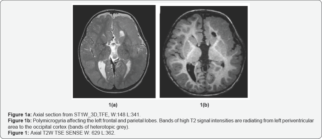
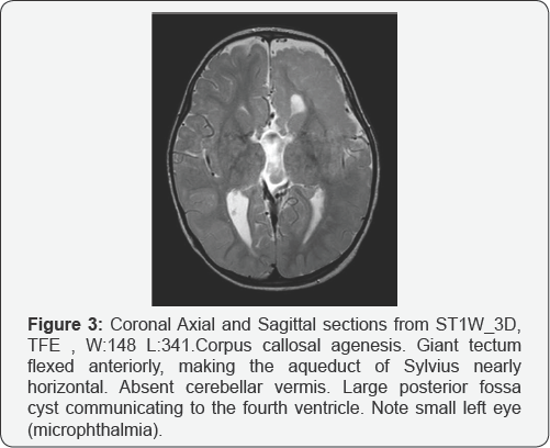
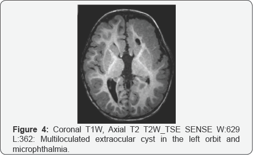
The patient had two MRI studies, at age of one month and age of 2 years. Both studies showed the same typical features of Delleman syndrome. He had polymicrogyria in the left frontal and parietal lobes (Figure 1a & 1b), Migrational anomalies in the form of heterotopic grey matter in the ependymal lining of left lateral ventricles, nodular heterotopias in the frontal and occipital lobes, more posteriorly (Figure 2a, 2b & 2c), and bands of heterotopic grey matter which were seen radiating from the periventricular area to the occipital cortex (Figure 3). The left lateral ventricle was slightly prominent. The above findings were confined to the left cerebral hemisphere. The right cerebral hemisphere was spared. Corpus callosum agenesis was also noted, without midline cyst or lipoma. The tectum was giant, slightly flexed anteriorly, making the aqueduct nearly horizontal. The vermis was absent and there was large posterior fossa cyst, communicating to the fourth ventricle (Figure 3). The left eye was small (microphthalmia) and there was a multiloculated cyst in the roof of left orbit anteriorly (Figure 4). The above findings are typical in a patient with the oculocerebrocutaneous syndrome [4-6].


Discussion
Delleman syndrome is a rare idiopathic congenital condition. Diagnosed is made by the characteristic skin, eye and brain malformation with the latter being pathognomonic for the OCCS [6]. All cases were sporadic, the striking male preponderance and the unusual mid-hind brain malformation suggest that OCCS is may be x-linked genetic disorder [6] while some suggested it is due to an autosomal dominant lethal somatic mutation that survives by mosaicism [4]. The eye malformations consist mainly of cystic anophthalmia or microphthalmia and colobomata, while the skin abnormalities consist of skin appendages and focal aplasia or hypoplasia seen as pink or pigmented areas where subcutaneous fat prolapses through atrophic dermis [7]. Sometimes OCCS associated with other features such as craniofacial clefts, skull or rib defects, and urogenital anomalies such as our patient [6,7]. Moog U. reviewed the brain malformation in 11 patients and found a consistent pattern in eight of his patients. These malformations include forebrain and Mid-Hind brain malformation.
The forebrain malformation includes frontal predominant polymicrogyria; periventricular nodular heterotopia; complete or partial agenesis of the corpus callosum, sometimes associated with interhemispheric cysts; and enlarged third and lateral ventricles, which can be complicated by hydrocephalus. The mid-hindbrain malformation consisted of a giant tectum, absent vermis, and large posterior fossa fluid collection. The midbrain tegmentum sometimes flexed forward, making the aqueduct nearly horizontal. The giant and dysplastic tectum was rotated upward well above the normal position and appeared to form an arch over the enlarged lower aqueduct in 7/8 patients. The cerebellar hemispheres were missing or hypoplastic with a dysplastic foliar pattern in 7/8 patients. The fourth ventricle communicated widely with a large posterior fossa fluid collection, sometimes associated with an occipital meningocele.
Differential diagnosis of Delleman syndrome includes Goldenhar and Gotlz syndromes as some of the congenital features can be seen in these syndromes. What makes OCCS different is the presence of brain malformation; also, the skin tags in Goldenhar syndrome are preauricular as opposed to periocular tags in OCCS [1,5]. Management of the Delleman syndrome is symptomatic. Surgical Intervention may include drainage of orbital cysts; excision of orbital cysts, hamartomas, or skin tags; or surgical repair of certain abnormalities, such as colobomas of the lids or the cleft palate or repair of undescended testis. Antiepileptic medications may be given as prophylactic or to stop the seizures [8].
Conclusion
Delleman syndrome is a rare congenital disorder of unknown causes. It is diagnosed by skin, eye and brain malformation. The characteristic brain malformation, as in our patient, is important in the diagnosis of this disease and in excluding the differential diagnosis.
References
- Billings JK, Milgraum SS, Rasmussen JE (1986) Multiple mesoectodermal defects in an infant. Focal dermal hypoplasia syndrome, or Goltz syndrome. Archives of Dermatology 122(10): 1200-1200.
- Bleeker WLM, Hamel BC, Hennekam RC, Beemer FA, Oorthuys HW (1990) Oculocerebrocutaneous syndrome. J Med Genet 27(1): 69-70.
- Cock RD, Merizian A (1992) Delleman syndrome: a case report and review. Br J Ophthalmol 76(2): 115-116.
- Happle R (1987) Lethal genes surviving by mosaicism: A possible explanation for sporadic birth defects involving the skin. J Am Acad Dermatol 16(4): 899-906.
- Mccandless SE, Robin NH (1998) Severe oculocerebrocutaneous (Delleman) syndrome: Overlap with Goldenhar anomaly. Am J Med Genet 78(3): 282-285.
- Moog U (2005) Oculocerebrocutaneous syndrome: the brain malformation defines a core phenotype. Journal J Med Genet 42(12): 913-921.
- Moog U, Smulders DC, Systermans JM, Cobben JM (2008) Oculocerebrocutaneous syndrome: report of three additional cases and aetiological considerations. Clinical Genetics 52(4): 219-225.
- Rizvi SW, Siddiqui M, Khan A, Siddiqui Z (2015) Delleman Oorthuys syndrome. Middle East Afr J Ophthalmol 22(1): 122-124.


