




 GJPPS Home



Abstract
Oral Squamous Cell Carcinoma (OSCC) remains a significant global health burden, with survival rates that have not substantially improved despite advances in treatment. Current therapeutic strategies rely heavily on established chemotherapy regimens; however, resistance to single agents remains a major limitation. Collateral sensitivity, in which adaptation to one therapeutic pressure induces increased vulnerability to a second, mechanistically distinct agent, represents a potential framework for identifying more effective combination therapies. The objective of this study was to evaluate the potential for collateral sensitivity–driven drug combinations to enhance cytotoxic efficacy in OSCC. Two OSCC cell lines were treated with a panel of clinically relevant chemotherapy agents and mechanistically distinct compounds to assess drug sensitivity and interaction effects. Cytotoxicity assays were used to determine single-agent activity, and selected drug combinations were subsequently evaluated to identify synergistic interactions. Select combinations demonstrated enhanced reduction in cell viability relative to single-agent treatments, with evidence of synergy across mechanistically diverse drug classes. These findings support the utility of combining agents that target complementary cellular processes, consistent with a collateral sensitivity framework. Overall, this study highlights the potential of collateral sensitivity–based approaches to guide rational design of combination therapies in OSCC. Further investigation is warranted to determine optimal combination strategies, treatment sequencing, and translational applicability in more complex models.
Keywords
Squamous Cell Carcinoma, Oral; Drug Synergism; Drug Resistance, Neoplasm; Antineoplastic Combined Chemotherapy Protocols; Antimetabolites, Antineoplastic; Methotrexate; Molecular Targeted Therapy; Gene Expression Regulation, Neoplastic; Cell Proliferation/drug effects; Collateral Sensitivity
Abbreviations
Oral Squamous Cell Carcinoma (OSCC); Oxaliplatin (OxPt); 5-Fluorouracil (5-FU), Cisplatin (Cis); Methotrexate (MTX); Capecitabine (CAP); Gemcitabine (GEM); Vincristine (VIN); Docetaxel (DTx); Hydroxyuracil (HDU); Mirdametinib (PD0); Molibresib (IBET-762); Venetoclax (ABT-199)
Introduction
Oral cancer is a subtype of head and neck cancer and is defined as a malignant tumor arising in the oral cavity, including the lips, tongue, gingiva, floor of the mouth, and salivary glands [1]. Worldwide, oral cancer is the sixth most common cancer and the third most prevalent in developing countries, with Oral Squamous Cell Carcinoma (OSCC) accounting for approximately 90% of all cases [2]. Major risk factors include tobacco and alcohol use; however, human papillomavirus infection, nutritional deficiencies, and genetic predispositions also contribute to disease development [2,3]. OSCC arises through a multifactorial process involving the accumulation of oncogenic alterations in epithelial cells. Changes in TP53, Epidermal Growth Factor Receptor (EGFR), and cyclin-dependent kinase inhibitor 2A disrupt DNA repair, cell differentiation, and cell cycle progression, ultimately promoting uncontrolled proliferation. As these alterations accumulate, transformed cells become increasingly independent of the surrounding oral epithelium, and EGFR overexpression further contributes to aberrant growth signaling and tumor progression [2,3].
Despite advances in understanding the molecular basis of oral cancer, disease prevalence and survival have remained relatively unchanged. Current treatment strategies for OSCC rely on surgery, chemotherapy, and radiotherapy [4], yet resistance to chemotherapy continues to limit durable responses [5]. Cisplatin-based therapy, particularly in combination with 5-fluorouracil (5-FU), remains a standard regimen, and additional agents such as gemcitabine and capecitabine have also been evaluated in combination settings [5-7]. However, although some regimens have improved efficacy and safety, overall survival has not substantially improved, underscoring the need for new therapeutic strategies. Preclinical studies in OSCC models offer an opportunity to identify more effective drug combinations, but the literature remains limited, with relatively few studies beyond early work demonstrating enhanced cytotoxicity for combinations such as taxol with cisplatin, carboplatin, or 5-FU [8].
Within this context, collateral sensitivity provides a useful conceptual framework for combination therapy. Collateral sensitivity refers to the phenomenon in which resistance or adaptation to one therapeutic pressure is accompanied by increased vulnerability to a second, mechanistically distinct agent, thereby creating an exploitable therapeutic weakness [9,10]. In cancer, this framework has been described as a form of acquired hypersensitivity that may emerge as resistant cells adapt to drug-induced stress, suggesting that resistance-associated liabilities can be therapeutically targeted rather than merely bypassed [10]. This concept is particularly relevant to OSCC, where resistance to standard cytotoxic agents remains a major barrier to treatment success. The most effective anticancer regimens often combine drugs that target distinct cellular dependencies, allowing lower concentrations of each agent to be used while achieving greater overall efficacy [11,12]. Such combinations may not only produce synergy but may also exploit collateral sensitivity by targeting vulnerabilities that emerge when cancer cells adapt to metabolic, replicative, or transcriptional stress. Accordingly, evaluating combinations of oral cancer drugs with mechanistically distinct nontraditional agents may identify therapeutic pairs that are both synergistic and capable of overcoming resistance-associated phenotypes [10].
In this study, we investigated drug synergy in OSCC using clinically relevant and repurposed agents selected to target complementary cellular processes. This framework provides a strong rationale for combinations such as Methotrexate (MTX) and JQ1. MTX disrupts folate-dependent purine and pyrimidine synthesis, thereby impairing nucleotide availability and DNA synthesis [13], whereas JQ1 inhibits BRD4-dependent oncogenic transcriptional programs that support proliferation and survival [14]. Simultaneous disruption of nucleotide biosynthesis and transcriptional regulation may impose dual stress on OSCC cells and may be especially effective in cells that have adapted to single-agent treatment. More broadly, repurposing approved or mechanistically characterized agents offers practical advantages, including lower development costs, greater accessibility, and a clearer understanding of pharmacologic behavior [2,9,10]. The findings of this study therefore aim not only to identify synergistic combinations in OSCC, but also to support a collateral sensitivity–based strategy for developing more effective therapeutic approaches.
Materials and Methods
Cell Culture
Cal-27 and OECM-1 cells were purchased from Millipore Sigma and ATCC, respectively. Cal-27 cells were cultured in Dulbecco's Modified Eagle Medium: High Glucose (DMEM) (Fisher Scientific, DMEM, 1:1, 1x) supplemented with 10% fetal bovine serum (Gemini Biological), 1% penicillin-streptomycin (Caisson Labs, 100x 1:1), and 1% L-alanine L-glutamine (Caisson Labs, 43.4 mg/mL, 0.85% saline). OECM-1 was cultured in Roswell Park Memorial Institute 1640 medium (RPMI) (Thermo Fisher, RPMI, 1:1, 1x) supplemented with 10% fetal bovine serum (Gemini Biological), 1% penicillin-streptomycin (Caisson Labs, 100x 1:1), and 1% L-alanine L-glutamine (Caisson Labs, 43.4 mg/mL, 0.85% saline).
Cells were cultured in a humidified incubator at 37°C with 5% carbon dioxide. Cells were split when they appeared to be between 70% and 100% confluent. Untreated cells were harvested to split using 0.25% trypsin solution (Fisher Scientific, 1x). When storage was necessary, cells were frozen in Bambanker (GC Lymphotec) and stored in a -80°C freezer.
Chemotherapy Drug Preparation
Fourteen drugs used in this study included: Oxaliplatin (OxPt), 5-Fluorouracil (5-FU), Cisplatin (Cis), Methotrexate (MTX), Capecitabine (CAP), TH278, Gemcitabine (GEM), Vincristine (VIN), Docetaxel (DTx), Hydroxyuracil (HDU), Mirdametinib (PD0), Molibresib (IBET-762), Venetoclax (ABT-199), and JQ1.
Each of the drugs were obtained in a powdered form and were dissolved in DMSO except for Oxaliplatin and Cisplatin, which were dissolved in water. The desired final concentration was identified for each drug based on availability and solubility, then the appropriate volume of DMSO or water for that concentration was added until all powder was dissolved. MTX stock was prepared at 20 mM, VIN stock was prepared at 12.5 mM, HDU stock was prepared at 180 mM, DTX stock was prepared at 0.5 mM, IBET and PD0 stocks were both prepared at 175 mM, and OXPT, 5-FU, CIS, CAP, TH278, GEM, ABT199, and JQ1 stocks were prepared at 10 mM. When not in use, prepared drugs were stored in a -40°C freezer.
Evaluating Cytotoxicity
Initial Drug Sensitivity
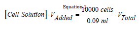
Confluent 25cm3 or 75cm3 flasks were used to conduct drug cytotoxicity assays. Cells were harvested using TrypLE express (Gybco) and transferred to a 15 mL centrifuge tube (CellTreat). The walls of the flask were rinsed well with media prior to transfer. Cells were centrifuged in a Sorvall ST40R Centrifuge at 1200x gravity for 5 minutes. Media was aspirated off and a single cell suspension in 1 mL of media was achieved. 10 µL of cell solution was then removed, placed in an Eppendorf tube, and added 10 µL of Trypan Blue solution (Sigma). The resulting solution was then counted in a Cell Drop Brightfield Cell Counter (DeNovix) to obtain the concentration of cells in the solution. Cells were seeded into white walled, clear bottom 96-well assay plates (Nest Scientific). Each well requires 10,000 cells in 90µL of media. To achieve the desired number of cells per well, a dilution of the cell solution was performed using Equation 1. The concentration of each solution, [Cell Solution], was obtained from the particle counter (cells/mL). VAdded was the volume of cell solution added to achieve a total volume, VTotal. The final volume was 10 mL to provide enough solution to fill the 96-well plate.
To treat cells, drug plates were made in 96-well clear V-bottom plates (Corning). A 1:10 serial dilution occurred down each column in the plate, giving a range of treatment dosages depending on the drug stock concentration. The initial concentration for MTX was 20 mM, VIN was 12.5 mM, HDU was 180 mM, DTX was 0.5 mM, IBET and PD0 were both 175 mM, and OXPT, 5-FU, CIS, CAP, TH278, GEM, ABT199, and JQ1 stocks were all 10 mM. A media plate was then generated by moving 5µL of solution from the drug plate to a corresponding well filled with 245µL of media. This resulted in a 1:50 dilution. The concentration of the selected drug in the media plate varied depending on the drug stock concentration. The resulting highest concentration in the media plate for MTX was 400µM, VIN was 250µM, HDU was 3.6µM, DTX was 10µM, IBET and PD0 were both 3.5 mM, and OXPT, 5-FU, CIS, CAP, TH278, GEM, ABT199, and JQ1 stocks were all 200µM. In addition to treatment dosages, each media plate also contained a column of solvent (DMSO or water) control wells and a column of non-treatment wells to act as negative controls.
After a 24-hour waiting period which allowed cells to adhere to the 96-well plate, cells were treated with 10µL of the selected drug solution from the media plate. This resulted in a second 1:10 dilution. The final dose given to cells at the highest concentration for MTX was 40µM, VIN was 25µM, HDU was 260µM, DTX was 1.0µM, IBET and PD0 were both 350µM, and OXPT, 5-FU, CIS, CAP, TH278, GEM, ABT199, and JQ1 stocks were all 20µM.
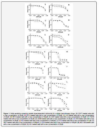
Cells were treated for 72 hours in a humidified 37°C incubator at 5% carbon dioxide. Next, a 15µL aliquot of Cell Titer Glo (CTG) reagent (Promega) was added to each well of the treated 96-well plate and stored in a dark cabinet for 10 minutes. The plate was then run on a EnSpire multimode plate reader (Perkin Elmer) to measure cell viability. All CTG assays were run in triplicate. A ratio of the luminescence between the solvent control well and each treated we then plotted against the concentration of drug in each well to generate GI50 plots [15]. GI50 values were calculated using the Absolute IC50 Analysis in GraphPad (Prism). All samples were run in triplicate, six separate times, and GI50 values averaged after few outliers were removed using a Grubbs analysis.
Drug Combination Cytoxicity
Drug concentrations to use in combination were chosen using the GI25, the concentration of drug needed to inhibit growth of 25% of the cells. GI25 values were interpolated from each initial drug cytotoxicity plot using a line of best fit drawn between the two points between which relative cell viability crosses 0.75.
Drug Synergy
To evaluate synergy, cells were pretreated for 24 hours with 5µL an oral cancer drug, the cells were incubated, then 5µL of the non-oral cancer drug was added at a 1:10 serial dilution for the remainder of the 72 hours to test for synergy at many concentrations. CTG was again used to examine cell viability after treatment.
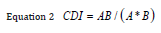
Drug synergy was calculated using the Coefficients of Drug Interaction (CDI) formula displayed in Equation 2, where AB is the cell average relative viability after combination treatment, A is the average relative viability after treatment with drug 1 alone, and B is the average relative viability after treatment with drug 2 alone [16,17].
Results and Discussion
Initial Drug Sensitivity
Two oral cancer cell lines, Cal24 and OECM-1, were selected to determine whether observed effects are cell line–specific or broadly applicable across oral cancers arising from different anatomical sites. Cal-27 cells are epithelial cells isolated from tissue taken prior to treatment from a 56-year-old, White male with a lesion in the middle of the tongue in 1982 [18]. The OECM-1 human oral cavity squamous cell carcinoma cell line was derived from surgical resection of a primary tumor of a Taiwanese male patient with a tumor in the gingiva [19]. A total of 14 drugs with distinct mechanisms of action were evaluated. Several agents target DNA synthesis and cell cycle progression. For instance, the antimetabolites 5-fluorouracil (5-FU) and gemcitabine (GEM) both interfere with nucleotide metabolism but through different mechanisms: 5-FU inhibits thymidylate synthase, reducing thymine and uracil production [20], whereas GEM functions as a cytidine analog [21]. Similarly, hydroxyurea (HDU) inhibits ribonucleoside reductase, thereby limiting the pool of deoxyribonucleotides [22], while methotrexate (MTX), a folate antagonist, disrupts both purine and pyrimidine synthesis [13]. In contrast, platinum-based agents such as cisplatin (CIS) and its derivative oxaliplatin (OXPT) are both platinum-based drugs but with distinct structures that directly damage DNA, interfering with replication and transcription [23,24]. Two other closely related drugs used were 5-FU and its prodrug Capecitabine, which was developed to improve tolerability and enhance intratumoral drug concentrations through tumor-specific conversion to the active compound [25]. Other drugs target mitotic processes through distinct effects on microtubules. Vincristine (VIN), a vinca alkaloid, inhibits microtubule polymerization [26], whereas docetaxel (DTX) stabilizes microtubules and disrupts their normal dynamics [27]. Additional agents modulate transcriptional regulation and apoptosis. IBET-762, a BET inhibitor, suppresses transcription of oncogenes such as MYC, BCL-2, and BCL-6 [28], while ABT-199 promotes apoptosis through selective inhibition of BCL-2 [29]. Finally, signaling pathway inhibitors such as PD0325901 (PD0), a non-ATP-competitive MEK inhibitor [30], and JQ1, a BRD4 inhibitor, reduce cell proliferation through distinct mechanisms involving suppression of oncogenic transcriptional programs [14]. Collectively, these drugs represent clinically relevant and mechanistically diverse agents, enabling evaluation of whether therapeutic responses are driven by shared pathways or drug-specific mechanisms of action.
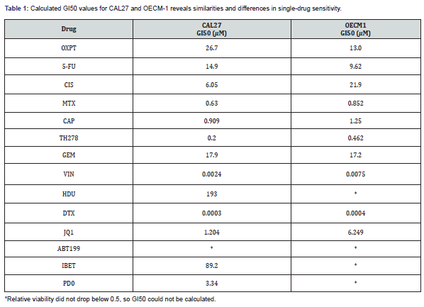
Both cell lines were treated with serial dilutions of each drug individually, and cell viability was assessed after 72 hours to evaluate relative drug sensitivity. Viability data were analyzed to determine the concentration required to reduce cell viability by 50% (GI50). Assays for each drug were performed in multiple independent experiments, and GI50 values were calculated by fitting semi-log plots of drug concentration versus percent viability (Figure 1) (Table 1). The two cell lines exhibited similar sensitivity to several agents, including 5-FU, methotrexate (MTX), capecitabine (CAP), and TH287; however, differential responses were observed for others, including oxaliplatin (OXPT), cisplatin (CIS), hydroxyurea (HDU), PD0325901 (PD0), and JQ1. Notably, Cal-27 cells were more sensitive to OXPT, whereas OECM-1 cells were more sensitive to CIS. Although both are platinum-based DNA-damaging agents, OXPT is a derivative of CIS with distinct pharmacologic properties. These differences in drug sensitivity may reflect variation in tumor origin within the oral cavity, as Cal-27 cells are derived from tongue epithelium and OECM-1 cells from gingival tissue. This observation suggests that oral squamous cell carcinomas may exhibit location-specific therapeutic responses. While surgical management of OSCC is clinically stratified by tumor location, chemotherapy selection is not routinely tailored in this manner [5]. These findings therefore support further investigation into site-specific therapeutic strategies and combination regimens for OSCC.
Evaluating Synergy
Four oral cancer drugs (DTX, VIN, CIS, and MTX) and three non–oral cancer agents (TH278, CAB, and JQ1) were selected for combination studies based on their initial cytotoxicity in Cal-27 and OECM-1 cells and relatively low GI50 values. Relative viability and combination drug index (CDI) values were calculated for each drug pair. A CDI > 1.0 indicates antagonism, CDI = 1.0 indicates an additive effect, and CDI < 1.0 indicates synergy, with CDI < 0.7 considered strongly synergistic [16] (Table 2). The observed synergistic interactions may be interpreted within the framework of collateral sensitivity, in which adaptation to one therapeutic pressure results in increased vulnerability to a second, mechanistically distinct agent. In this study, combinations involving antimetabolites or DNA-targeting agents with transcriptional or signaling inhibitors appear to exploit complementary cellular dependencies that may arise during stress adaptation. Notably, the MTX+JQ1 combination demonstrated the most pronounced synergy (CDI = 0.14), suggesting that inhibition of folate-dependent nucleotide biosynthesis in combination with suppression of BRD4-mediated transcription may impose dual, non-redundant stressors on OSCC cells. Under such conditions, cells attempting to compensate for nucleotide depletion may become increasingly dependent on transcriptional programs that are simultaneously disrupted by JQ1, thereby enhancing cytotoxic effects.
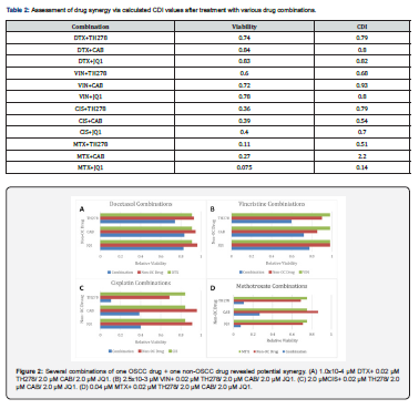
Similarly, combinations such as CIS+TH278 and VIN+TH278, which demonstrated moderate synergy, may reflect collateral sensitivity arising from stress imposed on DNA integrity or mitotic processes in the presence of additional mechanistic perturbation. These findings support the hypothesis that treatment-induced cellular adaptation does not solely confer resistance, but may also generate exploitable liabilities when paired with agents targeting distinct pathways. Importantly, the absence of synergy in certain combinations (e.g., MTX+CAB, CDI > 1.0) highlights that not all dual perturbations produce complementary stress. Instead, effective collateral sensitivity–based combinations likely require precise alignment of mechanistic vulnerabilities, reinforcing the need for systematic evaluation of drug pairs across multiple cellular contexts. Collectively, these results suggest that collateral sensitivity provides a useful conceptual framework for interpreting drug synergy in OSCC and may guide the rational design of combination therapies that not only enhance efficacy but also overcome or exploit resistance-associated phenotypes (Figure 2).
Conclusion and Future Directions
This study identifies several promising combinational strategies for the treatment of OSCC and provides mechanistic insight into how drug synergy may be leveraged to overcome therapeutic resistance. Of the fourteen agents initially evaluated, seven were advanced to combination studies based on favorable cytotoxic profiles, enabling focused assessment of synergistic interactions among mechanistically distinct drugs. Consistent with the collateral sensitivity framework described above, the most pronounced effect was observed for the methotrexate (MTX) and JQ1 combination, which produced substantial reductions in cell viability (0.075) and a strongly synergistic interaction (CDI = 0.14). These findings support the hypothesis that simultaneous disruption of nucleotide biosynthesis and BRD4-dependent transcriptional regulation imposes dual, non-redundant stressors on OSCC cells. In this context, adaptive responses to metabolic stress may increase reliance on transcriptional programs that are selectively vulnerable to BET inhibition, thereby exemplifying a collateral sensitivity–driven therapeutic vulnerability. More broadly, the identification of synergistic combinations involving DNA-targeting agents, antimetabolites, and transcriptional or signaling inhibitors suggests that collateral sensitivity may serve as a useful organizing principle for rational combination design in OSCC. Rather than viewing resistance solely as a barrier to treatment, these results support a model in which resistance-associated adaptations create new, targetable liabilities that can be exploited through carefully selected drug pairs.
Despite these promising findings, several limitations warrant consideration. Combination treatments were evaluated using a single treatment sequence, in which OSCC-directed agents were administered prior to addition of the second agent. Alternative dosing strategies-including simultaneous administration or reversed sequencing-may significantly influence the emergence of stress responses and thus the degree of collateral sensitivity observed. Future studies should therefore systematically evaluate treatment scheduling to better define optimal therapeutic regimens.
In addition, while the observed reductions in cancer cell viability are encouraging, the impact of these combinations on non-malignant cells remains undefined. Characterizing the therapeutic index in normal oral epithelial or stromal models will be essential to determine whether collateral sensitivity–based combinations selectively target tumor-specific vulnerabilities without exacerbating toxicity. Future work should also expand evaluation across a broader panel of OSCC models, including cells with defined genetic alterations, to determine whether specific molecular contexts predict responsiveness to collateral sensitivity–driven combinations. Integration of genomic or transcriptomic profiling may further enable identification of biomarkers associated with adaptive vulnerabilities, facilitating patient stratification and translational application.
Collectively, this study supports a strategy in which mechanistically complementary drug combinations are selected not only for their additive cytotoxic effects, but for their ability to exploit stress-adaptive dependencies in cancer cells. By leveraging collateral sensitivity as a guiding principle, such approaches have the potential to enhance therapeutic efficacy while addressing the persistent challenge of chemotherapy resistance in oral squamous cell carcinoma.
Acknowledgement
The authors would like to acknowledge the following Elon University’s Undergraduate Research Program and Department of Chemistry (MO and AW), and The Lumen Prize (MO). Additionally, High Point University, the Fred Wilson School of Pharmacy, and the Basic Pharmaceutical Sciences Department (VDGM).
References
- (2026) National Cancer Institute Head and Neck Cancers Fact Sheet.
- Kaur G, Sinha N, Vale N, Mendes RA (2025) Targeted Therapies in Oral and Oropharyngeal Cancer: An Overview of Emerging and Repurposed Agents. Cancers (Basel) 17(23): 3761.
- Mahmoudi AR, Davodpour AH, Ghodratizadeh S, Nikeghbal D, Sadeghzade A, et al. (2025) Recent knowledge on squamous cell carcinoma of the oral cavity: Contributing factors, underlying molecular pathways, and current attitudes in the therapeutic approaches. Int J Mol Cell Med 14(3): 928-948.
- Kitamura N, Sento S, Yoshizawa Y, Sasabe E, Kudo Y, et al. (2021) Current Trends and Future Prospects of Molecular Targeted Therapy in Head and Neck Squamous Cell Carcinoma. Int J Mol Sci 22(1): 240.
- Silva JPN, Pinto B, Monteiro L, Silva PMA, Bousbaa H (2023) Combination Therapy as a Promising Way to Fight Oral Cancer. Pharmaceutics 15(6): 1653.
- Housman G, Byler S, Heerboth S, Lapinska K, Longacre M, et al. (2014) Drug Resistance in Cancer: An Overview. Cancers (Basel) 6(3): 1769-1792.
- Ingole SG, Aher AA, Thitame SN (2025) Advancements in Targeted Therapy for Oral Cancer: A Mini Review. Journal of Pharmacy and Bioallied Sciences 17(Suppl 1): S49-S51.
- Huang GC, Liu SY, Lin MH, Kuo YY, Liu YC (2004) The Synergistic Cytotoxicity of Cisplatin and Taxol in Killing Oral Squamous Cell Carcinoma. Japanese Journal of Clinical Oncology 34(9): 499–504.
- Pluchino KM, Hall MD, Goldsborough AS, Callaghan R, Gottesman MM (2012) Collateral sensitivity as a strategy against cancer multidrug resistance. Drug Resist Updat 15(1-2): 98-105.
- Hall MD, Handley MD, Gottesman MM (2009) Is resistance useless? Multidrug resistance and collateral sensitivity. Trends Pharmacol Sci 30(10): 546-56.
- Chan SK, Chan SY, Tong CC, Lam KO, Kwong DLW, et al. (2021) Comparison of Efficacy and Safety of Three Induction Chemotherapy Regimens with Gemcitabine plus Cisplatin (GP), Cisplatin plus Fluorouracil (PF) and Cisplatin plus Capecitabine (PX) for Locoregionally Advanced Previously Untreated Nasopharyngeal Carcinoma: A Pooled Analysis of Two Prospective Studies. Oral Oncology 114: 105158.
- Shahsavani MB, Heidari M, Yousefi R, Moosavi MAA (2025) Platinum-Based Chemotherapeutics in the Modern Era: From Classical DNA-Targeting Mechanisms to Next-Generation Innovations in Cancer Therapy. Chem Biol Drug Des 106(6): e70208.
- Tian H, Cronstein BN (2007) Understanding the Mechanisms of Action of Methotrexate: Implications for the Treatment of Rheumatoid Arthritis. Bull NYU Hosp Jt Dis 65 (3): 168-173.
- Jiang G, Deng W, Liu Y, Wang C (2020) General Mechanism of JQ1 in Inhibiting Various Types of Cancer. Mol Med Rep 21(3): 1021-1034.
- Sebaugh JL (2011) Guidelines for Accurate EC50/IC50 Estimation. Pharmaceutical Statistics 10(2): 128-134.
- Tallarida RJ (2011) Quantitative Methods for Assessing Drug Synergism. Genes Cancer 2(11): 1003-1008.
- Chou TC, Talalay P (1984) Quantitative Analysis of Dose-Effect Relationships: The Combined Effects of Multiple Drugs or Enzyme Inhibitors. Adv Enzyme Regul 22: 27-55.
- (2021) CAL 27 ATCC ® CRL-2095TM Homo sapiens tongue squamous cell ca.
- (2021) OECM-1 Human Oral Squamous Carcinoma Cell Line SCC180. Sigma-Aldrich.
- Longley DB, Harkin DP, Johnston PG (2003) 5-Fluorouracil: Mechanisms of Action and Clinical Strategies. Nat Rev Cancer 3(5): 330-338.
- Ciccolini J, Serdjebi C, Peters GJ, Giovannetti E (2016) Pharmacokinetics and Pharmacogenetics of Gemcitabine as a Mainstay in Adult and Pediatric Oncology: An EORTC-PAMM Perspective. Cancer Chemother Pharmacol 78: 1-12.
- Agrawal RK, Patel RK, Shah V, Nainiwal L, Trivedi B (2014) Hydroxyurea in Sickle Cell Disease: Drug Review. Indian J Hematol Blood Transfus 30(2): 91-96.
- Dasari S, Tchounwou PB (2014) Cisplatin in Cancer Therapy: Molecular Mechanisms of Action. Eur J Pharmacol 740: 364-378.
- Arango D, Wilson AJ, Shi Q, Corner GA, Arañes MJ (2004) Molecular Mechanisms of Action and Prediction of Response to Oxaliplatin in Colorectal Cancer Cells. Br J Cancer 91(11): 1931-1946.
- Walko CM, Lindley C (2005) Capecitabine: A Review. Clin Ther 27(1): 23-44.
- Below J, Das M, Vincristine J (2023) In StatPearls; StatPearls Publishing: Treasure Island (FL).
- Pienta KJ (2001) Preclinical Mechanisms of Action of Docetaxel and Docetaxel Combinations in Prostate Cancer. Semin Oncol 28 (4 Suppl 15): 3-7.
- Doroshow DB, Eder JP, Lorusso PM (2017) BET Inhibitors: A Novel Epigenetic Approach. Annals of Oncology 28(8): 1776-1787.
- Bose P, Gandhi VV, Konopleva MY (2017) Pathways and Mechanisms of Venetoclax Resistance. Leuk Lymphoma 58(9): 2026-2039.
- Han J, Liu Y, Yang S, Wu X, Li H, et al. (2021) MEK Inhibitors for the Treatment of Non-Small Cell Lung Cancer. J Hematol Oncol 14: 1.

























