




 GJPPS Home



Abstract
Alzheimer’s disease (AD) is the most common cause of dementia. Nearly, 44 million people are suffering from AD or other dementias worldwide. Around 6.1 million of senior citizens suffer from AD in India. The exact cause of AD is not known. Alzheimer’s disease pathology is characterized by the formation and accumulation of misfolded proteins (beta amyloid and Tau) in the form of plaques and tangles, in the brain. AD mostly affects the aging population and the available pharmacological therapies are still not very much effective to prevent disease progression. In this study, we compared the effects of Zamzam water with memantine in AD induced rat model and found that it plays a neuroprotective role similar to memantine in AD induced rat brain tissues. We observed that beta amyloid, Tau, p53, and ApoE4 were downregulated in the hippocampus of AD-induced rats, which could be upregulated by pre-treatment with STZ. This study suggests a neuroprotective effect of Zamzam water in AD induced rats’ model. However, more studies are needed for further validation.
Keywords:Zamzam Water; Memantine; Alzheimer ’s Disease; Memory; ApoE4; p53
Abbreviations:AD: Alzheimer’s Disease; NCD: Neurocognitive Disorder; ADLs: Activities of Daily Living; CEIs: Cholinesterase Inhibitors; NMDA: N-methyl-D-aspartate; STZ: Streptozotocin; IAEC: Institutional Animal Ethics Committee; OFT: Open Field Test
Introduction
Alzheimer’s disease (AD) is widely recognized as the prototypical and most prevalent form of major neurocognitive disorder (NCD). It is a chronic, progressive neurodegenerative condition primarily affecting the elderly population and is characterized by a gradual decline in cognitive abilities, functional independence, and behavioral stability. The disease typically begins with subtle deficits in memory- particularly short-term memory- and over time progresses to involve more complex cognitive domains, including executive functions (such as reasoning, problem-solving, and planning), visuospatial skills, language abilities, and motor coordination. Other hallmark symptoms include impaired praxis (the ability to perform purposeful movements), making even routine tasks increasingly difficult [1].
As AD advances, individuals experience increasing difficulty in performing basic and instrumental activities of daily living (ADLs), such as cooking, managing finances, or personal hygiene. This erosion of independence significantly affects quality of life and imposes a growing burden on caregivers and healthcare systems. Importantly, AD is not limited to cognitive deterioration alone; neuropsychiatric symptoms are also common. These include depression, anxiety, apathy, agitation, hallucinations, and delusions, which not only exacerbate caregiver stress but may also appear before overt cognitive symptoms become apparent, complicating early diagnosis and management [2].
The global burden of major neurocognitive disorders is rising rapidly. Currently, an estimated 50 million people worldwide are living with some form of NCD, with AD accounting for the majority of cases. Notably, around 60% of these individuals reside in low- and middle-income countries, where access to diagnostic and treatment resources is often limited. Projections suggest a dramatic increase in prevalence, with the number of affected individuals expected to reach 78 million by 2030 and surpass 1.3 billion by 2050, primarily driven by aging populations and increased life expectancy [3].
Despite decades of research, there is still no curative treatment for Alzheimer’s disease. Current pharmacological strategies are focused on symptomatic relief and slowing disease progression rather than reversing or halting its course. Among these, cholinesterase inhibitors (CEIs)- such as donepezil, rivastigmine, and galantamine- are considered the first-line therapy for patients with mild to moderate AD. These agents function by enhancing cholinergic transmission, which is typically reduced in AD, thereby offering modest improvements in cognition and function [4].
For individuals with moderate to severe AD, the N-methyl- D-aspartate (NMDA) receptor antagonist Memantine has been approved. Memantine acts by regulating glutamatergic activity, which is often dysregulated in AD, thereby helping to protect neurons from excitotoxic damage. Despite their widespread use, the effectiveness of these medications is limited, and they do not alter the underlying neurodegenerative process. Moreover, most of the existing literature and clinical guidelines have predominantly focused on CEIs, with relatively less exploration into the usage patterns, efficacy, and long-term outcomes associated with Memantine [5].
In parallel with pharmaceutical interventions, interest in complementary and alternative therapies has grown, particularly in culturally significant remedies. One such substance is the Zamzam water, sourced from the Zamzam well in Makkah, Saudi Arabia. Revered by millions of Muslims for its religious significance, Zamzam water is also used for its purported medicinal properties [6]. What makes Zamzam water unique is its alkaline nature, along with the absence of biological contaminants, including bacteria and mold, which contributes to its long shelf life and lack of unpleasant taste or odor [7,8].
Scientific analyses have revealed that Zamzam water is naturally enriched with essential minerals, such as calcium and magnesium, which may confer antioxidant properties [9-11]. Moreover, concentrations of potentially harmful heavy metalssuch as arsenic (As), cadmium (Cd), lead (Pb), and selenium (Se)- have been found to be well below danger thresholds, supporting its safety and potability [12]. The presence of fluoride in Zamzam water also contributes to its potential germicidal and antibacterial activity [13].
Emerging studies have suggested that Zamzam water may have therapeutic benefits across a variety of health conditions, including diabetes mellitus, liver toxicity, and various forms of neuropathy. These observations have spurred scientific interest in investigating its role in neurodegenerative diseases such as AD [14]. Given the ongoing search for safer and more accessible treatment options, especially in low-resource settings, the current study aims to examine the potential neuroprotective effects of Zamzam water in a Streptozotocin (STZ)-induced rat model of Alzheimer’s disease, and to compare these effects with those of Memantine, a standard pharmacological treatment.
Material and Methods
Reagents
Streptozotocin (Cat no. 14653) and Ethidium Bromide (Cat no. E8751-1G) were procured from Sigma Aldrich, Inc., (MO, USA). cDNA synthesis kit (Cat no. 4368814) and PCR Master Mix (at no. K0171) were purchased from Thermo Scientific (India). Syber green Master Mix (Cat no. 04710924001) was procured from Roche. Primers were synthesized from Eurofin (India). All other general chemicals were acquired from Merck India Pvt Ltd.
Animals
Healthy wistar rats (450–500g) obtained from Central Animal House facility, Jawaharlal Nehru Medical College and Hospital (JNMCH), Aligarh Muslim University (AMU), Aligarh, India was used in this study. Rats were housed under controlled conditions (three rats per cage; 12 h light and dark; temperature at 22 ± 2 °C) and provided food and water ad libitum for 2 weeks prior to starting the experiments. Rats were divided in to four groups, each group contain six rats: Control, Streptozotocin (STZ), STZ + Memantine and STZ + Zamzam respectively. Control group received saline and STZ was used to induce AD in rats. Memantine and Zamzam water were used as protectant for 28 days.
Healthy wistar rats (450–500g) obtained from Central Animal House facility, Jawaharlal Nehru Medical College and Hospital (JNMCH), Aligarh Muslim University (AMU), Aligarh, India was used in this study. Rats were housed under controlled conditions (three rats per cage; 12 h light and dark; temperature at 22 ± 2 °C) and provided food and water ad libitum for 2 weeks prior to starting the experiments. Rats were divided in to four groups, each group contain six rats: Control, Streptozotocin (STZ), STZ + Memantine and STZ + Zamzam respectively. Control group received saline and STZ was used to induce AD in rats. Memantine and Zamzam water were used as protectant for 28 days.
Experimental design
Experiments of this study were divided in two sets:
1. Experiment I: AD was induced in rats using
Streptozotocin (STZ), in three groups (except control) for 28 days.
2. Experiment II: Rats with STZ induced AD were treated
with memantine and Zamzam water respectively for 28 days. All
rats received Zamzam water ad libitum.
Behavioral testing
All the behavioral assessments were carried out between 10:00 A.M. and 5:00 P.M.
Assessment of anxiogenic behavior by open field test (OFT): The OFT was performed as previously described with slight modification [20]. The OFT arena consisted of 60×60 cm black wooden apparatus surrounded by 30 cm high walls and equally divided into 15×15 cm squares on the floor by white-colored lines. It was a two-day procedure; on the first day 5 minutes trial was given, and the final test was conducted on the day after. The test was started by placing the rats at the center of the arena allowing it to explore freely for 5 min and recording their behavior by the overhead camera attached to ANY-Maze system V4.3 (Steolting, IL, USA). After each trial, the apparatus was cleaned using 70 percent ethyl alcohol.
Tissue sampling: Rats were sacrificed, and their brains were extracted immediately to dissect the hippocampus. The isolated tissue samples were kept in RNAlater (Qiagen, Hilden Germany) and stored at -200C for molecular analysis and in paraformaldehyde (PFA) for histopathological experiments.
Histopathological analysis of rat brain hippocampi by H&E: Rat brain samples (Hippocampus) were embedded in paraffin and then sectioned in to 4 μm thin layers and placed on slides. Slides were dewaxed in xylene for 20 minutes and rehydrated in a series of alcohol-distilled water solutions (100%- 50% ethanol) for a time interval of 5 minutes and then dipped in distilled water. Furthermore, the sections were stained with hematoxylin and eosin (H&E) for 15 and 5 minutes respectively. Sections were dehydrated by repeating the rehydration steps in the reverse order.
DAPI staining: For DAPI (4’.6-diamidino-2-phenylindole) staining, tissues were embedded in paraffin and cut in 4 μm paraffin sections. The paraffin sectioned samples dewaxed, rehydrated, stained with DAPI for 5 minutes and then washed with PBS buffer. The slides were mounted and images for DAPI were acquired by a fluorescent microscope (Nikon, Tokyo, Japan).
Immunohistochemical analysis of rat brain hippocampi: Rat brain samples (Hippocampus) were fixed in 4% PFA. Fixed tissue was dehydrated by xylene in series of 50% ethanol to 100% ethanol for 30 minutes and then tissues were embedded in paraffin wax. The paraffin- sectioned samples were dewaxed, rehydrated, and incubated with primary antibodies (0.1 μg/100 μL) against p53 transcription factor for 30 minutes (Novus mouse monoclonal). The non-covalently bound antibodies were then washed out with PBS-T buffer for four times (10 minutes) followed by incubation with secondary antibody (anti-mouse secondary antibody-fluorescein isothiocyanate tagged) 0.05 μg/100 μl per sample for 30 minutes and then the washing steps were repeated. The slides were cleaned, mounted with the ibidimounting medium. The images were acquired using a fluorescent microscope (Nikon, Tokyo, Japan).
Quantitative real-time PCR (qRT-PCR) for analysis of gene expression: The RNA from rat brain tissue samples was isolated and purified using the TRI-reagent (Sigma-Aldrich, St Louis, MO, USA). RNA concentration was determined spectroscopically (Shiamdzu, UV-1800, Germany) by measuring the absorption at A260/A280 ratio. The RNA integrity was assessed by employing agarose gel electrophoresis. 1 μg of total RNA was used to prepare cDNA using the High-capacity cDNA synthesis kit (Thermofisher). After cDNA synthesis, the reverse transcriptase PCR (RT-PCR) (Thermal Cycler TCS) and quantitative real-time PCR (qRT-PCR) (Applied biosystems) was done.
Beta amyloid, Tau, ApoE4 and P53 are known biomarkers for AD, so we investigated their expression by Quantitative PCR and Real-Time PCR. We used β-actin as the housekeeping gene and the target genes were Beta Amyloid, Tau, ApoE4 and p53. The expression of mRNA was first observed by RT-PCR followed by agarose gel electrophoresis. Real-time quantitative PCR was performed by Step-One (Applied Biosystems) with a SYBR green PCR master mix (Thermo-Scientific, Waltham, MA, USA). The reaction mixture contained 150 ng of a cDNA sample and appropriate PCR primers. The cycle profile included an initial denaturation at 95°C for 10 minutes, followed by a 40-cycle amplification consisting of denaturation at 95°C for 15 seconds, annealing at 60°C for 30 seconds and extension at 72°C for 30 seconds. Each sample was run in triplicate and the means and standard deviations were determined. Rat brain tissues were examined and compared for the expression of Beta Amyloid, Tau, ApoE4 and p53 genes.
Statistical analyses: All the results were analyzed by one-way ANOVA using Graph pad Prism 7 and the results were expressed as Mean ± SD.
Results
Open Field Test (OFT)
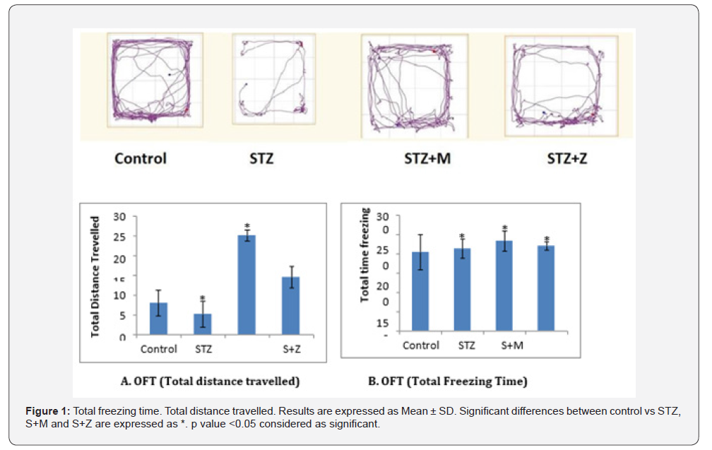
Open Field Test was carried out to assess the effects of STZ, Memantine and Zamzam on rats. Total distance travelled and freezing time were found significant in AD group compared to control group (Figures 1A & 1B).
Histopathological analysis of STZ induced AD rat brain tissue
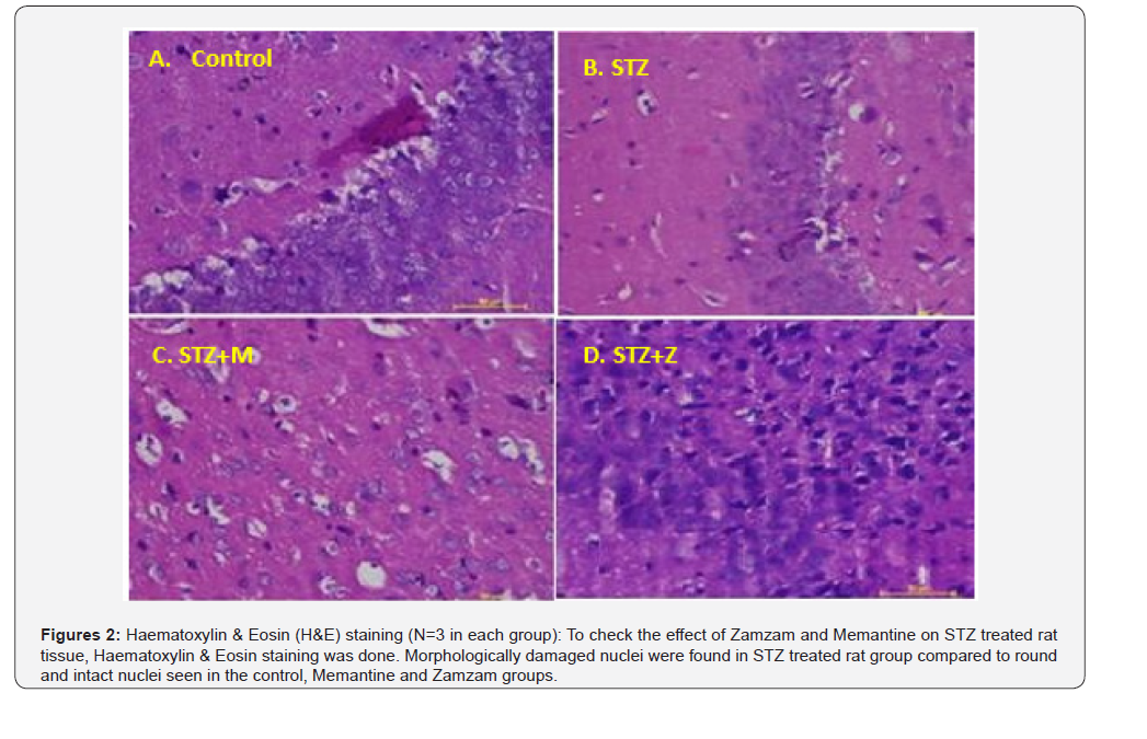
Paraffin sectioned samples were stained with haematoxylin and eosin (H&E) to determine the morphological changes in rat tissue samples (Figures 2A-2D).
DAPI Staining
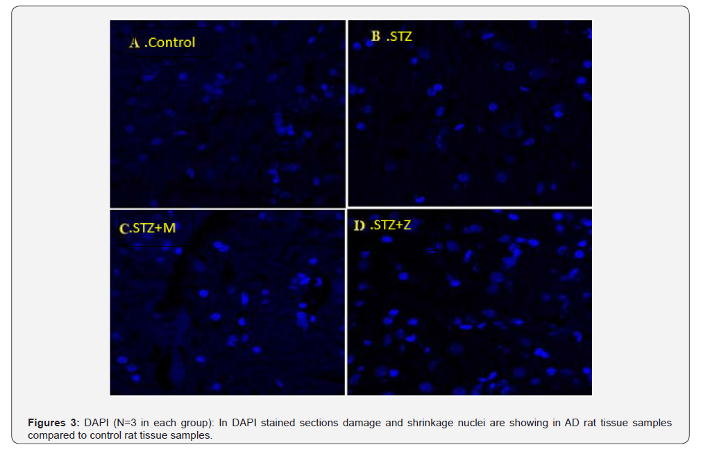
We stained 4μm paraffin sections of tissue samples by DAPI. DAPI is a fluorescent stain which is used to stain nuclei (Figures 3A-3D).
Immunohistochemical (IHC) analysis of AD rat brain tissue
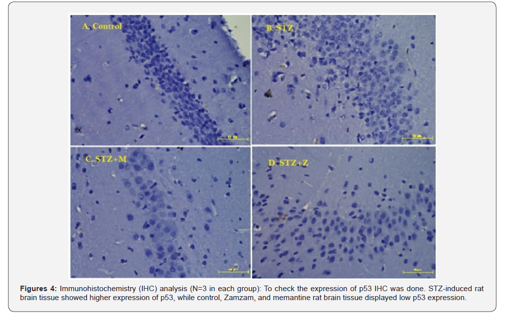
The IHC analysis revealed high expression of p53 in the induced AD rats’ group as compared to control rat group while S+M and S+Z groups showed lower expression of p53 compared to AD group (Figure 4A-4D).
Reverse transcriptase PCR and Real-Time PCR
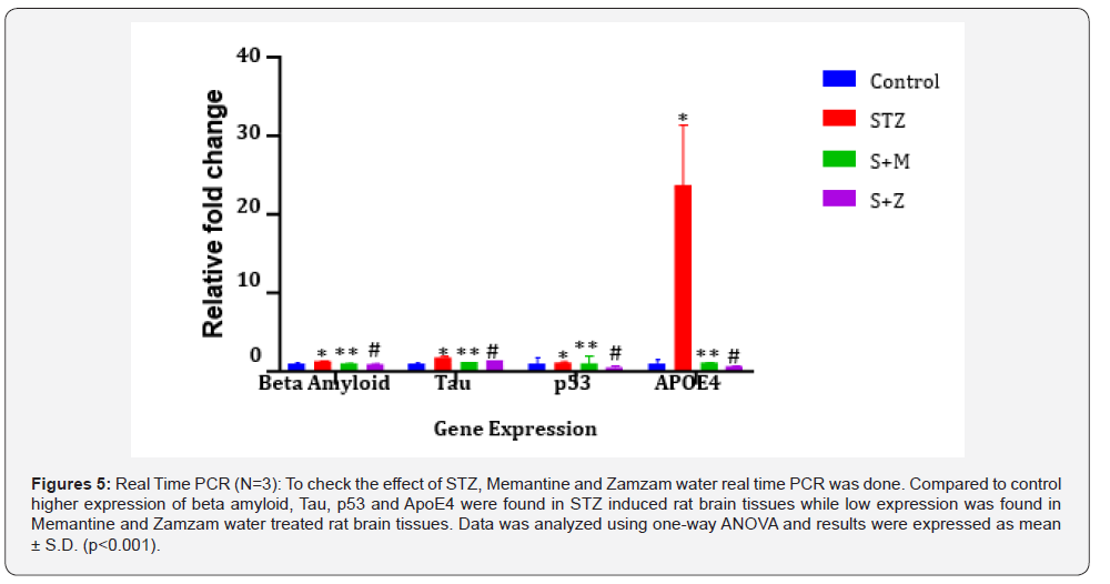
In RT-PCR analysis, as compared to control high expression of beta amyloid, Tau, p53 and ApoE4 were found in STZ induced rat brain tissues. However, we have found low expression was found in Memantine and Zamzam water treated rat brain tissues. Thereafter, we checked these genes expression by Quantitative real-time-PCR. qRT-PCR expression showed a significant upregulation of beta amyloid, Tau, p53 and ApoE4 in STZ induced rat brain tissues while low expression was found in Memantine and Zamzam water treated rat brain tissues as compared to control (Figure 5A-5D).
Discussion
Alzheimer’s disease (AD) is a devastating, irreversible neurodegenerative disorder that primarily affects older adults. It is the most common cause of dementia and poses significant personal, social, and economic challenges. AD is characterized by a progressive decline in cognitive function, memory, language, and the ability to perform daily activities. The pathological hallmarks of the disease include the accumulation of extracellular β-amyloid (Aβ) plaques and intracellular neurofibrillary tangles (NFTs) composed of hyperphosphorylated tau protein [21,22]. These abnormalities contribute to synaptic dysfunction, neuronal loss, and widespread brain atrophy, particularly in regions like the hippocampus and cerebral cortex that are critical for memory and learning.
Despite extensive research, the exact etiology of Alzheimer’s disease remains elusive. However, both genetic and environmental factors are believed to play a role in its development. Among the genetic contributors, the Apolipoprotein E4 (ApoE4) allele is the most prominent and well-established risk factor for late-onset AD [23]. ApoE is a multifunctional protein that regulates lipid metabolism, neuronal repair, and immune responses. The ApoE4 variant is associated with enhanced Aβ aggregation and impaired clearance, thus accelerating plaque formation and disease progression [24].
Another emerging biomarker of interest in AD is p53, a tumor suppressor protein traditionally known for its role in regulating cell cycle arrest and apoptosis. Recent evidence indicates that p53 expression is upregulated in response to Aβ toxicity and oxidative stress in neuronal cells. Moreover, misfolded or conformationally altered isoforms of p53 have been identified in peripheral blood cells of AD patients, suggesting a systemic component to the disease and the possibility of using p53 as a peripheral biomarker [25].
In this study, Streptozotocin (STZ) was employed to induce an AD-like state in rats. STZ is a glucosamine-nitrosourea compound that, when administered intracerebroventricularly, leads to oxidative stress, neuroinflammation, insulin resistance, and Aβ accumulation- phenomena closely resembling the pathological features of sporadic AD in humans. Following successful induction, the rats exhibited significant upregulation of β-amyloid, Tau, ApoE4, and p53, confirming the establishment of AD pathology at a molecular level [15-19].
Current pharmacological treatments for AD are primarily symptomatic and do not halt the progression of the disease. Memantine, a non-competitive NMDA receptor antagonist, is one such treatment approved for moderate to severe stages of AD [26]. It protects neurons by inhibiting glutamate-induced excitotoxicity, which occurs due to excessive calcium influx through overactivated NMDA receptors [27]. While several clinical trials have demonstrated Memantine’s efficacy in improving cognition and behavior, findings remain inconsistent. For instance, the MEM-MD-10 trial showed significant improvements in cognitive and global function as well as behavioral symptoms [28,29]. However, other studies, including a major European trial, did not find Memantine to be significantly better than placebo in all outcome measures [30].
Given the lack of a curative treatment and the limited efficacy of existing medications, there has been a growing interest in exploring nutritional, lifestyle-based, and complementary therapeutic interventions for AD. Nutritional approaches that enhance antioxidant defense mechanisms, reduce inflammation, and correct metabolic imbalances have shown potential in delaying or even preventing the onset of neurodegenerative diseases. These dietary strategies are thought to operate via mechanisms such as improving insulin sensitivity, correcting dyslipidemia, and most importantly, reducing oxidative stress- a central contributor to AD pathology.
In this context, Zamzam water presents a unique and culturally significant candidate for therapeutic exploration. Sourced from the Zamzam well in Makkah, Saudi Arabia, this water holds deep religious significance for Muslims and has traditionally been regarded as possessing healing and purifying properties [6]. Scientific investigations into its composition have revealed that Zamzam water is alkaline, free from biological contaminants, and naturally rich in essential minerals such as calcium, magnesium, selenium, zinc, and manganese- elements that are essential cofactors for various antioxidant enzymes including superoxide dismutase (SOD), glutathione peroxidase (GPx), and catalase [7-10,31].
Studies have highlighted the therapeutic potential of Zamzam water in a variety of disease models. For instance, it has shown significant antioxidant and Reno protective effects in patients with chronic kidney disease [32], slowed cellular aging by reducing oxidative DNA damage [33], and demonstrated antidiabetic effects by normalizing blood glucose levels in experimental models [34]. Moreover, in models of gentamicin-induced nephrotoxicity, Zamzam water enhanced antioxidant capacity and reduced oxidative stress markers [35-37].
Building on these promising findings, our study evaluated the comparative efficacy of Zamzam water and Memantine in mitigating Alzheimer’s-like pathology in STZ-induced rats. Realtime PCR analysis demonstrated that both treatments resulted in markedly reduced expression of β-amyloid, Tau, ApoE4, and p53 in rat brain tissues, especially within the hippocampal region, compared to untreated AD model rats. These results suggest that Zamzam water exerts neuroprotective effects, possibly through its antioxidant properties and mineral content, and performs comparably to Memantine in the context of early intervention.
While Memantine acts by directly modulating neurotransmission through NMDA receptors, Zamzam water’s effects are likely multifactorial, involving the neutralization of free radicals, restoration of redox balance, and improvement in mitochondrial function, all of which are pivotal in preventing neuronal damage in AD. Furthermore, the high levels of selenium and zinc in Zamzam water may contribute to downregulating inflammatory pathways and inhibiting the aggregation of Aβ peptides- a key pathological process in AD.
Importantly, Zamzam water may offer a safe, cost-effective, and culturally acceptable alternative or adjunct to pharmacological therapies, particularly in low-resource settings where access to medications is limited. It is also worth noting that Zamzam water is consumed widely by Muslim populations for spiritual and medicinal purposes, which enhances its acceptability and potential for integration into complementary therapeutic strategies.
Conclusion
To the best of our knowledge, this study represents the first attempt to observe the effect of Zamzam water in AD rat brain tissue samples. In conclusion, our study provides compelling preliminary evidence that Zamzam water possesses neuroprotective properties in an STZ-induced rat model of Alzheimer’s disease. Its ability to reduce the expression of key AD biomarkers such as β-amyloid, Tau, ApoE4, and p53, suggests that it may offer therapeutic benefits similar to those of Memantine. However, more extensive preclinical studies and controlled clinical trials are required to elucidate the precise mechanisms of action, validate efficacy, determine appropriate dosage, and ensure long-term safety in humans.
This investigation also highlights the broader potential of integrating culturally relevant and natural therapeutic agents into modern neurodegenerative disease management frameworks, especially where conventional pharmacotherapy may fall short or be inaccessible. With the growing global burden of Alzheimer’s disease and the limitations of existing treatments, such alternative strategies could pave the way for more holistic, inclusive, and effective care models in the future.
References
- Apostolova LG (2016) Alzheimer’s disease. Continuum 22(2): 419-434.
- Sandeep G, Aditya S (2016) Etiologies and risk factors for dementia. Journal of Geriatric mental health 3(2): 100-107.
- WHO (2019).
- Constantine GL (2006) Position statement of the American Association for Geriatric Psychiatry regarding principles of care for patients with dementia resulting from Alzheimer disease. Am J Geriatr Psychiatry 14(7): 561-572.
- Zhu G, Chen Y, Huang Y, Li Q, Behnisch T, et al. (2011) MPTP-meditated hippocampal dopamine deprivation modulates synaptic transmission and activity-dependent synaptic plasticity. Toxicol Appl Pharmacol 254(3): 332-341.
- Shihri AZ (2005) Makkah Al-Mukarramah in the Old Period: A Reading in History and Poetry. Scientific J of King Faisal University. Makkah Capital of Islamic Culture 2: 473-479.
- Koshak YH, Zam Z (1983) First edition, Dar Alelm for Publications. Jeddah 19: 126.
- Mashat BH (2010) The microbiological quality of sabil free drinking water in Makkah Al-Mukarramah. JKAU Met Env & Arid Land Agric Sci 21: 87-100.
- Nassini R, Andrè E, Gazzieri D, Siena DG, Zanasi A, et al. (2010) A bicarbonate-alkaline mineral water protects from ethanol-induced hemorrhagic gastric lesions in mice. Biol Pharm Bull 33(8): 1319-1323.
- Shomar B (2012) Zamzam water: concentration of trace elements and other characteristics. Chemosphere 86(6): 600-605.
- Khalid N, Ahmad A, Khalid S, Ahmed A, Irfan M, et al. (2014) Mineral composition and health functionality of zamzam water: a review. Int J Food Prop 17(3): 661-677.
- Naeem N, Alsanussi H, Almohandis (1983) A Multi elemental and hydro chemical study of Holy Zamzam water. Journal new England water works Association 47: 158.
- Zuhair AN, Khounganian RA (2006) comparative study between the chemical composition of potable water and zamzam water and its effect on tooth structure in Saudi Arabia. Saudi Dental Journal 18: 843-855.
- Badar A, Bamosa A, Salahuddin M, Meheithif AA (2019) Effect of zamzam water on blood methemoglobin level in young rats, J. Fam. Community Med 26(1): 30-35.
- Correia SC, Santos RX, Santos MS, Casadesus G, Lamanna JC, et al. (2013) Mitochondrial abnormalities in a streptozotocin-induced rat model of sporadic Alzheimer’s disease. Curr Alzheimer Res 10(4): 406-419.
- Kamat PK (2015) Streptozotocin induced Alzheimer’s disease like changes and the underlying neural degeneration and regeneration mechanism. Neural Regen Res 10(7): 1050-1052.
- Deeds MC, Anderson JM, Armstrong AS (2011) Single Dose Streptozotocin Induced Diabetes: Considerations for Study Design in Islet Transplantation Models. Lab Anim 45(3): 131-140.
- Attila G, Barbara H, Aliz JE, Brigitta TT (2022) Performance of the intracerebroventricularly injected streptozotocin Alzheimer’s disease model in a translationally relevant, aged and experienced rat population. Scientifc Reports 12(1): 20247.
- Onesimus M, Mohamad THB, Nurul HMN (2019) Chemicals used for the induction of Alzheimer’s disease-like cognitive dysfunctions in rodents. Biomedical Research and Therapy 6(11): 3460-3484.
- Villemagne VL, Burnham S, Bourgeat P, Brown B, Ellis KA, et al. (2013) Amyloid β deposition, neurodegeneration, and cognitive decline in sporadic Alzheimer's disease: a prospective cohort study. Australian Imaging Biomarkers and Lifestyle (AIBL) Research Group. Lancet Neurol 12(4): 357-367.
- Strittmatter WJ, Saunders AM, Schmechel D, Pericak VM, Enghild J, et al. (1993) Apolipoprotein E: high avidity binding to b-amyloid and increased frequency of type 4 allele in late-onset familial Alzheimer disease. Proc Natl Acad Sci U S A 90(5): 1977-1981.
- Weller J, Budson A (2018) Current understanding of Alzheimer's disease diagnosis and treatment. F1000Research 7: 1000-1161.
- Castellano JM, Kim J, Stewart FR, Jiang H, Mattos DRB, et al. (2011) Human apoE isoforms differentially regulate brain amyloid- β peptide clearance. Sci Transl Med 3(89).
- Frieden C, Garai K (2012) Structural differences between apoE3 and ApoE4 may be useful in developing therapeutic agents for Alzheimer’s disease. Proc Natl Acad Sci USA 109(23): 8913-8918.
- Levine AJ (1997) p53, the cellular gatekeeper for growth and division. Cell 88(3): 323-331.
- Winblad B, Poritis N (1999) Memantine in severe dementia: results of the 9M-best study (benefit and efficacy in severly demented patients during treatment with memantine). International journal of geriatric psychiatry 14(2): 135-146.
- Jiang J, Jiang H (2015) Efficacy and adverse effects of memantine treatment for Alzheimer’s disease from randomized controlled trials. Neurological Sciences 36(9): 1633-1641.
- Areosa SA, Sherriff F, McShane R (2005) Memantine for dementia. The Cochrane database of systematic reviews 20(3): 3154.
- Peskind ER, Potkin SG, Pomara N, Ott BR, Graham SM, et al. (2006) Memantine MEM-MD-10 Study Group. Memantine treatment in mild to moderate Alzheimer disease: a 24-week randomized, controlled trial. The American Journal of Geriatric Psychiatry 14(8): 704-715.
- Bakchine S, Loft H (2007) Memantine treatment in patients with mild to moderate Alzheimer's disease: results of a randomized, double-blind, placebo-controlled 6-month study. Journal of Alzheimer's Disease 11(4): 471-479.
- Matthew WS, Katherine H, Jaclyn LC (2016) Nutritional interventions for Alzheimer’s prevention: a clinical precision medicine approach. Ann N Y Acad Sci Mar 1367(1): 50-56.
- Hofer TEM, Xu J, Seo AY, Gulec S, Knutson MD, et al. (2008) Increased iron content and rna oxidative damage in skeletal muscle with aging and disuse atrophy. Exp Gerontol 43(6): 563-570.
- Jin D, Ryu SH, Kim HW, Yang EJ, Lim SJ, et al. (2006) Anti-diabetic effect of alkaline-reduced water on oletf rats. Biosci Biotechnol Biochem 70(1): 31-37.
- Bamosa A, Elnour A, Kaatabi H, Meheithif AA, Aleissa K, et al. (2013) Zamzam water ameliorates oxidative stress and reduces hemoglobinA1c in Type 2 diabetic patients. J Diabetes Metab 4(3).
- Abdullah AM, Abdelsalam E, Abdullah B, Khaled A (2012) Antioxidant effects of zamzam water in normal rats and those under induced-oxidative stress. J Med Plants Res 6: 5507-5512.
- Khalid N, Ahmad A, Khalid S, Ahmed A, Irfan M, et al. (2014) Mineral composition and health functionality of zamzam water: a review. Int J Food Prop 17(3): 661-677.
- Huang KC, Yang CC, Lee KT, Chien CT (2003) Reduced hemodialysis-induced oxidative stress in end-stage renal disease patients by electrolyzed reduced water. Kidney Int 64 (2): 704-714.

























