Indian Medicinal Plants, A Possible Cure For COVID-19- A Review
Pushpalatha HG1, Manasa R Walmiki2, Shrinivas3 and Shobha MS4*
1Department of Botany, Maharani’s Science College for Women (Autonomous), India
2Department of Biotechnology, Teresian College, India
3Department of Dentistry, Koppal Institute of Medical Sciences, India
4Department of Microbiology, Maharani’s Science College for Women (Autonomous), India
Submission: September 18, 2021; Published: October 28, 2021
*Corresponding author: Shobha MS, Department of Microbiology, Maharani’s Science College for Women (Autonomous), India
How to cite this article:Pushpalatha HG, Manasa R Walmiki, Shrinivas, Shobha M. Indian Medicinal Plants, A Possible Cure For COVID-19- A Review. Glob J Pharmaceu Sci. 2021; 9(1): 555754. DOI: 10.19080/GJPPS.2021.09.555754.
Abstract
Viral diseases are widespread and continue to emerge as a significant concern and a serious threat to humankind. Viral infections occur when a virus enters the body of an individual and invades the cells to multiply. Unlike bacterial infections, viral infections cannot be treated using antibiotics. It requires the body to develop a strategy to strengthen the immune system and attack the virus. Though there are several antiviral drugs, they are not recommended often. If a body’s immune system is weak or unable to fight the virus for any reason, the virus multiplies and spreads to neighbouring cells leading to a widespread infection. Although not all viral diseases with cause life-Threatening conditions, some respiratory viral diseases such as Parainfluenza virus infection (PVI), Respiratory syncytial virus (RSV), severe acute respiratory syndrome (SARS), and similar to SARS, a novel infection has emerged in recent days which is known as Coronavirus-2019 (COVID 19) have known to pose a severe threat to humans. Viral diseases result in a wide variety of symptoms which may vary in severity depending on the type of viral infection, cells or organ affected, the presence of any secondary infection, and age. In the present review, the outbreak of COVID-19, its clinical findings, the medicinal properties of few plants possible for its prevention has been elaborated. Recently delta variant SARS-CoV-2 B.1.617.2 was first noticed in Maharashtra state, India. Though various advanced methods have been used from a scientific and therapeutic point of view, it is of utmost importance to be knowledgeable on the medicinal importance of specific plant-based sources are efficacy against highly fit and immune evasive B. 1.617.2 delta variant assures continued infection control measures against COVID-19.
Keywords: COVID-19; Transmission; Pathogenesis; Treatment; Medicinal plants
Abbreviations: PVI: Parainfluenza Virus Infection; RSV: Respiratory Syncytial Virus; SARS: Severe Acute Respiratory Syndrome; WHO: World Health Organization
Introduction
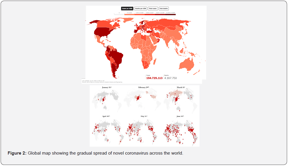
Coronaviruses are related enveloped viruses with a single-stranded RNA as their genetic material, infecting mammals, and birds. The severity of infection may range from mild to lethal depending on the species causing disease, the immunity of the host, and the environmental factors. A novel coronavirus called SARS-CoV (severe acute respiratory syndrome - Corona Virus) has created huge havoc recently, causing an infectious disease named COVID-19 (an acronym for coronavirus disease 2019), which is a new strain, which was not identified in humans earlier. It was recorded only by early December 2019 in China’s Wuhan province and has since been widespread, causing a global pandemic Walls et al. [1] On December 31, 2019, it was reported by the World Health Organization’s (WHO) Country Office. Earlier, this was labeled ‘Pneumonia of unknown etiology’ since the causative agent was not identified (Features). It was declared an outbreak and a Public Health Emergency of International Concern by WHO, on January 30, 2020, Tang et al. [2]. The potential of this species of the virus to become pandemic globally seems to pose a severe threat to humankind. Recently delta variant SARS-CoV-2 B.1.617.2 was first noticed in Maharashtra state, India and spreads faster than other variants. As of June 28, 2021, over 190,233,278 cases have been recorded, of which over 175,584,289 have recovered, and 4,211,875 people died. In India alone, there are over 31,700,456 cases recorded, resulting in over 418,584 deaths (world-o-meter website). The mortality rate reported is different among regions because of various factors such as environmental factors, accurate and fast diagnosis, preventive measures, etc. World governments are rushing to establish countermeasures to reduce the devastating effects. Health organizations are coordinating the information flow across the globe and issuing guidelines and safety measures to relieve the impact of the threat and avoid panic amongst people. Although at the moment, there are several uncertainties regarding virus transmission, the clinical spectrum of the disease, diagnosis, prevention, and cure, work has been going on around the world at a rapid pace to contain the infection. Since there are limited therapeutic strategies available currently and are just supportive, preventing the infection by maintaining social distancing and abiding by the safety rules set by WHO are the best weapons for now. Aggressive isolation measures in many countries, such as the UK, India, Germany, etc., have led to a progressive reduction of cases Guo et al. [3].
The Virus: Origin
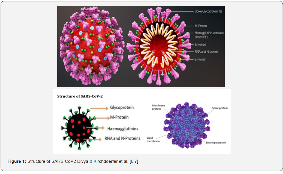
Coronavirus derives its name from the Latin term coronam, which means ‹crown› when observed under an electron microscope; it appears crown-like structure due to spiked glycoproteins present on the envelope. They have a Positive-Stranded RNA within their nucleocapsid, which ranges approximately 27-30 kb in length. Coronavirus is among the most significant known RNA viruses with a 3`-poly-A-tail and a 5`- cap. There are several gene sequences of SARS-CoV published in international gene banks, which allow the researchers to trace out the virus’s phylogenetic tree and understand the different strains and mutations. According to studies, a spike mutation in November 2019 could have triggered the virus to transfer to humans and cause infection. SARS-CoV belongs to the family Corona viridae, order Nidovirales and kingdom Riboviria which is further divided into four genera namely, Alpha coronavirus, Beta coronavirus, Gamma coronavirus and Delta coronavirus Harapan et al. [4]. Alphacoronavirus consists of a human coronavirus known as HCoV-229E and HCV-NL63, Beta coronavirus contains SARSHCoV and MERS-CoV (Middle Eastern Respiratory Syndrome Coronavirus), Gamma coronavirus comprises of viruses isolated from Birds and Whales, and Delta coronavirus consists of viruses of Pigs and Birds. Common Human coronaviruses such as HCVOC43 and HCV-HKU1 cause common cold and upper respiratory tract infections in immune-competent individuals. Still, Immunecompromised individuals can suffer from lower respiratory tract infections as well. Other human coronaviruses such as SARS-CoV and MERS-CoV can cause pandemics with unpredictable clinical severity, including extra-respiratory infections. Mortality rates for these viruses are 10% and 35%, respectively. Phylogenetic analysis of this viral genome suggests that the virus appears approximately 88% identical to the two bat-derived coronaviruses collected in eastern China in 2018 [4]. Along with this, several other studies were conducted, which showed that the virus genome was similar to the viruses isolated from bats. Because of the said studies, the researchers considered the possibility that bats appearing to be the original host of the virus cannot be ruled out. However, bats are considered unlikely to transmit the viral particles to human beings because the seafood market did not sell bats. SARS-CoV-2 and its closely related bat viruses have relatively long branches. Although bats are the natural reservoirs of SARSCoV and MERS-Cov, the other animals (possibly civets and camels) have been the intermediate hosts. It is found to be sensitive to UV rays and heat and can be inactivated by lipid solvents such as ether, ethanol, chloroform, and chlorine-containing disinfectants Cascella et al. [5]. ARS-CoV-2 belongs to the Beta coronavirus category and is about 60-140 nm in diameter. The RNA molecule present in the Corona virus is the largest among the RNA viruses, and the genome consists of ~30,000 nucleotides, twice that of the influenza A genome. To effectively package the viral RNA, it is encoded by four structural proteins, the N protein (Nucleocapsid protein), the M protein (Membrane), the S (Spike) protein, and the E (Envelop) protein, as shown in Figure 1 [6,7].The S protein is also known as the spike protein, which is organized in groups of three on the external surface with almost a ‘crown’ or ‘corona’ like appearance and named as “corona virus,” likely to be observed through Cryo-electron microscopy [4]. In SARS-CoV-2, the lipid membrane S protein is the most significant protein required for viral attachment to the uninfected cells. Membrane protein (M protein) is the most abundant of all the proteins embedded in the outer lipid membrane, giving the virus particle a particular shape and integrity. Membrane protein plays a significant part in the terminal steps of infection, wherein the new viral proteins are compacted into particles before their release and infection to new cells Gupta et al. [8]. Envelope proteins (E protein) appear to be present in a minimum amount, which accounts for the growth and the ability to cause disease. The E-protein is a small membrane protein, consisting of ~76 to 109 amino acids. It forms minute pores, which can alter the host cell membrane characteristics. Still, it plays a significant role in preventing the M protein from clanking at the time of virus particle assemblage within the host [8]. Nucleocapsid protein (N) protein in multiple copies attaches with the RNA to form a spiral structure that tightly wraps and coils the RNA, which makes the long RNA molecule get attached into a small virus particle, to form a protein coat around the RNA to protect from damage. N protein functions at the primary stages of the infection, during the release of RNA molecules into the host cell, which brings down the cell’s natural (Figure 2) defences against the virus [8].
Transmission
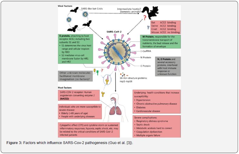
Since the first few cases of COVID-19 were linked to the Huanan Seafood market and presumed that animal to human transmission was the main mechanism, the later cases concluded that the virus could be transmitted from human to human well. Symptomatic people were found to be the source of transmission. Furthermore, subsequent cases and studies confirmed asymptomatic individuals, could also transmit the virus and the possible reason to be asymptomatic could be immunocompetence of those individuals. The transmission occurs through respiratory droplets from coughing and sneezing, aerosol transmission, and prolonged exposure in closed spaces. Based on the investigations and studies conducted, incubation time could usually be within 3 days to 2 weeks (Features). Pathogenic mechanisms were responsible for causing pneumonia to seem to be more complex than thought. Few studies show that when compared to SARS, SARS-CoV-2 leads to less Extra-respiratory involvement. However, it is difficult to draw definite clinical information because of the lack of extensive data and very little knowledge about the molecular aspect of the virus. The virus’s reproductive number (R0) has always varied and probably depends on several factors such as environment, host immunity, and viral load Wan et al. [9]. Studies also show that the SARS-CoV-2 infection is most commonly seen in adult males aged between 24 and 59 years and is likely to infect people who are already suffering from cardiovascular diseases, cerebrovascular diseases, and diabetes. The severity of the infection could also increase due to secondary infections, in infants and in immunocompromised individuals. There are still studies to understand the pathogenetic mechanisms, the genes involved in pathogenesis, the mutation (Figure 3) rate, the vaccine development, and the immunology of the infection Shen et al. [10].
Pathogenesis and immune response
The Beta coronaviruses are known to express high species specificity. However, certain changes in the genome can change their host range, pathogenicity, tissue tropism and the most similar example is the emergence of SARS-CoV and MERS-CoV. These viruses have bats as their reservoirs, intermediate hosts were civet and camels, whereas humans were the terminal hosts. Intermediatory hosts play an important role in cross-species transmission since they allow ample time for the virus to adjust to a new host due to increased contact between the virus and the new host [4]. Several molecular interactions play a role in governing the host range of the virus. It is observed that SARSCoV- 2 may enter the host cell through a receptor known as angiotensin-converting enzyme 2, to infect the airway epithelium and the alveolar type-2 pneumocystis. The spike protein present in the coronavirus consists of two domains namely, S1 responsible for binding to the receptor and S2 responsible for the fusion of the virus with the host cell membrane Yu et al. [11]. According to studies, S1 domain of SARS-CoV-2 and SARS-CoV share about 50 conserved amino acids, while bat derived viruses show several variations. Key residues such as, Gln493 and Asn501 govern the binding of the receptor binding domain of SARS-Cov-2 with ACE2 (Found in lower respiratory tract of humans), which supports the fact that this virus has acquired the capability of human-human transmission. After binding to the receptor and the cell membrane fusion, the virus releases its genome into the host cell’s cytoplasm and the RNA further translates two polyproteins namely, pp1a and pp1ab, which are responsible for the formation of replication transcription complex (RTC) that, replicates continuously and synthesizes sub genomic RNAs. There are RNA codes for the accessory as well as the structural proteins to form viral buds and the virion containing vesicles, which merge with the plasma membrane to release the virus Chu et al. [12]. Although the sequence of the spike protein of SARS-CoV-2 is almost the same as SARS-CoV, the whole genome sequencing shows that it is more closely related to the bat-derived viruses [4]. Viruses can mutate frequently. This ability of the Covid-19 or SARS-CoV-2 has led to various mutants like Alpha, Beta, Gamma, and Delta. The Delta (B.1.617.1 and B.1.617.2) variant was first detected in India in December 2020 and became the most frequently reported variant during mid-April 2021 Bernal et al. [13]. Based on the CDC (Centre for Disease Control) and GISAID (Global Initiative on Sharing Avian Influenza Data) report, as of May 19, 2021, the variant had been detected in 43 countries across six continents (Public Health England, 2021). The UK also reported the spike in delta variant cases which could be associated with travel from India. The delta variant is 60% more transmissible than the highly infectious alpha variant, which was detected in the UK in 2020 [13]. The most recent Delta variant shows two mutations E484Q (glutamic acid E substituted by glutamine Q) and L452R (leucine L altered by arginine R), identified in India’s second COVID-19 wave. Excluding the two mutations, the Delta variant also harbors a unique mutation of T478K (threonine T replaced by lysine K) Manigandan et al. [14]. The S1 mutations considerably increase the binding affinity to ACE2 while showing lower affinity to neutralizing antibodies [15,16], signifying a possible explanation for their increasing higher transmissibility and virulence [17,18].
Clinical Manifestations
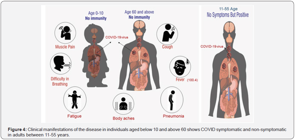
The clinical manifestations of SARS-CoV-2 are almost the same as SARS-CoV in which the primary symptoms include fever, chest pain, fatigue, dry cough, dyspnoea, and myalgia. Although less common, headache, dizziness, sputum production, abdominal pain, haemoptysis vomiting, and nausea are also seen in few individuals. About 75% of the patients have bilateral pneumonia. The difference between SARS and COVID-19 patients is that in COVID-19, the lower respiratory tract infection is more prominent than the upper respiratory tract infection [19]. Other complications include hypoxaemia, acute ARDS, cardiac injury, shock, and kidney injury. The symptoms may worsen and become life-threatening if the patient has been suffering from other diseases such as cancer, bacterial infections, respiratory diseases, autoimmune disorders, etc. The incubation period ranges from 2 to 14 days. Fever and respiratory symptoms tend to appear about 3 to 7 days after exposure to the virus [16]. Children below 10 years and adults above 60 years shows most of the symptoms such as muscle pain, cough, high fever, pneumonia, body aches, fatigue, and difficulty in breathing, but these symptoms are not seen among 11-55, year-old people, as shown in Figure 4.
Diagnosis
Several measures are taken to check the spread of the disease, such as institutional quarantine, home isolation measures, social distancing, and thorough sanitization of public places. Nevertheless, the spread of SARS-CoV-2 cannot be contained, and the cases are still on the steady rise around the globe. According to the standards set by the WHO, the suspected cases of individuals suffering from COVID-19 should have severe acute respiratory infection apart from most of the symptoms such as fever and dry cough. For SARS-CoV-2 diagnosis, the specimens are collected from the upper respiratory tract of the patient,either nasopharyngeal swab or oropharyngeal swab. If further tests are required, the lower respiratory tract, such as sputum or tracheal aspirate, is also collected to detect the RNA virus that, includes several clinical trials such as the CT scan of the chest, blood counting, the affected person’s medical history and the exposure to the specific symptoms Wang et al. [20]. Currently, the preferred protocol for testing coronavirus is the real-time reverse transcriptase (RT-PCR) detection method, which has been diagnosed as effective in preliminary infections Han, et al. [21]. RT-PCR makes the COVID-19 testing rapid and also less expensive. This essay appears to be an important method in detecting all types of coronaviruses Noh et al. [22].
Current treatments
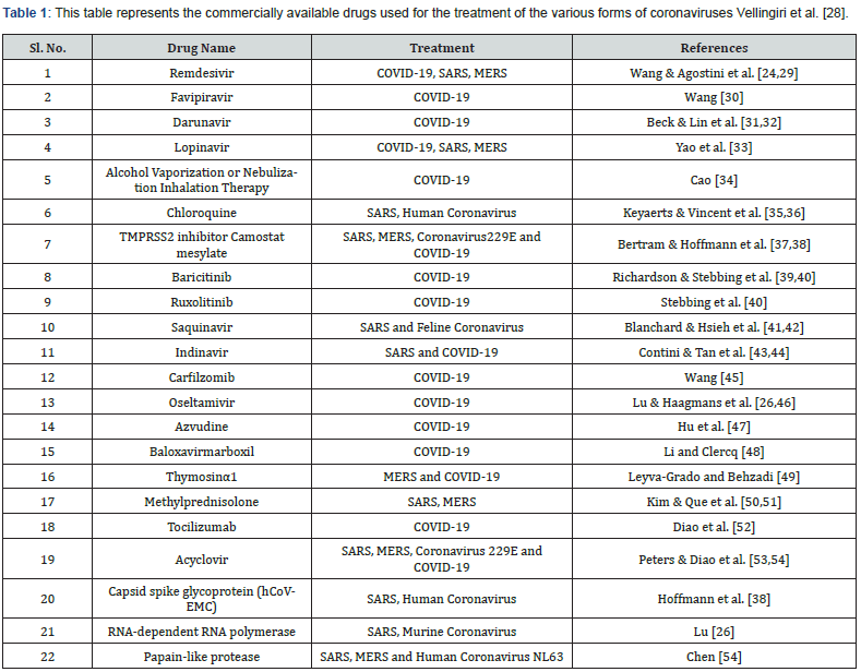
Similar to SARS-CoV and MERS-CoV, there are no treatments available for SARS-CoV-2, and the current treatments are targeted to reduce the symptoms and provide respiratory support. A majority requires oxygen therapy of the patients, and WHO has recommended extracorporeal oxygen therapy for the patients suffering from refractory hypoxemia. Treatments with convalescent plasma and immunoglobulin G are provided to rescue critical patients. At the first-time antiviral treatments such as Ganciclovir, Acyclovir, Ribavirin, etc., which were used to treat SARS are not recommended for COVID-19. Based on the response of the infected patients, more than 15 drugs all over the world were initially identified as the potential drugs for the treatment of the COVID-19 namely, chloroquine-hydroxychloroquine, lopinavir-Ritonavir, nafamostat-camostat, famotidine, umifenovir, nitazoxanide, ivermectin, corticosteroids, tocilizumab-sarilumab, bevacizumab and fluvoxamine Shaffer [23]. Besides, based on the recent study dexamethasone and remdesivir drugs are widely considered as the life saving drug. However, use of the remdesivir in the developing countries like India was questionable due to its cost and procurement from other countries. Dexamethasone is a cheap steroid used to fight the inflammation in the moderate and severely infected patient. In the other end, the studies predicted use of the remdesivir, and dexamethasone drugs are safe since it carries fewer side effects based on the short exposure. On the contrary, the long-term side effects of the drug are still under progress Manigandan et al. [14]. A combination of chloroquine and remdesivir is also proved to be effective in treating COVID-19 Wang et al. [24]. The broad-spectrum antiviral medicine exhibiting the nucleoside analogues and HIV protease inhibitors could attenuate the infectious virus in anticipating the accessible antiviral befalls Shen et al. [25]. Further, a clinical agent, EIDD-2801, exhibits maximum therapeutic activity against the infectious virus for treating COVID-19 Lu et al. [26]. Preliminary clinical analysis has proven chloroquine to treat COVID-19 and its safety for long term usage Toots et al. [27]. The list of drugs is provided in Table 1.
Control and prevention strategies
Now, COVID-19 is the highest international concern due to its rapid spread. According to some reports, SARS-CoV-2 has a higher reproductive number and fatality rate than MERS and SARS. Different strategies are being implemented globally by the health care sectors, which is, unfortunately, an essential source of transmission of the virus. Applying triage, following safety measures and correct infection control measures, isolating affected individuals and their families, and contact tracing are important strategies that are being implemented in almost all countries. Suspected cases should wear protective masks and contain the virus to prevent its spread and strictly adhere to the triage procedures. Patients require ventilated rooms for isolating 2 meters away from other patients. They should have easy access to respiratory hygiene supplies. If a COVID-19 patient has to be put on hospitalization, they have to be provided with a separate individual room and to be provided with negative air pressure with a minimum of 6 air changes/hour, provided that the air exhausted has been subjected to filtration via HEPA filters. Further, the medical personnel accessing the room have to be provided with the necessary protective cover. Since there is a lack of space, in several cities, to isolate the infected individuals, isolating them in their homes is the best option, if they are not symptomatic or with bearable symptoms, so that the hospitals can focus on patients with worse conditions Vellingiri et al. [28]. World Health Organization has suggested restricted travel to high-risk areas, to avoid the entry of people from affected regions, to wear a face mask, thus, preventing the access of virus and social distancing for at least 2 meters from one person to another in all areas (Centers for Disease Control). Washing the hands using soaps and daily use of sanitizers may minimize the viral risk on the skin’s surface. The virus can also be washed out on frequent cleaning of hands using soaps and sanitizers Sohrabi et al. [55].
Role of Indian medicinal plants in the prevention of COVID-19
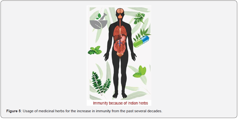
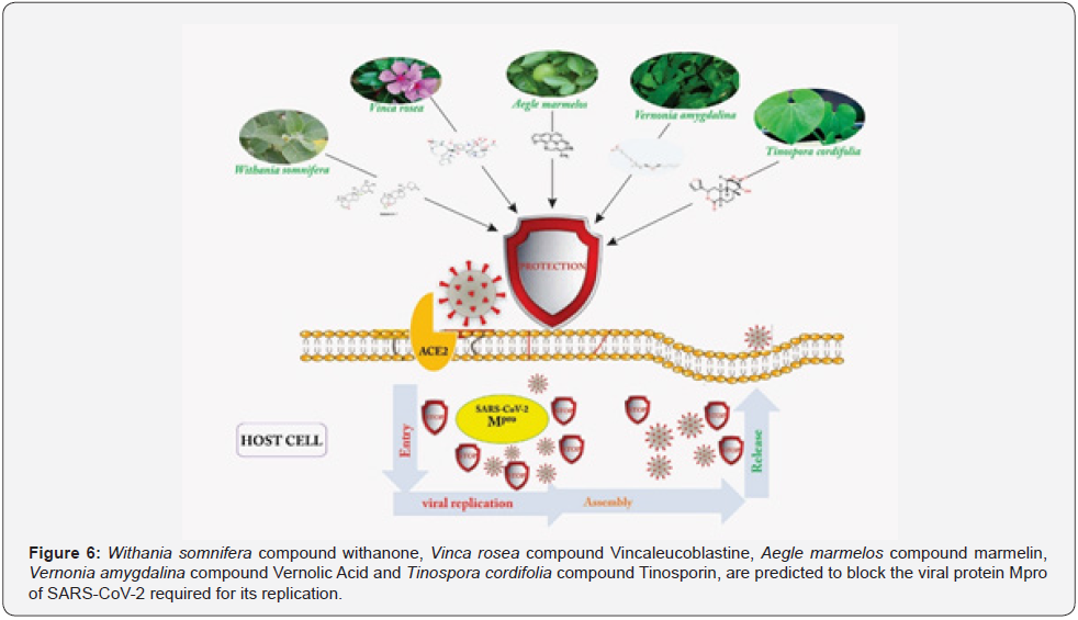
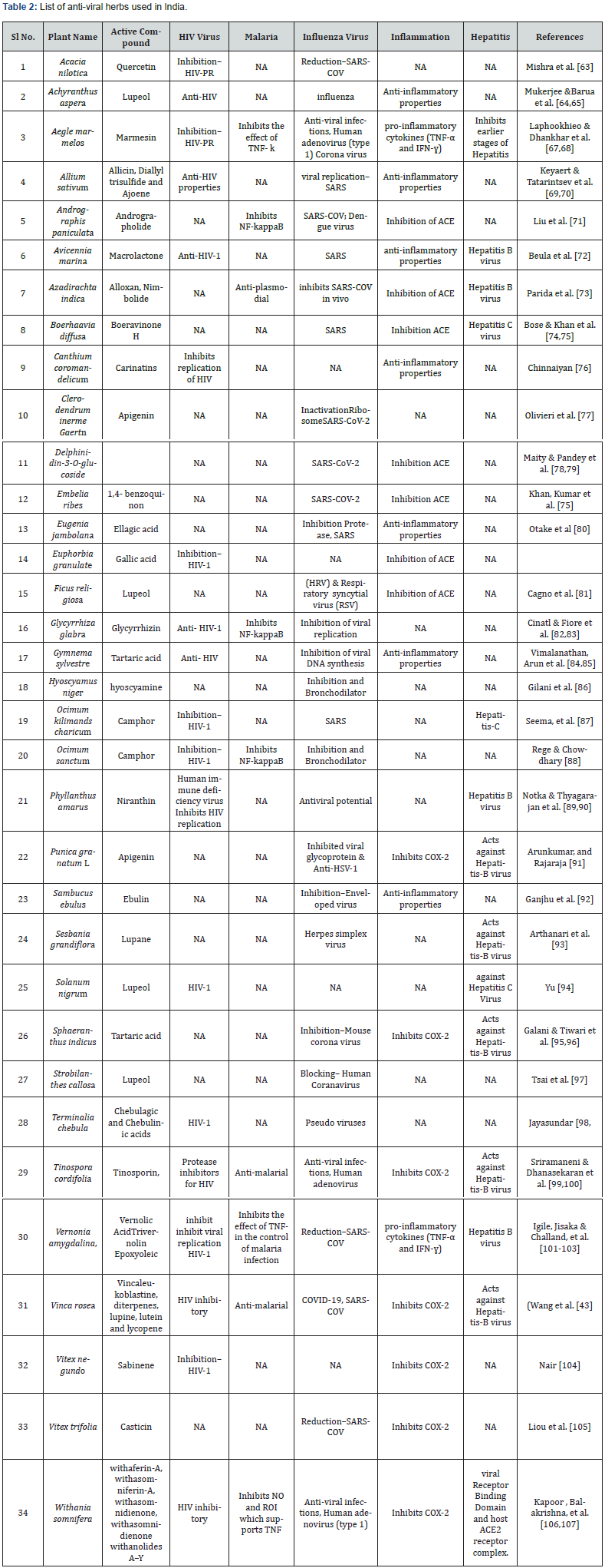
The traditional medicinal systems in India are one of the ancient treatments in human history. They play an important position in encountering global health care needs [56]. Ayurveda, Siddha, Unani, Yoga, Naturopathy, and Homeopathy are the traditional medicinal practices successfully carried out in India to treat various diseases [57]. The traditional medicinal preparations existed for 2000 years and have been witnessed and further scripted in the ancient literature, as shown in the illustrated Figure 5. The Indian culture is dependent on traditional plants for several diseases throughout the history. The Indian medicinal plants are also used across the globe, both by the developed and developing countries, which emphasize on their primary healthcare [58]. Clinical trials for influenza, hepatitis B, tuberculosis, malaria, HIV etc. have been evaluated with the use of plant-based vaccines, [59]. India has a vast collection of medicinal plants, estimated to be around 45,000 species, which are frequently used in the traditional medicine systems [60]. Several plants have been demonstrated to exhibit antiviral activities and have been used against severe diseases such as HIV and COVID-19 [61]. At present, there is no specific treatment available for COVID-19. Treatments using drugs isolated from medicinal plants, consisting of potent compounds, which include zinc, chloroquine, hydroxychloroquine and many more, are observed [62]. In this review, some medicinal plants showing antiviral properties are listed in Table 2. Furthermore, the plants showing all the activities against HIV, malaria, influenza, inflammation and hepatitis are discussed in brief and more research and clinical trials should be done to confirm the potential cure to COVID-19. Studies using medicinal plants for coronavirus prevention and treatment are very scarce in India. A study is available on the activity of coronavirus (a surrogate of SARS-CoV) using Indian medicinal plants, which could (Figure 6) prevent the symptoms apparent of COVID-19. There are certain medicinal plants, which are widely used for respiratory diseases and are listed in Table 2. Only V. amygdalina, W. somnifera C. roseus T. cordifolia and A. marmelos plants have been demonstrated for the development of the drug, specific (Table 3) to SARS-CoV-2.

Withania somnifera
Withania somnifera (L.) Dunal (Solanaceae), belongings to the Solanaceae family and commonly known as ‘Ashwagandha’, is used in more than 100 formulations of Ayurvedic products and is thought to be therapeutically equivalent to Ginseng. Hence it is also known as Indian Ginseng [108]. W. somnifera is a small shrub usually found in India and North Africa. W. somnifera contains multiple compounds such as alkaloids, steroidal lactones, saponins, etc. Therefore, it is commonly used for the treatment of genital disease (caused by Herpes Simplex Virus), influenza, malaria, HIV, stress, tumor, inflammation, etc. [109]. Several classes of metabolites have been reported, including alkaloids and steroidal compounds, which are found to be very effective remedies for various diseases. The ability of the aforesaid ayurvedic herb as a therapeutic drug against COVID-19, caused by the novel coronavirus (110). It has been reported that the Withanone compound isolated from W. somnifera reduced the binding interface of AEC2-RBD complex. The withanone compound significantly decreased the binding free energies of ACE2-RBD complex. Based on this evidence, the authors have interpreted that the electrostatic interactions between ACE2 and RBD could block or weaken the COVID-19’s entry into the infected persons. Kumar et al. [111] reported that a new bioactive compound, with none (Wi-N) derived from W. somnifera, and another compound Caffeic Acid Phenethyl Ester (CAPE), derived from an active ingredient of the New Zealand Propolis, collected from the honey bees, which have potential against COVID-19 disease. Furthermore, the authors have targeted the main SARS-CoV-2’s enzyme known to be the Main protease (Mpro), which exhibits its major function to mediate the viral replication. This Mpro is less toxic and an attractive drug target against the virus (Singh and Gilca [108].
Catharanthus roseus
Catharanthus roseus G. Don, commonly known as Vinca rosea, belongs to the Apocynaceae family. C. roseus is well known for its drugs vincristine and vinblastine used to treat cancer patients. Leaves of C. roseus have high alkaloids, which are utilized in the treatment of cancer, high sugar levels, hypertension, and various viral diseases [112]. Vinca alkaloids are organic compounds made up of carbon, hydrogen, nitrogen, and oxygen. These alkaloids are generally used in combination with chemical drugs in various medical therapies. Alkaloids such as Vinblastine and Vincristine are used to treat testicular cancer, Hodgkin Lymphoma, and Non-Hodgkin Lymphomas. The alkaloids have been proven to show potential antitumor activity against mammary carcinoma and bone tumor cells. Leurocristine is used against the poliovirus [113]. Vincaleukoblastine possesses a high virucidal activity against the influenza virus.
Aegle marmelos
Aegle marmelos (L) Correa, commonly known as “Indian Bael,” belongs to the family Rutacaece. Its leaves have been extensively used in traditional systems of Indian medicine from ancient times. A. marmelos is a native of Northern India but is also found in Burma, Ceylon, Bangladesh, Pakistan, Thailand, Nepal, and China [114]. A. marmelos proved to be a good antidiarrhoeal, anti-microbial, antiviral, radioprotective, anti-cancer, chemo-preventive, anti-influenza, anti-HIV, antipyretic, antimalarial, anti-inflammatory, ulcer healing, and anti-hepatis-B activities and is used in fertility treatments [115]. A marmelos has been proven to contain numerous phytochemical constituents mainly, marmin, marmenol, marmelide, marmelosin, psoralen, rutaretin, aegelin, scopoletin, fagarine, marmelin, limonene, anhydromarmelin, betulinic acid, â-phellandrene, imperatorin, marmesin, luvangentin, and auroptene [116] Rahman and Parvin, 2014 reported the presence of hexanal, limonene, isoamyl acetate, β-phellandrene, acetoin, p-cymene, dehydro-p-cymene, linalool oxide, citronellal, β-caryophyllene, pulegone, hexadecane, α-Humulene, β-cubebene, carvone, verbenone, caryophyllene oxide, humulene oxide and hexadecanoic acid in A. marmelos.
Vernonia amygdalina
Vernonia amygdalina, commonly known as “Bitter leaf” due to its bitter taste, appears as a small shrub belonging to the Asteraceae family. This plant is used to treat HIV/AIDS, malaria, pneumonia, etc. Several researchers have proved Vernonia amygdalina as an anti-HIV drug to treat HIV positive patients compared to the commercially available tablet Immunace [117]. Studies are available on the effect of V. amygdalina leaf extract on patients infected with HIV and undergoing antiviral therapy alone to evaluate the effect of the leaf extract on CD4 + cell number. The results suggest the increase in the number of CD4+ cells in patients consuming leaf extract of V. amygdalina whereas the control patients remained. Furthermore, the patients with skin rashes, continuous fever, headache, and joint pain were recovered due to its enhanced nutritional values and high health improving properties [118]. Vernolic Acid, Trivernolin, and Epoxyoleic compounds present inV. amygdalina could be maximizing the vulnerability to malaria and other infections
Tinospora cordifolia
Tinospora cordifolia commonly known as ‘Guduchi’ belongs to the Menispermaceae family. Traditionally, it is known for its extensive use in the treatment of various viral diseases in Indian medicine T. cordifolia contains several biologically active compounds, Tinosporin, alkaloids, steroids, diterpenoid lactones, aliphatics sesquiterpenoid, phenolics, aliphatic compounds, and glycosides extracted from its root, stem and also from the whole plant [119]. Several researchers have reported its medicinal properties, including anti-inflammatory, anti-allergic, antiarthritic, antioxidant, anti-stress, anti-leprotic, anti-malarial, anti-neoplastic, and immunomodulatory activities. [120]. Studies have reported the decrease in the resistance of HIV, which was confirmed through the decrease in the number of eosinophils, macrophage stimulation, B-cell stimulation, stimulation of polymorphonuclear leucocytes, and elevation in the percentage of haemoglobin. The study suggested this plants’ promising role and application in HIV disease management [121]. Alkaloids, glycosides, lactones, and steroids present in T. cordifolia, decrease the resistance of HIV to antiretroviral therapy [122]. The crystal structures current on the membrane receptors, the activation of the downstream signaling cascades, and the changes in the intermediate action site can help stop the SARS-COV-2 virus. The future scope should exploit the signaling pathways of the components of T. cordifolia. These plants can genuinely act as an incredible source to counter the virus.
Conclusion
Historical evidence on SARS, H1N1, influenza and, hepatitis prevention proves that Indian medicinal plants could act as an alternative for preventing COVID-19 in a high-risk population. From a pharmacological point of view, medicinal plants especially, W. somnifera, A. marmelos, C. roseus, V. amygdalina, and T. cordifolia, should be further explored to investigate the production of a vaccine for COVID-19 disease. Furthermore, importance should be given to produce plant-coated products such as plant-based face masks, soaps, sanitizer, and also plant-based decoction instead of coffee and tea, which will improve the immunity against COVID-19 naturally. Moreover, it is of utmost importance to maintain social distancing in public places and to always sanitize the hands thoroughly, using a 70% Alcohol-based hand sanitizer, which will keep the virus in check. Moreover, frequent touching of the nose, mouth, and eyes should be avoided. It’s high time for the human community to come forward in fighting the pandemic coronavirus disease through the maintenance of self-hygiene and social distancing strategies.
References
- Walls A C, Park Y J, Tortorici M A, Wall A, McGuire A T, et al. (2020) Structure, function, and antigenicity of the SARS-CoV-2 spike glycoprotein. Cell 181(2): 281-292.
- Tang JW, Tambyah PA, Hui DS (2020) Emergence of a novel coronavirus causing respiratory illness from Wuhan, China. Journal of Infection 80(3): 350-371.
- Guo YR, Cao QD, Hong ZS, Tan YY, Chen SD, et al. (2020) The origin, transmission and clinical therapies on coronavirus disease 2019 (COVID-19) outbreak–an update on the status. Military Medical Research 7(1): 1-10.
- Harapan H, Itoh N, Yufika A, Winardi W, Keam S, et al. (2020) Coronavirus disease 2019 (COVID-19): A literature review. J Infect Public Health 13(4): 667-673.
- Cascella M, Rajnik M, Cuomo A, Dulebohn SC, Di Napoli R (2020) Features, evaluation and treatment coronavirus (COVID-19). In: Statpearls. Stat Pearls Publishing, London, US.
- Divya M, Vijayakumar S, Chen J, Vaseeharan B, Durán LEF (2020) A review of South Indian medicinal plant has the ability to combat against deadly viruses along with COVID-19? Microbial Pathogenesis: 104277.
- Kirchdoerfer RN, Cottrell CA, Wang N, Pallesen J, Yassine HM (2016) Pre-fusion structure of a human coronavirus spike protein. Nature 531(7592): 118-121.
- Gupta MK, Vemula S, Donde R, Gouda G, Behera L, et al. (2020) In-silico approaches to detect inhibitors of the human severe acute respiratory syndrome coronavirus envelope protein ion channel. Journal of Biomolecular Structure and Dynamics 39(7): 2617-2627.
- Wan Y, Shang J, Graham R, Baric RS, Li F (2020) Receptor recognition by novel coronavirus from Wuhan: an analysis based on decade-long structural studies of SARS Coronavirus. J Virol 94(7): 00127-00120.
- Shen KL, Yang YH (2020) Diagnosis and treatment of 2019 novel coronavirus infection in children: a pressing issue. World J Pediatr 16(3): 219-221.
- Yu W, Tang G, Zhang L, Corlett R (2020) Decoding the evolution and transmissions of the novel pneumonia coronavirus (SARS-CoV-2) using whole genomic data. Zool Res 41(3): 247-257.
- Chu DKW, Pan Y, Cheng SMS, Hui KPY, Krishnan P, et al. (2020) Molecular diagnosis of a novel coronavirus (2019-nCoV) causing an outbreak of pneumonia. Clin Chem 66(4): 549-555.
- Bernal JL, Andrews N, Gower C, Gallagher E, Simmons R, et al. (2021) Effectiveness of Covid-19 Vaccines against the B.1.617.2 (Delta) Variant. The New England Journal of Medicine 385(7): 585-594.
- Manigandan S, Wu MT, Ponnusamy VK, Raghavendra VB, Pugazhendhi A, et al. (2020) A systematic review on recent trends in transmission, diagnosis, prevention and imaging features of COVID-19. Process Biochemistry 98: 233-240.
- Piccoli L, Park YJ, Tortorici MA, Czudnochowski N, Walls AC, et al. (2020) Mapping neutralizing and immunodominant sites on the SARS-CoV-2 spike receptor-binding domain by structure-guided high-resolution serology. Cell. 183(4): 1024-1042.e21.
- Fratev F (2020) The SARS-CoV-2 S1 spike protein mutation N501Y alters the protein interactions with both hACE2 and human derived antibody: a Free energy of perturbation study.
- Nelson G, Buzko O, Patricia S, Niazi K, Rabizadeh S, et al. (2021) Molecular dynamic simulation reveals E484K mutation enhances spike RBD-ACE2 affinity and the 1 combination of E484K, K417N and N501Y mutations (501Y.V2 variant) induces conformational 2 change greater than N501Y mutant alone, potentially resulting in an e. bioRxiv.
- Starr TN, Greaney AJ, Hilton SK, Ellis D, Crawford KHD, et al. (2020) Deep mutational scanning of SARS-CoV-2 receptor binding domain reveals constraints on folding and ACE2 binding. Cell 182(5): 1295-1310.e20.
- Lu H, Stratton CW, Tang YW (2020) Outbreak of pneumonia of unknown etiology in Wuhan China: the mystery and the miracle. J Med Virol 92(4): 401-402.
- Wang J Tang, F Wei (2020) Updated understanding of the outbreak of 2019 novel coronavirus (2019-nCoV) in Wuhan, China. J Med Virol 92(4): 441-447.
- Han Q, Lin S, Jin L (2020) Coronavirus 2019-nCoV: a brief perspective from the front line. J Infect 80(4): 373-377.
- Noh, SW Yoon, DJ Kim (2017) Simultaneous detection of severe acute respiratory syndrome, Middle East respiratory syndrome, and related bat coronaviruses by real-time reverse transcription PCR. Arch Virol 162(6): 1617-1623.
- Shaffer L (2020) 15 drugs being tested to treat COVID-19 and how they would work. Nat Med
- Wang M, Cao R, Zhang L, Yang X, Liu J, et al. (2020) Remdesivir and chloroquine effectively inhibit the recently emerged novel co-ronavirus (2019-nCoV) in vitro. Cell Res 30(2): 269-271.
- Shen M, Zhou Y, Ye J, Al-Maskri, AAA Kang, et al. (2020) Recent advances and perspectives of nucleic acid detection for coronavirus. Journal of Pharmaceutical Analysis 10(2): 97-101.
- Lu H (2020) Drug treatment options for the 2019-new coronavirus (2019-nCoV). Biosci Trend 14(1): 69-71.
- Toots M, Yoon J J, Cox R M, Hart M, Sticher Z M, et al. (2019) Characterization of orally efficacious influenza drug with high resistance barrier in ferrets and human airway epithelia. Sci Transl Med 11(515): eaax5866.
- Vellingiri B, Jayaramayya K, Iyer M, Narayanasamy A, Govindasamy V, et al. (2020) COVID-19: A promising cure for the global panic. Sci Total Environ 725: 138277.
- Agostini ML, Andres EL, Sims AC, Graham RL, Sheahan TP, et al. (2018) Coronavirus susceptibility to the antiviral remdesivir (GS-5734) is mediated by the viral polymerase and the proofreading exoribonuclease.
- Wang Q, Zhang Y, Wu L, Niu S, Song C, et al. (2020) Structural and functional basis of SARS-CoV-2 entry by using human ACE2. Cell 181(4): 894-904.
- Beck BR, Shin B, Choi Y, Park S, Kang K (2020) Predicting commercially available antiviral drugs that may act on the novel coronavirus (2019-nCoV), Wuhan, China through a drug-target interaction deep learning model.
- Lin S, Shen R, He J, Li X, Guo X (2020) Molecular Modeling Evaluation of the Binding Effect of Ritonavir, Lopinavir and Darunavir to Severe Acute Respiratory Syndrome Coronavirus 2 Proteases. BioRxiv.
- Yao X, Ye F, Zhang M, Cui C, Huang B, et al. (2020) In vitro antiviral activity and projection of op-timized dosing design of hydroxychloroquine for the treatment of severe acute respi-ratory syndrome coronavirus 2 (SARS-CoV-2). Clin Infect Dis 71(15): 732-739.
- Cao B, Wang Y, Wen D, Wen L, Jingli W, et al. (2020) A trial of lopinavir-ritonavir in adults hospitalized with severe COVID-19. N Engl J Med 382(19): 1787-1799.
- Keyaerts E, Li S, Vijgen L, Rysman E, Verbeeck J, et al. (2009) Antiviral activity of chloroquine against human coronavirus OC43 infection in newborn mice. Antimicrob. Agents Chemother 53: 3416-3421.
- Vincent M J, Bergeron E, Benjannet S, Erickson B R, Rollin P E, et al. (2005) Chloroquine is a potent inhibitor of SARS coronavirus infection and spread. Virol J 2: 69.
- Bertram S, Dijkman R, Habjan M, Heurich A, Gierer S, et al. (2013) TMPRSS2 activates the human coronavirus 229E for cathepsin-independent host cell entry and is expressed in viral target cells in the respiratory epithelium. J Virol 87(11): 6150-6160.
- Hoffmann M, Kleine Weber H, Krueger N, Mueller MA, Drosten C, et al. (2020) The novel coronavirus 2019 (2019-nCoV) uses the SARS-coronavirus receptor ACE2 and the cellular protease TMPRSS2 for entry into target cells. BioRxiv.
- Richardson P, Griffin I, Tucker C, Smith D, Oechsle O, et al. (2020) Baricitinib as potential treatment for 2019-nCoV acute respiratory disease. Lancet 395(10223): 30-31.
- Stebbing J, Phelan A, Griffin I, Tucker C, Oechsle O, et al. (2020) COVID-19: combining antiviral and anti-inflammatory treatments. Lancet Infect. Dis 20(4): 400-402.
- Blanchard JE, Elowe NH, Huitema C, Fortin PD, Cechetto JD (2004) High-throughput screening identifies inhibitors of the SARS coronavirus main proteinase. Chem Biol 11(10): 1445-1453.
- Hsieh LE, Lin CN, Su BL, Jan TR, Chen CM, et al. (2010) Synergistic antiviral effect of Galanthus nivalis agglutinin and nelfinavir against feline coronavirus. Antivir. Res. 88(1): 25-30.
- Contini A (2020) Virtual screening of an FDA approved drugs database on two COVID-19 coronavirus proteins. Chem Rxiv.
- Tan EL, Ooi EE, Lin CY, Tan HC, Ling AE, et al. (2004) Inhibition of SARS coronavirus infection in vitro with clinically approved antiviral drugs. Emerg Infect Dis 10(4): 581-586.
- Wang L, He H P, Di Y T, Zhang Y, Hao X J (2012) Catharoseumine, a new monoterpenoid indole alkaloid possessing a peroxy bridge from Catharanthus roseus. Tetrahedron Letters 53(13): 1576-1578.
- Haagmans BL, Kuiken T, Martina BE, Fouchier RA, Rimmelzwaan GF, Van Amerongen G, et al. (2004) Pegylated interferon-α protects type 1 pneumocytes against SARS coronavirus infection in macaques. Nat Med 10(3): 290-293.
- Hu F, Jiang J, Yin P (2020) Prediction of Potential Commercially Inhibitors Against SARSCoV-2 by Multi-task Deep Model. arXiv : 2003.00728.
- Li G, Clercq E (2020) Therapeutic options for the 2019 novel coronavirus (2019-nCoV). Nat. Rev. Drug Discov 19: 1449-14150.
- Leyva Grado VH, Behzadi MA (2019). Overview of current therapeutics and novel candidates against influenza, respiratory syncytial virus and Middle East respiratory syndrome coronavirus infections. Front Microbiol 10: 1327.
- Kim I, Lee JE, Kim KH, Lee S, Lee K, et al. (2016) Successful treatment of suspected organizing pneumonia in a patient with Middle East respiratory syndrome coronavirus infection: a case report. J Thorac Dis 8 (10): E1190-E1194.
- Que T, Wong V, Yuen K (2003) Treatment of severe acute respiratory syndrome with lopinavir/ritonavir: a multicentre retrospective matched cohort study. Hong Kong Med J 9(6): 399-406.
- Diao B, Wang C, Tan Y, Chen X, Ying L, et al. (2020) Reduction and Functional Exhaustion of T Cells in Patients with Coronavirus Disease 2019 (COVID-19). medRxiv.
- Peters HL, Jochmans D, de Wilde AH, Posthuma CC, Snijder EJ (2015) Design, synthesis and evaluation of a series of acyclic fleximer nucleoside analogues with anti-coronavirus activity. Bioorg Med Chem Lett 25(15): 2923-2926.
- Chen J (2020) Pathogenicity and transmissibility of 2019-nCoV - a quick overview and comparison with other emerging viruses. Microbes Infect 22(2): 69-71.
- Sohrabi C, Alsafi Z, O Neill N, Khan M, Kerwan A, et al. (2020) World Health Organization declares global emergency: A review of the 2019 novel coronavirus (COVID-19). International Journal of Surgery 67: 71-76.
- Ravishankar B, Shukla VJ (2007) Indian systems of medicine: a brief profile. African Journal of Traditional, Complementary and Alternative Medicines 4(3): 319-337.
- Gomathi M, Padmapriya S, Balachandar V (2020) Drug studies on Rett syndrome: from bench to bedside. J Autism Dev Disord 50(8): 2740-2764.
- Munuswamy, T Thirunavukkarasu, S Rajamani, EK Elumalai, D Ernest (2013) A review on antimicrobial efficacy of some traditional medicinal plants in Tamilnadu. J Acute Dis 2(2): 99-105.
- Kumar SV, Navaratnam V (2013) Neem (Azadirachta indica): Prehistory to contemporary medicinal uses to humankind, Asian Pacific. J Trop Biomed 3(7): 505-514.
- Bespoke (2020) Bebot Launches Free Coronavirus Information Bot.
- Salazar-González, C Angulo, S Rosales-Mendoza (2015) Chikungunya virus vaccines: Current strategies and prospects for developing plant-made vaccines. Vaccine 33(31): 3650-3658.
- Zumla, A, Hui D S, Azhar E I, Memish Z A, Maeurer M (2020) Reducing mortality from 2019-nCoV: host-directed therapies should be an option. Lancet 395(10224): 35-36.
- Mishra S, Aeri V, Gaur PK, Jachak SM (2014) Phytochemical, therapeutic, and ethnopharmacological overview for a traditionally important herb: Boerhavia diffusa Linn. Biomed Res Int.
- Mukherjee H, D Ojha, P Bag, HS Chandel, S Bhattacharyya, et al. (2013) Anti-herpes virus activities of Achyranthes aspera: an Indian ethnomedicine, and its triterpene acid. Microbiol Res 168(4): 238-244.
- Barua CC, Begum SA, Talukdar A, Pathak DC, Barua AG (2010) Effect of Achyranthes aspera Linn on modified forced swimming in rats. Pharmacologyonline 1: 183-191.
- Laphookhieo S, Phungpanya C, Tantapakul C, Techa S, Tha-in S, et al. (2011) Chemical constituents from Aegle marmelos. Journal of the Brazilian Chemical Society 22(1): 176-178.
- Laphookhieo S, Phungpanya C, Tantapakul C, Techa S, Tha-in S, et al. (2011) Chemical constituents from Aegle marmelos. Journal of the Brazilian Chemical Society 22(1): 176-178.
- Dhankhar S, Ruhil S, Balhara M, Dhankhar S, Chhillar A (2011) Aegle marmelos(Linn.) Correa: A potential source of phytomedicine. J Med Plant Res 5(9): 1497-1507.
- Keyaerts E, Vijgen L, Maes P, Neyts J, Van Ranst, et al. (2004) In vitro inhibition of severe acute respiratory syndrome coronavirus by chloroquine. Biochem Biophys Res Commun 323: 264-268.
- Tatarintsev A, PV Vrzhets, DE Ershov, AA Shchegolev, AS Turgiev, et al. (1992) The ajoene blockade of integrin-dependent processes in an HIV-infected cell system. Vestn Ross Akad Med Nauk 11: 6-10.
- Liu YT, Chen HW, Lii CK, Jhuang JH, Huang CS, et al. (2020) A diterpenoid, 14-deoxy-11, 12-didehydroandrographolide, in Andrographis paniculata reduces steatohepatitis and liver injury in mice fed a high-fat and high cholesterol diet. Nutrients 12(2): 523.
- Beula M, Gnanadesigan PB, Rajkumar S, Ravikuma MA (2012) Antiviral, antioxidant and toxicological evaluation of mangrove plant from South East coast of India. Asian Pacific J Tropical Biomed 2(1): S352-S357.
- Parida MM, Upadhyay C, Pandya G, Jana AM (2002) Inhibitory potential of neem (Azadirachta indica Juss) leaves on dengue virus type-2 replication. Journal of ethnopharmacology 79(2): 273-278.
- Bose M, Kamra M, Mullick R, Bhattacharya S, Das S, et al. (2017) A plant‐derived dehydrorotenoid: a new inhibitor of hepatitis C virus entry.Febs Letters 591(9): 1305-1317.
- Khan MY, Kumar V (2019) Mechanism& inhibition kinetics of bioassay-guided fractions of Indian medicinal plants and foods as ACE inhibitors. J Tradit Complement Med 9(1): 73-84.
- Chinnaiyan, MR, Subramanian SV, Kumar AN, Chandu K (2013) Deivasigamani, Antimicrobial and anti-HIV activity of extracts of Canthium coromandelicum (Burm. f.) Alston leaves, J Pharma Res 7: 588-594.
- Olivieri, F, Prasad V, Valbonesi P, Srivastava S, Ghosal-Chowdhury P, et al. (1996) A systemic antiviral resistance-inducing protein isolated from Clerodendrum inerme Gaert Is a polynucleotide: adenosine glycosidase (ribosome-inactivating protein). FEBS Lett 396(2-3): 132-134.
- Maity N, Nema NK, Sarkar BK, Mukherjee PK (2012) Standardized Clitoria ternatea leaf extract as hyaluronidase, elastase and matrix-metalloproteinase-1 inhibitor. Indian. J pharmacol 44(5): 584-587.
- Pandey A, Bigoniya P, Raj V, Patel KK (2011) Pharmacological screening of Coriandrum sativum for hepatoprotective activity. J Pharm Bioallied Sci 3: 435.
- Otake T, Mori H, Morimoto M, Ueba N, Sutardjo S, et al. (1995) Screening of Indonesian plant extracts for anti-human immunodeficiency virus-type 1 (HIV-1) activity. Phytother Res 9(1): 6-10.
- Cagno A, Civra R, Kumar S, Pradhan M, Donalisio BN, et al. (2015) Ficus religiosa bark extracts inhibit human rhinovirus and respiratory syncytial virus infection in vitro. J Ethnopharmacol 176: 252-257.
- Cinatl J, Morgenstern B, Bauer G, Chandra P, Rabenau H (2003) Treatment of SARS with human interferons. Lancet 362: 293-294.
- Fiore C, Eisenhut M, Krausse R, Ragazzi E, Pellati D, et al. (2008) Antiviral effects of Glycyrrhiza species. Phytother Res 22(2): 141-148.
- Vimalanathan S, Ignacimuthu S, Hudson J (2009) Medicinal plants of Tamil Nadu (southern India) are a rich source of antiviral activities. Pharm Biol 47(5): 422-429.
- Arun LB, Arunachalam AM, Arunachalam KD, Annamalai SK, Kumar KA (2014) In vivo anti-ulcer, anti-stress, anti-allergic, and functional properties of Gymnemic acid isolated from Gymnema sylvestre R Br. BMC Compl Alternative Med 14: 70.
- Gilani AH, Khan A, Raoof M, Ghayur MN, Siddiqui BS (2008) Gastrointestinal, selective airways and urinary bladder relaxant effects of Hyoscyamus niger are mediated through dual blockade of muscarinic receptors and Ca2+ Fundam Clin Pharmacol 22(1): 87-99.
- Seema TM, Thyagarajan S (2016) Methanol and aqueous extracts of Ocimumckilimandscharicum (Karpuratulasi) inhibits HIV-1 reverse transcriptase in vitro. Int. J. Pharmacogn. Phytochem. Res 8: 1099-1103.
- Rege A, Chowdhary AS (2014) Evaluation of Ocimum sanctum and Tinospora cordifolia as probable HIV protease inhibitors. Int J of Pharm Sci Rev Res 25: 315-318.
- Notka, F, Meier G, Wagner R (2004) Concerted inhibitory activities of Phyllanthus amarus on HIV replication in vitro and ex vivo. Antiviral research. 64(2): 93-102.
- Thyagarajan SP, Thirunalasundari T, Subramanian S, Venkateswaran PS, Blumberg BS (1988) Effect of Phyllanthus amarus on chronic carriers of hepatitis B virus. The Lancet 332(8614): 764
- Arunkumar J, Rajarajan S (2018) Study on antiviral activities, drug-likeness and molecular docking of bioactive compounds of Punica granatum to Herpes simplex virus-2 (HSV-2). Microbial pathogenesis 118: 301-309.
- Ganjhu RK, Mudgal PP, Maity H, Dowarha D, Devadiga S (2015) Herbal plants and plant preparations as remedial approach for viral diseases. Virusdisease 26(4): 225-236.
- Arthanari SK, Vanitha J, Ganesh M, Venkateshwaran K, Clercq D (2012) Evaluation of antiviral and cytotoxic activities of methanolic extract of grandiflor Walls et al a (Fabaceae) flowers. Asian Pacific Journal of Tropical Biomedicine 2(2): S855-S858.
- Yu Y B (2004) The extracts of Solanum nigrum for inhibitory effects on HIV-1 and its essential enzymes. Korean J Orient Med 10: 119-126.
- Galani VJ, Patel BG, Rana DG (2010) Sphaeranthus indicus: a phytopharmacological review. Int J Ayurveda Res 1(4): 247-253.
- Tiwari B K, Khosa R L (2009) Hepatoprotective and antioxidant effect of Sphaeranthus indicus against acetaminophen-induced hepatotoxicity in rats. J Pharm Sci Res 1: 26-30.
- Tsai Y C, Lee C L, Yen H R, Chang Y S, Lin Y P, et al. (2020) Antiviral action of Tryptanthrin isolated from Strobilanthes cusia leaf against human coronavirus NL63. Biomolecules 10: 366.
- Jayasundar R, Ghatak S, Makhdoomi MA, Luthra K, Singh A, et al. (2019) Challenges in integrating component level technology and system level information from Ayurveda: Insights from NMR phytometabolomics and anti-HIV potential of select Ayurvedic medicinal plants. Journal of Ayurveda and Integrative Medicine 10(2): 94-101.
- Sriramaneni RN, Omar AZ, Ibrahim SM, Amirin S, Zaini Mohd A (2010) Vasorelaxant effect of diterpenoid lactones from andrographis paniculata chloroform extract on rat aortic rings. Pharmacognosy Res 2: 242–246.
- Dhanasekaran M, Baskar AA, Ignacimuthu S, Agastian P, Duraipandiyan V (2009) Chemopreventive potential of Epoxy clerodane diterpene from Tinospora cordifolia against diethylnitrosamine-induced hepatocellular carcinoma. Invest New Drugs 27(4): 347-355.
- Igile GO, Oleszek W, Burda S, Jurzysta M (1995) Nutritional assessment of Vernonia amygdalina leaves in growing mice. J. Agr. Food Chem 43: 2162–2166.
- Ohigashi H, Jisaka M, Takagaki T, Nozaki H, Tada T, et al. (1991) Bitter principle and a related steroid glucoside from Vernonia amygdalina, a possible medicinal plant for wild chimpanzees. Agr Biol Chem 55(6): 1201-1203.
- Challand S, Willcox M (2009) A clinical trial of the traditional medicine Vernonia amygdalina in the treatment of uncomplicated malaria. J Altern Com Med 15(11): 1231-1237.
- Nair R (2012) HIV-1 reverse transcriptase inhibition by Vitex negundo L. leaf extract and quantification of flavonoids in relation to anti-HIV activity. J Cell.Mol Biol 10: 53-59.
- Liou CJ, Cheng CY, Yeh KW, Wu YH, HuangWC (2018) Protective effects of casticin from vitex trifolia alleviate eosinophilic airway inflammation and oxidative stress in a murine asthma model. Front Pharma col 9: 635.
- Kapoor LD (2001) Handbook of Ayurvedic Medicinal Plants. CRC Press London, UK. pp. 337-338.
- Balkrishna A, Pokhrel S, Singh J, Varshney A (2020) Withanone from Withania somnifera may inhibit novel Coronavirus (COVID-19) entry by disrupting interactions between viral S-protein receptor binding domain and host ACE2 receptor.
- Singh N, Gilca M (2010) Herbal Medicine-Science embraces tradition-a new insight into the ancient Ayurveda. Germany: Lambert Academic Publishing p. 51-67.
- Mishra LC, Singh BB, Dagenais S (2000) Scientific basis for the therapeutic use of Withania somnifera. (Ashwagandha): A review. Alternative Medicine Reviews 5: 334-346.
- Huang C, Wang Y, Li X, Ren L, Zhao J, et al. (2020) Clinical features of patients infected with 2019 novel coronavirus in Wuhan, China. Lancet 395(10223): 497-506.
- Kumar V, Dhanjal JK, Kaul SC, Wadhwa R, Sundar D (2020) Withanone and caffeic acid phenethyl ester are predicted to interact with main protease (Mpro) of SARS-CoV-2 and inhibit its activity. Journal of Biomolecular Structure and Dynamics, (just accepted) : 1-13.
- Schutz FA, Bellmunt J, Rosenberg JE, Choueiri TK (2011) Vinflunine: Drug safety evaluation of this novel synthetic vinca alkaloid. Expert Opin Drug Saf 10(4): 645-653.
- Almagro L, Fernández PF, Pedreño MA (2015) Indole alkaloids from Catharanthus roseus: bioproduction and their effect on human health. Molecules 20(2): 2973-3000.
- Das B, Das R (1995) Medicinal properties and chemical constituents of Aegle marmelos Indian Drugs 32(3): 93-99.
- Rahman S, Parvin R (2014) Therapeutic potential of Aegle marmelos (L.)-An overview. Asian Pacific journal of tropical disease 4(1): 71-77.
- Laphookhieo S, Phungpanya C, Tantapakul C, Techa S, Tha-in S, et al. (2011) Chemical constituents from Aegle marmelos. Journal of the Brazilian Chemical Society 22(1): 176-178.
- Erasto P, Grierson DS, Afolayan AJ (2006) Bioactive sesquiterpenes lactones from the leaves of Vernonia amygdalina. J Ethnopharmacol 106(1): 117-120.
- Oyeyemi IT, Akinlabi AA, Adewumi A, Aleshinloye AO, Oyeyemi, OT (2018) Vernonia amygdalina: A folkloric herb with anthelminthic properties. Beni-Suef University Journal of Basic and Applied Sciences, 7: 43-49.
- Saha S, Ghosh S (2012) Tinospora cordifolia: One plant, many roles. Ancient science of life 31(4): 151-159.
- Sharma P, Dwivedee BP, Bisht D, Dash AK, Kumar D (2019) The chemical constituents and diverse pharmacological importance of Tinospora cordifolia. Heliyon 5(9): e02437.
- Kalikar MV, Thawani VR, Varadpande UK, Sontakke SD, Singh RP, et al. (2008) Immunomodulatory effect of Tinospora cordifolia extract in human immuno-deficiency virus positive patients. Indian journal of pharmacology 40(3): 107-110.
- Akhtar S (2010) Use of Tinospora cordifolia in HIV infection. Indian Journal of Pharmacology 42(1): 57.
- Centers for Disease Control and Prevention (2019) Novel Coronavirus.
- Ewen C (2021) Delta coronavirus variant: scientists brace for impact. Nature 595(7865): 17-18.
- Galani VJ, Patel BG, Rana DG (2010) Sphaeranthus indicus: a phytopharmacological review. Int J Ayurveda Res 1(4): 247-253.
- Hu F, Jiang J, Yin P (2020) Prediction of Potential Commercially Inhibitors Against SARSCoV-2 by Multi-task Deep Model. arXiv: 2003.00728.
- Lin S, Shen R, He J, Li X, Guo X, et al. (2020) Molecular Modeling Evaluation of the Binding Effect of Ritonavir, Lopinavir and Darunavir to Severe Acute Respiratory Syndrome Coronavirus 2 Proteases. BioRxiv.
- https://assets.publishing.service.gov.uk/government/uploads/system/uploads/attachment_data/file/986380/Variants_of_Concern_VOC_Technical_Briefing_11_England.pdf. opens in new tab).
- Liu Z, Xiao X, Wei X, Li J, Yang J, et al. (2020) b. Composition and divergence of coronavirus spike proteins and host ACE2 receptors predict potential intermediate hosts of SARS-CoV-2. J Med Virol 92(6): 595-610.
- Tan EL, Ooi EE, Lin CY, Tan HC, Ling AE, et al. (2004) Inhibition of SARS coronavirus infection in vitro with clinically approved antiviral drugs. Emerg Infect Dis 10(4): 581-586.


