Herlyn-Werner-Wunderlich Syndrome: About an Uncommon Case Report
Mariam Mahtate*, Khaoula Lakhdar, Soukaina Cherradi, Aziz Slaoui, Najia Zeraidi, Amina Lakhdar and Aziz Baydada
Department of Gynecology-Obstetrics and Endoscopy, University Mohammed V, Morocco
Submission: March 20, 2023; Published:April 19, 2023
*Corresponding author: Mariam Mahtate, Department of Gynecology-Obstetrics and Endoscopy, University Mohammed V, Rabat, Morocco
How to cite this article: Mariam M, Khaoula L, Soukaina C, Aziz S, Najia Z, et al. Herlyn-Werner-Wunderlich Syndrome: About an Uncommon Case Report. Glob J Reprod Med. 2023; 9(5): 555774. DOI: 10.19080/GJORM.2023.09.555774.
Abstract
Herlyn-Werner-Wunderlich syndrome (HWWS), defined by the triad of uterus didelphys, obstructed hemivagina and ipsilateral renal agenesis, is a rare Mullerian duct malformation, usually diagnosed after menarche, when symptoms related to haematocolpos arise. HWW generally occurs at puberty and exhibits variable symptoms including pelvic pain shortly following menarche, dysmenorrhoea, palpable mass due to the associated haematocolpos or haematometra or haemoperitoneum due to retrograde menstruation.
Ultrasonography and MRI are extremely useful in the diagnosis and classification of Müllerian duct anomalies. 3D ultrasound is more effective in the diagnosis of uterine malformation. Early detection of this relatively rare syndrome can lead to the provision of immediate treatment to preserve future fertility. We present a rare case report on Herlyn-Werner-Wunderlich syndrome from Morocco.
Keywords: Herlyn-werner-wunderlich syndrome; Postmenarche; Malformations; Uterus didelphys
Introduction
HWW syndrome is an extremely rare congenital anomaly of the urogenital system due to a congenital developmental defect of the Mullerian ducts characterized by a triad, didelph uterus, obstructed hemi-vagina and ipsilateral renal agenesis [1]. The most common symptomatology of this syndrome in postmenarche is pelvic pain, pelvic mass, hematocolpos, primary amenorrhea, menstrual cycle disorder, abortion and prematurity [2]. The diagnosis of this syndrome is essentially radiological based on ultrasound combined with MRI for a better evaluation of associated malformations [1].
A proper diagnosis and prompt treatment can prevent the possible complications associated with this syndrome; pyocolpos, endometriosis and infertility [3]. We hereby present an uncommon case of a 13-year-old female patient referred to our department for an episode of acute urine retention associated with a pelvic pain after the onset of puberty in whom the diagnosis of HWW syndrome was made on pelvic MRI.
Case Presentation
We hereby report the uncommon case of A 13-year-old patient was referred to our structure for an episode of acute retention of urine associated with pelvic pain that increases in intensity at the same period of the menstrual cycle, during menstruation since her menarche. The patient reported urinary disorders such as dysuria and mictional imperiosis of progressive aggravation without associated digestive disorders before this episode of retention. The gynecological history indicates menarche at the age of 12 years with a regular cycle with minimal periods. The clinical examination found a pelvic curvature reaching the umbilicus associated with abdominal sensitivity, the gynecological examination found no external genital anomaly. In front of this atypical clinical presentation, a tumoral origin was suspected after bladder drainage.
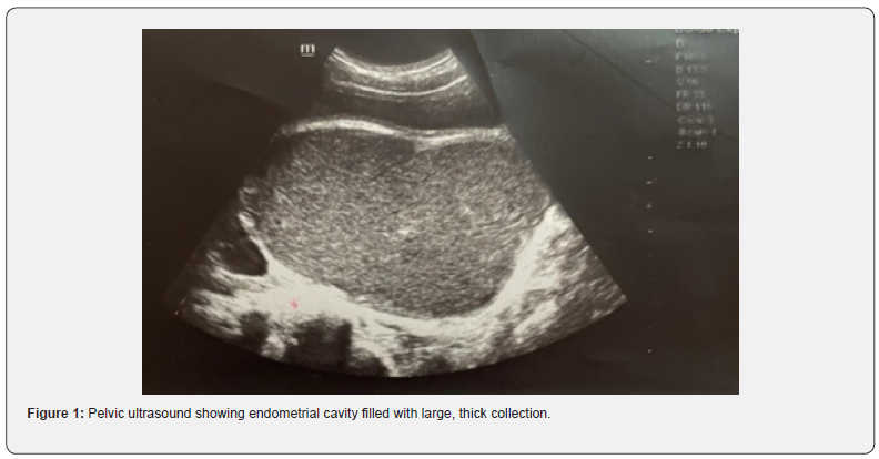
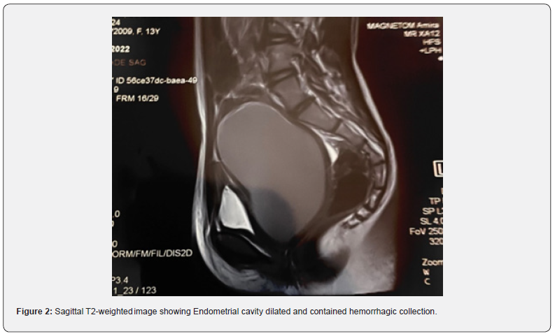
The pelvic ultrasound showed a hypoechogenic median retro vesical liquid formation (Figure 1), associated with a uterine anomaly that was difficult to distinguish with the absence of the right kidney; a pelvic MRI was requested for a better evaluation of the urinary tract. Parental consent was given for the MRI with injection of the contrast product. Pelvic MRI performed in three planes in T2, T1, diffusion and gadolinium injection sequence in favor of a didelphic bicervical uterus with hematocolps at the level of the right hemivagina which extends to the homolateral hemi uterus, the left hemi uterus, ovaries of normal size and morphologies with right renal agenesis, without signs of deep endometriosis (Figure 2).


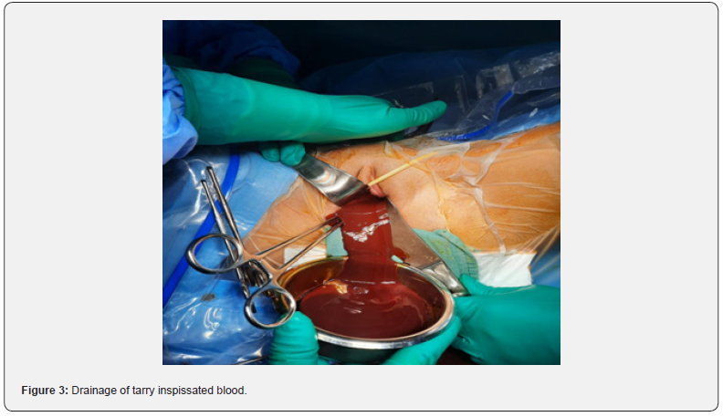
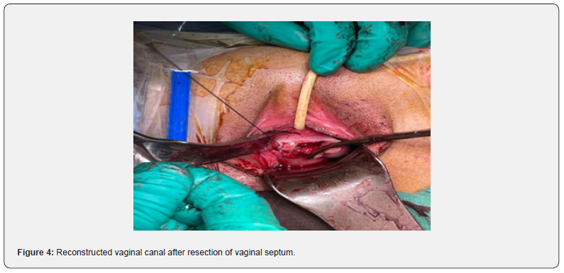
On the basis of the MRI radiological images in front of this bicornuate bi-cervical uterus associated with a hematocolpos of the right hemovagina in favor of an obstructed hemivagina and a right ipsilateral agenesis the diagnosis of HWW syndrome was made. The surgical management was done by Identification and resection of the vaginal septum and reached up to the right cervix for the drainage of tarry blood (Figure 3). Thus, vaginal canal was reconstructed (Figure 4). There were no perioperative or postoperative complications. She was discharged 5 days after surgery. The patient had regular clinical and echocardiographic check-ups. The patient has been symptom free for 6 months after her surgery.


Discussion
Purslow in 1922 was the pioneer in presenting a case of didelphic uterus with obstructed hemivagina, Miller in the same year presented the same case with ipsilateral renal anomaly [4]. Known as Herlyn-Werner-Wünderlich syndrome or with the acronym OHVIRA (Uterus didelphys associated with Obstructed Hemi-Vagina and Ipsilateral Renal Anomaly), is a rare malformation of the Müllerian ducts. The association with renal agenesis was reported in 1971 [5]. Recently, many cases of morphological diversity such as other uterine abnormalities, varying degrees of vaginal obstruction, and various renal/ureteral abnormalities have been noted [6]. Didelphic uterus accounts for 5% of Müllerian malformations, with an incidence of renal agenesis of 1/1,000. 25- 50% of her affected, get genital abnormalities [7].
The common embryological development of the urinary and genital systems explains the associated malformations of both systems. The female internal organs derive from the Müllerian ducts, which give their origin to the tubes, uterus and upper 2/3 of the vagina, the Wolffian ducts give the origin to the kidneys and induce proper fusion of the Müllerian ducts [8].
The aetiology and the pathogenesis of SHWW is unknown. It can be caused by exposure to environmental factors, teratogens, radiation, drugs, but most have a multifactorial polygenic basis. It is not associated with chromosomal abnormalities, so the Karyotype is normal, other genital, urological, rectal malformations or skeletal dysplasia may be associated, exceptionally, it is acquired [8]. our patient presented the congenital malformation generating a didelphic bi cervical uterus with associated hematocolpos with right renal agenesis classified as U3b C2 V2 according to the American society of reproductive medicine (ASMR) [9].
The diagnosis is rarely made antenatally or in childhood and coincides with the menarche around the age of 14 [10]. The symptoms are essentially attributed to menstruation by dysmenorrhea, hematocolpos, oligomenorrhea as is the case in our patient or primary amenorrhea. As well as symptoms related to hematocolpos by compression of the surrounding organs causing acute retention of urine, or pyelocaliceal dilatation. several symptoms can confuse the clinician nonspecific. As we can see the patients at the stage of complications, pyohematocolpos, pyosalpinx, pelviperitonitis, endometriosis, pelvic adhesions thus affecting the fertile prognosis of the patient hence all the interest to think about the diagnosis in any young patient presenting with gynecological symptoms. our patient was diagnosed at the stage of complication by an acute retention of urine thus discovering the syndrome via ultrasound and more exactly by MRI.
Diagnosis is based on clinical history and imaging exams, specifically ultrasound and MRI [11]. Ultrasound is accessible and assesses the renal system, 3D transvaginal ultrasound more accurately evaluates the uterus and adnexa and MRI is more precise [12]. The differential diagnosis is made with abnormalities of uterine development, unicornuate uterus with contralateral corne, bicornuate uterus, imperforate hymen, and hypoplasia or agenesis of the cervix. The management relieves symptoms, avoids complications and conserves fertility. Surgery is usually conservative, resection of the vaginal septum, marsupialization of the obturated hemivagina as in our case, and drainage of collections if present.
Efforts should be made to preserve the affected hemiuterus because pregnancy can be achieved there. Some studies show that 87% have successful pregnancies, 23% abortions, 15% preterm deliveries and intrauterine growth retardation [13-15]. It’s recommended a therapeutic abstinence in renal agenesis; it is important to prevent urinary tract infections and to monitor renal function [15].
Conclusion
HWWS is a rare condition relatively unknown to medical professionals, and associated dysmenorrhea is often misdiagnosed as another, more common cause of dysmenorrhoea in juvenility. The correct and accurate diagnosis of female reproductive system disorders, including HWW syndrome, is necessary to avoid complications. Early detection of this relatively rare syndrome can lead to the provision of immediate treatment to preserve future fertility.
Guarantor of Submission
The corresponding author is the guarantor of submission.
Provenance and Peer Review
Not commissioned, externally peer reviewed.
Funding
There are no funding sources to be declared.
Availability of Data and Materials
Supporting material is available if further analysis is needed.
Consent for Publication
Written informed consent was obtained from the patient for publication of this case report and any accompanying images. A copy of the written consent is available for review by the Editorin- Chief of this journal.
Ethics Approval and Consent to Participate
Ethics approval has been obtained to proceed with the current study. Written informed consent was obtained from the patient for participation in this publication.
Credit Authorship Contribution Statement
Mahtate Mariam and Lakhdar Khaoula: Study concept and design, data collection, data analysis and interpretation, writing the paper.
References
1. Guillán Maquieira C, Sánchez Merino JM, Méndez Díaz C (2012) Obstructed hemivagina and ipsilateral renal anomaly (OHVIRA) associated to uterus didelphys. Prog obstet ginecol 55(6): 281-284.
4. Suárez Chulián C, González Soria, Sánchez de la Vega R, Bueno Rodríguez MG, Morillo Gutiérrez B (2016) Renal agenesis and Mullerian anomalies regarding two cases. Vox Pediatric 23(2): 47-50.
9. Navarro Ballester A, Pérez Caballero FA, Salelles Climent P, Díaz Ramos C (2023) Anomalías de los conductos de Müller: conceptos básicos e imagen.
11. Fuentes Rozalen A, Gómez García MT, López del Cerro E, Belmonte Andújar LL, González de Merlo G (2015) Síndrome de Herlyn Werner Wunderlich. Prog Obstet Ginecol 58(1): 20-24.


