Tinnitus as a Symptom of Transotic Schwannoma
P Krasnodębska1, M Furmanek2, D Raj Koziak*1 and H Skarżyński3
1Audiology and Phoniatrics Clinic, Institute of Physiology and Pathology of Hearing, Poland
2Department of Radiological and Imaging Diagnostics, The Medical Centre of Postgraduate Education, Poland
3Otorhino!aryngo!ogic Clinic, Institute of Physiology and Pathology of Hearing, Poland
Submission: March 06, 2018; Published: April 10, 2018
*Corresponding author: Danuta Raj Koziak, Audiology and Phoniatrics Clinic, The Institute of Physiology and Pathology of Hearing, Mochnackiego 10, 02-042 Warsaw, Poland, Tel: +48 22 356 03 SI; Email: d.koziak@ifps.org.pl
How to cite this article: P Krasnodebska, M Furmanek, D Raj K, H Skariynski. Tinnitus as a Symptom of Transotic Schwannoma. Glob J Oto 2018; 14(4): 555894. DOI: 10.19080/GJO.2018.14.555894
Abstract
Middle ear is an extremely rare but possible place of occurrence of the acoustic schwannoma. Tinnitus is one of its first symptoms, frequently ignored by the patient and undiagnosed by physicians. This case study report of a fourth case in the literature of the neoplasm extending from the inner ear to the middle ear, and not present in the cerebellopontine angle. Detailed audio logic and radiologic features of transonic schwannoma are presented.
Abbreviations: AS: Acoustic schwannoma; lCA : lnternal Auditory Canal; CPA: Cerebellopontine Angle; THS: Tinnitus and Hearing Survey; THl: Tinnitus Handicap lnventory; SOT: Sensory Organization Test; ClSS: Constructive lnterference Steady State; CT: Computed Tomography
Introduction
Acoustic schwannoma (AS) is the most common benign neoplasm of the internal auditory canal (ICA) and the cerebellopontine angle (CPA) [1]. Not frequently observed extension of the lesion is through the labyrinth into the lAC [2]. Tinnitus is one of its first symptoms, frequently ignored by the patient and undiagnosed by physicians. Slow growth of the tumour results in late diagnosis most commonly after sudden hearing loss. The above facts explain the rare diagnosis of intercanal AS. Middle ear is an extremely rare but possible place of occurrence of the neoplasm. Transotic schwannoma have been previously reported in the literature only in ten patients [3,4]. Only in three of the subjects AS extended from the inner ear to the middle ear, and was not present in the CPA. This case study describes a fourth case.
Aim
The aim of the study was to present audio logic features of transotic schwannoma in a 72-year-old man.
Material and methods
72-year-old man underwent audio logic and radiologic diagnostic procedure in the Audiology and Phoniatrics Clinic of the lnstitute of Physiology and Pathology of Hearing. Audio logical measurements are being listed below. Two tinnitus questionnaires were used in the study: Tinnitus and Hearing Survey (THS) and Tinnitus Handicap Inventory (THI) [5,6]. THS shows how many complains about tinnitus are due to hearing problems, and how many are caused by the tinnitus itself [7]. The questionnaire is composed of three sections: A-containing questions related to tinnitus, B-hearing loss and C-hyperacusis. THI shows the level of influence of tinnitus on quality of life.
Results
Medical interview
The patient presented tinnitus and a history of sudden rightsided hearing loss. He reported bilateral tinnitus from 40 years and both sided hearing impairment. Patient reported an episode of right-sided sudden hearing loss 30 years ago, left sided progressing hearing loss. The patient was using a hearing aid from 15 years on his left year. Accompanying complaints were balance disorders. The results of THS questionnaire showed, that hearing loss was a dominant problem. The patient's result was 12 points in part B, 0 points in part A. THI questionnaire results showed mild handicap because of tinnitus (26 points).
Physical examination
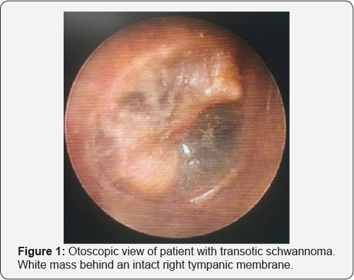
Otoscopy revealed a white, not pulsatile mass behind the tympanic membrane (Figure 1). No deficits of cranial nerves were evident in the physical examination.
Audiometric examination
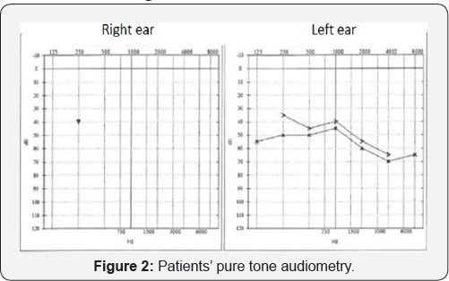
Hearing threshold was determined in both ears for air and bone conduction. Air conduction was measured for 0,125 kHz; 0,5 kHz; 1 kHz; 2 kHz; 4 kHz; 8 kHz and bone conduction for 0,5 kHz; 1 kHz; 2 kHz and 4 kHz. Pure tone audiometry showed complete left-ear deafness and moderate/severe sensor neural hearing loss according to WHO criteria on the left side (Figure 2). In tympanometry in both ears tympanogram type A was measured. In the right ear - ipsi reflex was absent for all frequencies, contra reflex was present only for 0, 5 kHz. In the left ear for all frequencies - ipsi reflexes were present, contra reflexes were absent. Psychoacoustic measurement of tinnitus pitch and loudness - patient compared his tinnitus in the right ear to the sound of frequency of 1 kHz and loudness of 60dB NBN. Minimal Making Level of tinnitus was 70dB.

a. Patient undergone hearing aid consultation: Hearing aid, CROS system and Active Middle Ear implants were proposed [8,9]. The patient decided to continue using hearing aid on the left year. Free field audiometry at 65dB showed 0% recognition threshold without hearing aid. Discrimination threshold with hearing aid at 65dB was 85%.
b. Sensory Organization Test (SOT): showed vestibular deficit, without visual and proprioceptive function deficits. Patient fell twice during the fifth trial (eyes closed, sway referenced surface).
Imaging examinations
Magnetic resonance examination was performed on Siemens Trio A Tim 3T unit. The protocol included Constructive Interference Steady State (CISS) and 3D T1 before and after intravenous gaudolinium contrast administration (Figure 3). The exam showed typical anatomical features of neurinoma/ schwannoma [10]. MRI revealed a strongly enhancing tumour within an internal auditory canal (IAC), labyrinth and tympanic cavity. Presence of fluid in an anterior part of IAC, direction of a facial nerve displacement, and partial atrophy of ipsilateral vestibulo-cochlear nerve as well as marked widening of a superior vestibular nerve suggested the vestibule and/or the superior vestibular nerve as starting point of the tumour. The tumour filled and widened the vestibule, entered into an anterior part of superior semicircular duct and filled % of the basal turn of the cochlea (supero-posterior quarter of the basal turn as well as mid and apical turns were preserved from tumour). The tumour filled: round window niche, oval window niche and posterior part of the tympanic cavity adhering to postero- inferior quadrant of a tympanic membrane.

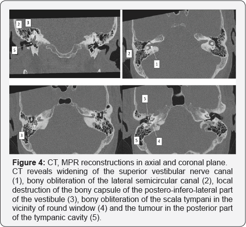
Computed tomography (CT) revealed bony destruction localized at postero-latero-inferior part of the vestibule i.e. near the posterior semicircular canal ampoule and at the posterior edge of the round window. CT confirmed segmental bony obliteration of scala tympani, total bony obliteration of the lateral semicircular canal as well as partial obliteration of superior and posterior semicircular canals (Figure 4). The patient was referred to the neurosurgery consultation. Due to the lack of progress in tumour growth during two-year checkups, watchful waiting strategy is continued.

Discussion
Schwannoma of the vestibulocochlear nerve typically arise from its vestibular branch and grow toward the CPA [11]. However, it may follow a very unusual, lateral course of extension. The bone of the otic capsule is very hard. That leads to conclusion, that transotic schwannoma extends via the round window into the middle ear [4,12]. Extension through the oval window is less likely due the mechanical barrier of the stapes [4,13]. In our study CT scan showed extension of the described lesion through the round window, without bone destruction. As mentioned before, among intralabirynthine schwannoma, transotic subtype is most rare. The development of sophisticated T2-weighted and gadolinium-enhanced T1 protocols has facilitated early diagnosis and anatomic delineation [14]. The Institute of Physiology and Pathology of Hearing is a highly specialized unit in otolaryngology, treating patients mainly with hearing impairment. Institutes Bio imaging Research Centre specializes in temporal bone imaging. Since 2009, more than 10,000 gaudolinium-enhanced MRI of head were conducted and only one patient was diagnosed with transotic schwannoma.
This highlights the unusual rarity of the tumour. The presented patient is the fourth case reported in the worldwide literature of AS extended from the inner ear only to the middle ear. According to Tran Ba Huy transotic schwannoma must be suspected in the presence of any atypical picture of ear disease [15]. CT scan and MRI are tests which enable to define tumour extension and the very nature of the lesion [16]. Proper anatomic localization by the radiologist is crucial in the assessment of patients with schwannoma [2]. In our study the diagnosis was based on the clinical evaluation and characteristic MRI features. Patient's complaints during two year observation in IFPS were stable. We did not observe any progression in audio logical or radiological features. Saltzman reviewed 645 patients with the diagnosis of acoustic neuroma [2].
Only 45 of them had intralabyrinthine schwannoma, from whom 2 had transotic location. 16 out of 45 underwent surgical intervention. The authors agreed, that surgery is indicated only in cases of intractable vertigo, extension of tumour to the CPA, or evidence of neoplasm growth with progressive symptoms [17]. Nikolopoulos reports that the expected annual growth of vestibule schwannoma is 1-2mm, but up to 75% of tumours show no expansion [18]. Tinnitus is one of the possible symptoms of acoustic neuroma. When accompanying hearing loss, the hearing disorders are always the dominant patient problem. In the present study results obtained in THS questionnaire confirmed that hearing loss was a main patient problem. Additional results of THI revealed mild handicap because of tinnitus. However in any case of one sided tinnitus it is a red flag symptom which requires further diagnosis including MRI in order to exclude acoustic schwannoma.
Conclusion
Transotic schwannoma is one of the rarest causes of tinnitus, always accompanied by sudden hearing loss. The diagnosis of transotic schwannoma is established on the radiologic evaluation. Transotic schwannoma should be included in differential diagnosis of middle ear tumours.
References
- LEE SK, CHOE MS (2013) The CT and Magnetic Resonance Imaging Features of Transotic Schwannoma: A Case Report. Journal of the Korean Society of Radiology 68(4): 281-284.
- Salzman K, Childs A, Davidson H, Kennedy R, Shelton C, et al. (2012) Intralabyrinthine schwannomas: imaging diagnosis and classification. American Journal of Neuroradiology, 33(1): 104-109.
- Amoils C, Michael J, Robert K (1992) Acoustic neuroma presenting as a middle ear mass. Otolaryngology- Head and Neck Surgery 107(3): 478-482.
- Ebmeyer J, Gehl H, Upile T, Sudhoff H (2011) Vestibular schwannoma presenting as a white middle ear mass behind an intact tympanic membrane. Otology & Neurotology 32(5): e32-e33.
- Skarzynski PH, Raj Koziak D, Rajchel J, Pilka A, Wlodarczyk A, et al. (2017) Adaptation of the Tinnitus Handicap Inventory into Polish and its testing on a clinical population of tinnitus sufferers. International Journal of Audiology 56(10): 711-715.
- Raj Koziak D, Rajchel J, Piika A, Skarzynski H, Rostkowska J, et al. (2017) Tinnitus and Hearing Survey: a Polish Study of Validity and Reliability in a clinical population. Int J of Audiol 22: 197-204.
- Henry JA, Griest S, Zaugg TL, Thielman E, Kaelin C, et al. (2015) Tinnitus and hearing survey: a screening tool to differentiate bothersome tinnitus from hearing difficulties. American journal of audiology 24(1): 66-77.
- Skarzynski H, Szkieikowska A, Olszewski t, Mrowka M, Porowski M, et al. (2015) Application of middle ear implants and bone anchored implants in treatment of hearing impairments. Nowa Audiofonologia 4(1): 9-23.
- Olszewski t (2013) The Middle Ear Implants - an overview 2(5): 1522.
- Tieleman A, Casselman JW, Somers T, Delanote J, Kuhweide R, et al. (2008) Imaging of intralabyrinthine schwannomas: A retrospective study of 52 cases with emphasis on lesion growth. AJNR Am J Neuroradiol 29(5): 898-905.
- Barrett G, Rock B, Prior M (2015) Soft tissue middle ear mass. Eur Ann Otorhinolaryngol, Head Neck dis 132: 295-296.
- Yamada S, Alba T, Takada K, Takemori S, Kumakawa K (1998) A rare case of acoustic neuroma extending from the cerebellopontine angle to the external auditory canal. J Clin Neurosci 5: 94-97.
- Sone M, Mizuno T, Nakashima T, takimoto I (2007) Middle ear schwannoma extending from the cerebellopontine angle in a patient with neurofibromatosis type 2. Otolaryngol Head Neck Surg 137: 511512.
- Shupak A, Holdstein Y, Kaminer M, Braverman I (2016) Primary solitary intralabyrinthine schwannoma: A report of 7 cases and a review of the literature. ENT Journal.
- Huy P, Hassan J, Wassef M, Mikol J, Thurel C (1987) Acoustic Schwannoma Presenting as a Tumor of the External Auditory Canal Case Report. Annals of Otology, Rhinology & Laryngology 96(4): 415418.
- Roig J, Roig Ocampos J, Serafini D, Filho O (2010) Middle ear schwannoma. BJORL 76(5): 673.
- Kennedy R, Shelton C, Salzman K, Davidson H, Harnsberger H (2004) Intralabyrinthine schwannomas: diagnosis, management, and a new classification system. Otology & Neurotology 25(2): 160-167.
- Nikolopoulos TP, Fortnum H, O Donoghue G, Baguley D (2010) Acoustic neuroma growth: a systematic review of the evidence. Otol Neurotol 31: 478-85.


