Temporal Bone Carcinoma Mimicking As Malignant Otitis Externa
Seema Monga, Arun Parkash Sharma, Junaid Nasim Malik*, Shahid Rasool, Ratna priya and Khaja Naseeruddin
Department of Otorhinolaryngology and Head-Neck surgery, Hamdard Institute of Medical Sciences & Research and HAHC Hospital, Jamia Hamdard University, India
Submission: August 14, 2017; Published: August 21, 2017
*Corresponding author: Junaid Nasim Malik, Assistant Professor, Department of Otorhinolaryngology and Head-Neck surgery, Hamdard Institute of Medical Sciences & Research and HAHC Hospital, Jamia Hamdard University, Delhi-110062, India, Tel: 91 9811710433; Email: drjunaidnasim@yahoo.co.in
How to cite this article: Seema M, Arun P S, Junaid N M, Shahid R, Ratna p, Khaja N. Temporal Bone Carcinoma Mimicking As Malignant Otitis Externa. Glob J Otolaryngol. 2017; 9(5): 555773. DOI: 10.19080/GJO.2017.09.555773
Abstract
Any elderly patient presenting with severe otalgia, profuse otorrhea and associated facial palsyhaving predisposing factors such as Diabetes mellitus, immunocompromised states should be worked up for the diagnosis malignant otitis externa or skull base osteomyelitis. Carcinoma of the temporal bone is rare but it also has a similar presentation. Cases of malignant otitis externa refractory to treatment should be suspected for possibility of squamous cell carcinoma of the temporal bone and biopsy should be taken for confirmation by histopahological examination so as to avoid undue delay in requisite treatment. We present our experience of such a case of temporal bone carcinoma mimicking as malignant otitis externa.
Keywords: Malignant otitis externa; Skull base osteomyelitis; Temporal bone squamous cell carcinoma
Introduction
Malignant otitis externa is a rare and potentially fatal invasive infection of external canal,mastoid and skull base seen predominantly in diabetics and immunocompromised patients. Predominant organism responsible for it is Pseudomonas aeruginosa although a variety of other organisms can cause it including fungal species like Aspergillus [1]. Both squamous cell carcinoma and malignant otitis externa are characteristically seen in sixth decade of life and can present with similar signs and symptoms (otalgia, ear discharge, granulations) in external auditory canal. These two diseases not only masquerade each other they can also coexist in rare situations [2]. We present an unusual case of temporal bone carcinoma and malignant otitis externa presenting synchronously in an elderly, nondiabetic, immunocompetent host. This case highlights that surgeons and physicians must now be cognizant of these two virtually indistinguishable and virtually fatal clinical entities thus taking an aggressive approach to make a clear cut diagnosis which can speed up the treatment.
Clinical Case
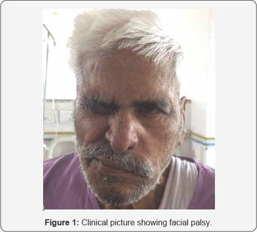
A 73 Year old male, resident of Delhi presented with asymmetry of face on left side and left ear discharge since one and a half month (Figure 1). He had no history of diabetes, tuberculosis or any significant prolonged illness. He had been on oral antibiotics including ciprofloxacin, amoxycillin-clavulanate from a local practitioner but was refractory to treatment. Physical examination revealed profuse discharge with blood in left EAC. On cleaning the ear external canal, it was found to be inflamed with polypoid tissue obstructing whole of the canal with only minimal tenderness.

Patient was admitted and all investigation was done. Audiometry revealed moderate to profound mixed hearing loss in left ear and normal hearing in right ear. He had left side grade IV facial palsy and absent gag reflex with left vocal cord palsy suggesting IX and X cranial nerve palsies rest of the cranial nerves were normal. His blood investigations, including viral markers, were all normal and did not suggest an immunocompromised state. ESR was slightly raised (46mm/hr).
Culture & Sensitivity of the pus from ear canal revealed Pseudomonas aeruginosa, sensitive to piperacillin/tazobactum, carbapenems, aztreonam, ceftazidime, ciprofloxacin, amikacin, polymyxin-B. CECT Temporal bone was done, showing heterogeneously enhancing soft tissue density mass lesion involving left external auditory canal, tympanic cavity, mastoid air cells with destruction of petrous part of temporal bone, inner ear structure, tympanic cavity and mastoid air cells and causing extension to nasopharynx and resultant attenuation of fossa of Rossenmullar on left side and same side loss of normal configuration of ossicular chain, bony canal, facial nerve canal and inner ear structure (Figure 2).

Provisional diagnosis of malignant otitis externa was made and patient was started on piperacillin-tazobactum and biopsy was taken from polypoidal tissue in external canal. Till the time biopsy report was awaited (7 days) patient was given intravenous antibiotics and there was only partial improvement in ear pain and ear discharge but no improvement in cranial nerve palsies. Final histopathology report came out to be Squamous cell carcinoma external auditory canal. Patient was referred to radiotherapy department in view of his age and stage of disease.
Discussion
Malignant otitis externa is a rare potentially fatal form of ear canal infection which typically start as a chronic infection of external auditory canal, middle ear and sinuses or may be as a result of skull base surgery [3]. Spread of disease outside of external auditory canal occurs through fissures of Santorini and osseo-cartilaginous junction. It was described as pyocutaneous osteomyelitis of temporal bone by Meltzer in 1959 although it is also believed that Toul Mouche was the one who first described this condition as early as 1838 [4,5]. Chandler further discussed the clinical features of malignant otitis externa and defined it as malignant otitis externa [6]. He described this entity as malignant because he observed an aggressive clinical behavior, poor treatment outcome and high mortality rate for these patients this aggressive infection is commonly seen in immunocompromised people such as those with diabetes mellitus, HIV, chemotherapy induced aplasia, refractory anemia, chronic leukemia. Pseudomonas Aeruginosa has been isolated from virtually all reported cases. Anaerobic organisms may also be involved as has been suggested by response to metronidazole in some cases [7].
Facial palsy is commonly present and paralysis of IX, X, XI and XII cranial nerves may also occur. Other complications include mastoiditis, meningitis, thrombosis of sigmoid sinus and septic arthritis of TM joint [8,9]. Examination of external auditory canal reveals edema of its walls, profuse discharge and granulations of its floor. Main differential diagnosis is carcinoma of temporal bone. Our patient was elderly non-diabetic who presented with otalgia, ear discharge and facial palsy. He also had cranial nerves IX, X nerve palsy. His ESR was 46mm/h. ESR is invariably elevated with an average of 87 mm/h. Levenson's criteria are useful for diagnosis of malignant otitis externa. The criteria include refractory otitis externa, severe nocturnal otalgia, purulent otorrhea, presence of granulation tissue in the external auditory canal, growth of pseudomonas in the culture from ear discharge and presence of diabetes or immunocompromised state [10].
Imaging studies are mandatory to differentiate the two diseases, to see extent of disease and response to therapy for malignant otitis externa. Gallium 67 scanning, Tc 99 methylate diphosphonate bone scan are mandatory. Ga 67 scan is more advantageous than Tc 99 in diagnosis and also in observing the response to treatment and detecting recurrence [11]. Our patient could not be evaluated for these scans due to lack of equipment and financial restraints. This case highlights that squamous cell carcinoma of the ear should be suspected in the presence of prolonged symptoms despite appropriate antibiotics and early biopsy is warranted. It cannot be determined what developed first in this patient, squamous cell carcinoma or malignant otitis externa but simultaneous treatment of both is the key to cure.
References
- JR Grandis, BF Branstetter IV, VL Yu (2004) The changing face of malignant (necrotising) external otitis: clinical, radiological, and anatomic correlations. Lancet Infectious Diseases 4(1): 34-39.
- SA Moody, BE Hirsch, EN Myers (2000) Squamous cell carcinoma of the external auditory canal: an evaluation of a staging system. American Journal of Otology 21(4): 582-588.
- WH Slattery III, DE Brackmann (1996) Skull base osteomyelitis: malignant external otitis. Otolaryngologic Clinics of North America 29(5): 795-806.
- Meltzer PE, Kelemen G (1959) Pyocutaneous osteomyelitis of the temporal bone, mandible and zygoma. Laryngoscope 169: 1300-1316.
- Bhandary S, Karki P, Sinha BK (2000) Malignant otitis externa: a review. Pac Health Dialog 9: 64-67.
- Chandler JR (1968) Malignant external otitis. Laryngoscope 78: 12571294.
- John AC, Cheeseman AD (1979) Malignant otitis externa. Hospital Update 5: 589-599.
- Aldous EW, Shin JB (1973) Far advanced malignant external otitis: report of a survival. Laryngoscope 83: 1810-1815.
- Chandler JR (1977) Malignant otitis: further considerations. Ann Otol Rhinol Laryngol 86: 417-428.
- Thiagarajan B (2000) Malignant otitis externa a review of current literature: Difficult to diagnose and troublesome to treat.
- Carfrae MJ, Kesser BW (2008) Malignant otitis externa. OtolaryngolClin North Am 41: 537-549.


