




 GJAA Home



Tarsal coalition: A case of Symphalagism in Ancient Macedonia
Cockerill Samuel James1, Garoufi Nefeli1 , Dimakis Nikolaos2 and Chovalopoulou Maria-Eleni1*
1Department of Animal and Human Physiology, Faculty of Biology, School of Sciences, National and Kapodistrian University of Athens, Panepistimiopolis, GR 157 01 Athens, Greece
2Department of History and Archaeology, School of Philosophy, National and Kapodistrian University of Athens, Panepistimiopolis, GR 157 01 Athens, Greece
Submission:April 26, 2026; Published:October 15, 2026
*Corresponding author: Chovalopoulou Maria-Eleni, Department of Animal and Human Physiology, Faculty of Biology, School of Sciences, National and Kapodistrian University of Athens, Panepistimiopolis, GR 157 01 Athens, Greece
How to cite this article: Cockerill Samuel J, Garoufi N , Dimakis N, Chovalopoulou Maria-E. Tarsal coalition: A case of Symphalagism in Ancient Macedonia. Glob J Arch & Anthropol. 2026; 15(1): 555901. DOI: 10.19080/GJAA.2026.15.555901
Abstract
The excavation in 2021 and 2023 Grevena, Western Macedonia, Greece presented four single graves, each with skeletal remains. The buried individuals were likely buried sometime after 148 b.C. as concluded by a dated coin found in the 2nd burial. The 3rd burial presented human remains of a male over the age of 20 years old showing a case of symphalangism in the fourth or fifth tarsal phalange. The phalange presents a porous surface with a deviated fused intermedial phalange that could have possibly fused during early childhood. Symphalangism is an important inherited condition that could aid kinship analysis and biodistance research in the future.
Keywords: Anatomical Anomalies; Ankylosis; Kinship; Biodistance; Ancient Greece
Introduction
Symphalangism is described as a congenital condition that results in partial or total ankylosis of the interphalangeal joints of the fingers or/and toes [1]. The first authors to describe this congenital fusion were Mercier in 1838 and Moutard-Martin & Pissavy in 1895 [2,3]. However, it was Harvey Cushing who named the condition symphalangism in 1916. This author described a case of a family in Virginia that had previously arrived in North America from Scotland in the 1700s. The trait does appear to follow a Mendelian transmission and found frequently among families. In Cushing’s case, a family of seven individuals were reported to present the congenital trait [4]. Symphalangism has been associated with syndactyly, brachydactyly, hearing loss and other syndromes [1].
Congenital anomalies such as symphalangism may be used to identify kinship groups in archaeological contexts. Kinship analysis and the process of identifying relationships in past populations is of utmost importance if we are to learn about how ancient societies lived. All the components of past populations worked due to the relationships that existed between social groups and family members. It is important to note that ancient societies did not perceive the concept of “family” or kinship in the same way we do today, and this has been observed in a variety of studies. Furthermore, kinship may reveal information on existing hierarchies, social structure, family structures, and other details on social groups within a cemetery [5,6]. Anatomical anomalies, such as non-metric traits, may be used as genetic proxies to help identify kinship groups when DNA analysis is not possible. Anatomical traits found among adults have shown developmental processes that occurred during childhood, providing insights into early biological development of past populations [7]. The process of identifying relationships in skeletal remains is also important in forensic contexts, where identifying anatomical anomalies could lead to a correct identification of a victim [8].
In archaeological literature, there is an absence of reporting of symphalangism in the fifth toe despite the common frequency of the fifth toe symphalangism in modern populations and this could possibly be due to the scarce attention of archaeologists on the minor bones of the foot [9].
In September 2021 and 2023 a total of 4 individuals were discovered during the excavation at Tsigodendro in Grevena, Western Macedonia, Greece which was once Ancient Macedonia. Individual II was found clutching an ancient coin dated from 148 B.C. Thessaloniki which gave the archaeologists a rounded time period from which the skeletal remains were buried (Dimakis et al., in press). These individuals who resided in the area of Grevena around the late Hellenistic period were likely associated with the Molossian tribes of this area or early Macedonian farmers or herders who settled in the area following the Macedonian expansion in 370 b.C. (Hammond, 1986). Among the skeletal remains, Individual III presented a proximal fourth or fifth phalange with symphalangism.
To our knowledge there have been no previously known archaeological cases showing proximal and intermediate symphalangism. Our objective is to show a novel case of tarsal symphalangism in the fourth or fifth proximal phalange and to describe the finding indicating its importance in ancient Macedonia.
Materials and Methods
In this work, the osteological findings of the archaeological excavations carried out in 2021 and 2023 at Tsigodentro in the area of Agios Georgios Grevena (see Figure 1) by the Ephorate of Antiquities of Grevena in collaboration with the Department of History and Archaeology of the National and Kapodistrian University of Athens, were analyzed. The rescue excavation began in 2021, following a report from a resident of a nearby village about antiquities on their plot.
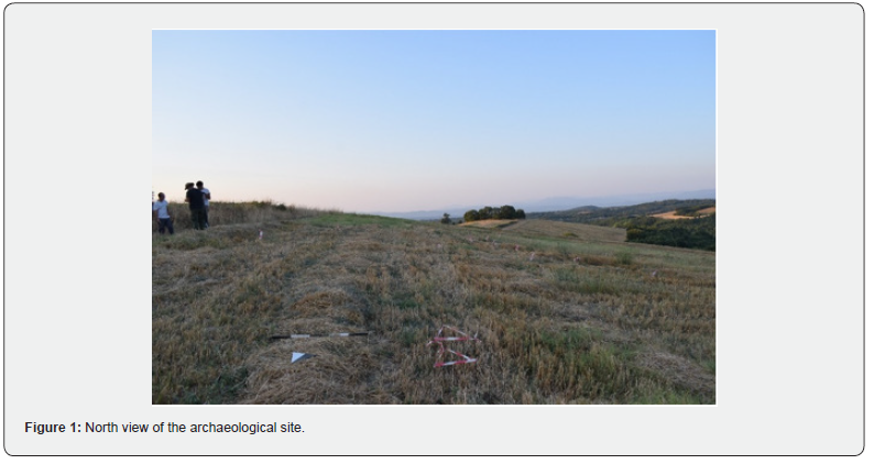
The phalange bone originated from a rescue excavation in Tsigodendro, Agios Georgios, Grevena 2021. This excavation revealed a total of 4 graves. The individual presented in this study was recovered from Grave III. All graves were orientated east-west, with the skull eastwards and their tarsal bones pointing westwards. The exception was Grave IV which was orientated northwest-southeast. All the osteological remains were transported and are currently stored in the Faculty of Biology in the Kapodistrian University of Athens. The age and sex of the individual were estimated using standard methods [10-12]. For age estimation, the AgeEst tool was also used using the standard morphological features [13]. Stature was estimated according to the equations developed by Lee et al. (2024) in cases of fragmented bones and Nikita and Chovalopoulou’s work (2017) in cases of whole long bones.
Results
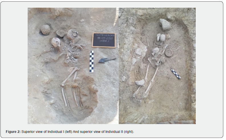
The skeletal analysis showed some fauna bones among the human remains. Individual I (see Figure 2) presented remains that were sufficiently preserved to estimate sex. Using the standard methods [10-12], the individual was likely female and due to findings of a 3rd molar, and the epiphyseal fusion, the individual was an adult. Despite the poor condition of the epiphysis of most of the long bones, height estimation was calculated from the maximum diameter of the deltoid tuberosity, presenting a height of 1.50m. No pathologies were detected in Individual I, although pre-mortem tooth loss was identified in the 2nd premolar, and the 1st and 2nd molars.
Regarding Individual II (see Figure 2), the remains were highly damaged but staining on one of the phalanges coincided with the presence of a coin dated to Thesaloniki 148 b.C. No pathologies were observed, except for an abscess in the maxilar. However, no evidence of spreading infection was found. Notably, osteophytes were found on the right clavicle and on the vertebrae joints on the ribs which provided evidence of possible lifting and carrying. The existence of alveolus of the third molar showed evidence of adulthood and the AgeEst tool was used for an age estimation of 77.2±19.0 years old, while using a neural network the result was 79.2±20.4 years old [13]. Sex was estimated as male using the standard methods and the stature was calculated using various long bone measurements resulting in a height of approximately 1.62m.
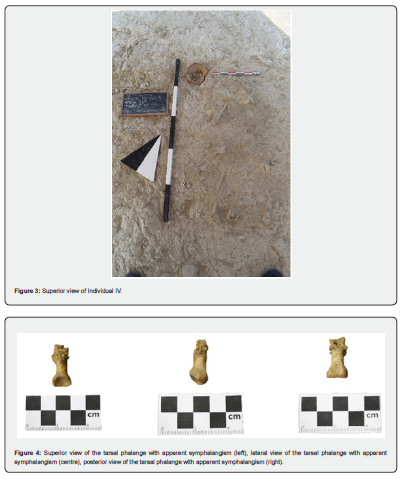
The remains of Individual IV (see Figure 3) were of a subadult with badly preserved osteological remains, likely due to the fragility of the bones. Age estimation showed the individual was between 4 and 5 + 1.3 years old using dental metrics and having found a fused clavicle [12]. Additionally, no pathologies were detected for this individual.
Individual III was estimated to be a male over the age of 20 years old. The individual presented ostephytes on the tibial tuberosity and soleus line, possibly indicating the presence of high activity on lower limbs during daily life. In Figure 4, a fourth or fifth proximal tarsal phalange with an intermediate phalange fused to its surface with slight side deviation can be observed. Due to its small overall size compared to the broad epiphyses, it is more probable that it is a fourth phalange. Unfortunately, it is impossible to confirm whether the bone was a fourth or fifth phalange due to the damaged remains of the individual. However, the phalange presents osteophytes on the distal epiphysis which are located on the attachment surface of the intermediate phalange ligaments. The fused intermediate phalange presents a porous surface and is slightly deviated.
In Figure 4, the typical shape of the proximal epiphysis of a tarsal phalange can be observed. However, regarding the distal epiphysis, a slightly deformed surface is observed, due to the synostosis of the two phalanges. As shown, there are no sutures or impartial fusion. Instead, the connection is complete, stable and unmovable.
Regarding Figure 4, we can see the posterior side of the phalange of interest which shows a porous surface in between the two phalanges.
Discussion
The development of symphalangism has been studied in previous studies and shows how the fusion of both phalanges originates from cartilaginous symphalangism which is an earlier stage of the condition [1]. This process is thought to occur during childhood, which suggests that our case may have also developed during childhood. The development of symphalangism from cartilaginous to bone also coincides with the porous surface of the bone in our case (see Figure 4) which can be observed among other anomalies which share similar hyperostotic development, such as os trigonum [14].
According to Harle & Stevenson [15] cases of symphalangism can occur symmetrically and in both carpal and tarsal bones. However, in our case we only found one phalange presenting symphalangism. It is possible that the individual presented other cases of symphalangism in other phalanges, but we did not identify any other cases due to the state of preservation of the material. The development of symphalangism seems to occur during early development.
In our case, the posterior face of the fusion shows porous bone which is consistent with the origin proposed by previous studies. According to Nightingale et al. [1], the development of symphalangism can occur as early as the first weeks of life after birth. These authors and others [16] also show how the first stage of development (Grade 1-2) is cartilaginous fusion of the interphalangeal joint, which could develop into the second stage of symphalangism in which bone fusion occurs (Grade 3-5) during early childhood. This suggests that Individual III likely developed symphalangism at a very early stage of life which could’ve transformed into bone fusion of the interphalangeal joint. Many authors mention that this trait is highly inheritable [1,4,17] and due to the context of the burial of the four individuals together, it is very possible they were family. However, no other cases of symphalangism were found among the other individuals. Likewise, due to the preservation of the human remains, we were unable to detect non-metric traits or anatomical variants shared between the group of individuals. Nevertheless, this cannot eliminate the possibility that these individuals did not present symphalangism or a related pathology such as ankyloses among bones that were badly preserved.
In previous studies, no reported age or sex differences have been reported [17,18]. Frequencies of the fifth toe presenting interphalangeal symphalangism showed very high percentages among modern Japanese , lower in modern Euro-Americans and modern African-Americans in previous research [17]. However, it was shown that almost half the population sample presented some form of symphalangism on the fifth toe. On the other hand, frequencies of symphalangism on the fourth toe are much rarer, showing percentages lower than 5.0% with the exception of the Japanese sample [17,18]. These low frequencies of the fourth toe and the very high inheritable characteristic of symphalangism highlight the trait’s relevance in kinship analysis, as it could be used in biodistance studies if it found numerous times within a sample.
A previous study showed that fused phalanges were present in less than 5% in Spain, while fused phalanges were present in 25% of modern sample of Spain showing how modern footwear can influence the presence of this trait [19]. Furthermore, the low presence of fused phalanges in past populations likely shows inheritable cases of symphalangism in contrast with the high frequency of fused phalanges in modern populations due to footwear which significantly affects phalange development. Thus, the discovery of symphalangism in past populations is rare and should be regarded as an important find and a trait that could be used in kinship analysis due to its scarcity in ancient populations. Unfortunately, in our case it could not be used as a trait in kinship analysis due to the state of preservation of the skeletal remains. However, symphalangism could likely be used in kinship analysis on future research and archaeologists should give attention to the presence of this anomaly in archaeological collections.
Conclusion
Individual III from the Tsigodendro, Grevena excavation of 2021 presents a case of symphalangism of the fourth or fifth phalange and this finding in Ancient Macedonia has historical and archaeological meaning. The anomaly is rare in the fourth phalange but rather common in the fifth phalange. We hope to have showcased a clear case of this condition in an archaeological context, Good moand we hope to have highlighted the importance of this anomaly for its use in future research in kinship analysis or biodistance studies.
References
- Nightingale Craig, Katerina Kyprianou, and Mobin Syed (2020) A Strongly Inherited Familial Symphalangism of All the Proximal Interphalangeal Joints. Plastic Surgery Case Studies 6: 2513826X20970355.
- Mercier LA (1838) Absence Hereditaire d’uue Phalange Aux Doigts et Aux Orteils. Bull Soc Anal 13(35).
- Moutard-Martin and Pissavy H (1896) Malformations congénitales, multiples et héréditaires, des doigts et des orteils. Paris, France.
- Cushing H (1916) Hereditary Anchylosis of the Proximal Phalan-Geal Joints (Symphalangism). Genetics 1(1): 90-106.
- Cockerill Samuel James, Verónica Alberto-Barroso, Matilde Arnay-De-La-Rosa, and Emilio González-Reimers (2024) Intracemetery Relationships: Non-Metric Trait Combination Analysis of Maspalomas of Gran Canaria (Canary Islands, Spain). Anthropologischer Anzeiger; Bericht Uber Die Biologisch-Anthropologische Literatur 81(4): 433-447.
- Cvrček Jan, Petr Velemínský, Ján Dupej, Luboš Vostrý, and Jaroslav Brůžek (2018) Kinship and Morphological Similarity in the Skeletal Remains of Individuals with Known Genealogical Data (Bohemia, 19th to 20th Centuries): A New Methodological Approach. American Journal of Physical Anthropology 167(3): 541-556.
- Cockerill SJ, and Rodríguez Caraballo S (2022) El Astrágalo Como Un Indicador Para El Estudio de La Infancia En Las Poblaciones Prehispánicas de Las Islas Canarias. Rev Esp Antrop Fis 46: 40-50.
- Palamenghi Andrea, Daniele Gibelli, Debora Mazzarelli, Danilo De Angelis, Chiarella Sforza, and Cristina Cattaneo (2023) Rumor Has It: A Narrative Review on the Use of Skeletal Non-Metric Traits and Variants for Personal Identification. Legal Medicine 65: 102316.
- Capasso Luigi, and Gabriella Di Tota (1998) Fifth Toe Distal Inter-Phalangeous Synostosis: Paleopathological Description and Possible Evolutionary Significance. Anthropologie (1962-) 36(3): 231-233.
- Bass WM (1987) Human Osteology. A Laboratory Field Manual. 3rd Missouri Archaeological Society, United States.
- Buikstra JE, and Ubelaker DH (1994) Standards for Data Collection from Human Skeletal Remains. Proceedings of a Seminar at the Field Museum of Natural History Organized by Jonathan Haas. Arkansas Archaeological Survey, USA.
- Scheuer and Black (2000) Developmental Juvenile Osteology. Elsevier.
- Constantinou C, Chovalopoulou ME, and Nikita E (2023) AgeEst: An Open Access Web Application for Skeletal Age-at-Death Estimation Employing Machine Learning. Forensic Science International: Reports 7: 100317.
- Cockerill Samuel James, Matilde Arnay-de-la-Rosa, and Emilio González-Reimers (2025) An Atlas of Anatomical Variants of the Human Talus. Journal of Morphology 286(1): e70023.
- Harle Thomas S, and James R Stevenson (1967) Hereditary Symphalangism Associated with Carpal and Tarsal Fusions. Radiology 89(1): 91-94.
- Baek Goo Hyun, and Hyuk Jin Lee (2012) Classification and Surgical Treatment of Symphalangism in Interphalangeal Joints of the Hand. Clinics in Orthopedic Surgery 4(1): 58-65.
- Case DT, and Heilman J (2005) Pedal Symphalangism in Modern American and Japanese Skeletons. HOMO 55(3): 251-262.
- Nakashima Tamiji, Teruyuki Hojo, Katsumi Suzuki, and Masateru Ijichi (1995) Symphalangism (Two Phalanges) in the Digits of the Japanese Foot. Annals of Anatomy - Anatomischer Anzeiger 177(3): 275-278.
- Saldías Eduardo, Assumpció Malgosa, Xavier Jordana, and Albert Isidro (2019) Morphological and Biomechanical Implications of Cuboid Facet of the Navicular Bone in the Gait. International Journal of Morphology 37(4): 1397-403.

























