Eruption Status and Congenital Absence of Mandibular Third Molars from the Past to the Present in the Japanese Population
Hiroyuki Yamada*
Ph.D. Former Dentist, Dental Anthropologist, Department of Oral Anatomy, School of Dentistry, Aichi-Gakuin University, Japan
Submission: October 12, 2023; Published: October 20, 2023
*Corresponding author: Hiroyuki Yamada, Ph.D. Former Dentist, Dental Anthropologist, Department of Oral Anatomy, School of Dentistry, AichiGakuin University, 15-16 Obatanaka 2 Chome, Moriyama-Ku, Nagoya 463-0013, Japan
How to cite this article: Hiroyuki Yamada*. Eruption Status and Congenital Absence of Mandibular Third Molars from the Past to the Present in the Japanese Population. Glob J Arch & Anthropol. 2023; 13(3): 555861. DOI: 10.19080/GJAA.2023.13.555861
Abstract
The eruption status and congenital absence of mandibular third molars were studied over time from the Jomon era to the present. The data were radiographs of buried human skeletons and modern populations. Eruption status was classified as upright, inclined, horizontal and congenital absence. The relationship with the M2-retromolar space was also examined. The eruption style of the mandibular third molars depends on the size of the M2-retromolar space, and as the M2-retromolar gap becomes narrower, the eruption goes from upright to inclined and then to horizontal. In congenital absence, most of the M2-retromolar space is less than 1/2 the mesiodistal diameter of the second molar. Congenital absence of mandibular third molars has been strongly influenced by heredity from prehistoric times to the Edo era. However, from the 1930s to the present, it has been more influenced by the environment, particularly food habits, with congenital absence of mandibular third molars declining as dietary habits becomes more favorable.
Keywords: Mandibular Third Molar; Eruption Status; Congenital Absence; M2-Retromolar Space; Secular Change
Introduction
The third molars are the teeth with the most delayed ontogeny, the highest incidence of absence, the greatest variation in size and shape, and the most pronounced tendency to degenerate. Dental anthropologists, anatomists, taxonomists and forensic scientists have taken note and carried out numerous studies. Garn and others [1,2] have studied the absence rate and eruption time of third molars, as well as the degeneration in size of remaining teeth other than third molars. Brothwell et al. [3] described the worldwide distribution of the incidence of congenital third molar absence. The eruption status of third molars [4,5] and the frequency of eruption [6] have also been studied. Byahatti et al. [4] reported that in a study of emergence direction in South Indians, vertical position was the most common (56.7%), followed by inclined position (21.1%) and horizontal position (7%). Kumar et al. [7] addressed the relationship between mandibular third molars and mandibular angle fractures, and Tuteja et al. [8] provided age estimates based on the timing of third molar formation from a legal dentistry perspective. There have been reports of genetic factors that are now being used to explain a significant proportion of dental agenesis. To date, the MSX1, PAX9, AXIN2 and EDAR genes have been identified as causes of familial dental agenesis, and some specific variants of the PAX9 and EDAR genes have been associated with third molar agenesis [9].
Various factors such as nutritional condition, living environment, diet, region of residence, climate and age/time of birth are thought to play a role in the eruption status and congenital absence of third molars [10]. However, there are not many studies that have investigated the eruption alignment and frequency of missing mandibular third molars historically [11]. Only one quantitative analysis of the changes in the M2-retromolar space in the Japanese population from prehistoric times to the present has been reported [12]. The aim of this study was to determine the relationship between the congenital missing of mandibular third molars and the eruption condition based on chronological changes, and to investigate the influence of the M2-retromolar clearance on them.
Materials and Methods
The materials examined were intraoral radiographs of human bones [13] from archaeological sites in the Jomon, Yayoi, Kofun, Kamakura, Muromachi, and Edo eras, and panoramic radiographs of patients as modern people. The study was conducted on the eruption alignment of the mandibular third molars by dividing them into four categories: upright, inclined, horizontal positions, and congenital absence. Material observations were made on the left and right sides of the jaw in both sexes, and statistics were calculated based on the number of sides of the jaw. For modern humans, the left and right teeth of men and women born from the 1920s to 1980s were examined at 10-year intervals. Most of the material came from people who were over 20 years old at the time of the study.
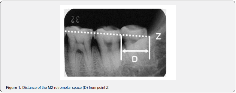
In examining the alignment status of the third molars, the horizontal position observed on the radiographs was defined as the case where the tooth axis was almost parallel to the occlusal plane, the inclined position was defined as the case where the third molars encroached on the distal cervical portion of the second molars and/or the tooth axis was inclined at approximately 45 degrees to the occlusal plane, and the upright position was defined as the case where the tooth axis was almost perpendicular to the occlusal plane. Congenital absence was defined as the absence of tooth embryos in the alveolar bone on radiographs and no history of tooth extraction in the past. Both the inclined and horizontal positions were difficult to erupt with ease, and both were combined as eruption difficulty (inclined + horizontal). On the molar radiographs, the intersection point (Z) was defined as the point of intersection between the extension of the straight line connecting the most mesially protruding point of the first molar and the most distally protruding point of the second molar and the internal oblique line. This point (Z) was used as a reference for the end point of the retromolar space. The distance (D= M2-retromolar space) from the distal surface of the mandibular second molar to the Z point (Figure 1) was measured and compared with the mesiodistal diameter of the mandibular second molar [12]. First, 30 individuals in each category were examined according to the following criteria. The frequency of this category was calculated as 〇 if the distance (D) was greater than the mesiodistal diameter of the second molar, △ if it was within half of this distance from the mesiodistal diameter of the second molar, and ✕ if it was less than half, and then the relationship between the frequency of this category and eruptive status was compared.
Results
The relationship between eruption status and D-values is shown (Table 1). The most common conditions in each eruption alignment were upright (76.7%: 〇), inclined (53.3%: △) or horizontal (53.3%: ✕) and congenital absence (76.7%: ✕).

Table 2 and Figure 2 summarise the number of jaw sides examined and the eruption style (horizontal, inclined and upright) for each era. The eruption status of the mandibular third molars shows that in the Jomon era, 1 out of 132 molars were horizontal, 3 were inclined, and 128 were vertical, with the majority in the upright position. The percentage of those with eruption difficulty was extremely low at 3.1%. The frequency of the eruption problem in the Kofun era was almost the same as in the Jomon era. Subsequently, from the Kamakura era until the 1940s, the difficulty fluctuated between 17% and 23%, with the exception of the Muromachi era. In modern times, the difficulty of eruption increased rapidly to over 40% in the 1950s, and this percentage continued to rise, reaching 76.6% in the 1980s. In particular, from the Jomon era until the 1960s, less than 16% of cases involved the inclination, but this increased to 23% in the 1970s and rose rapidly to 51% in the 1980s. Today, the majority of people with eruption difficulties are in the inclined position.
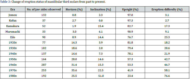
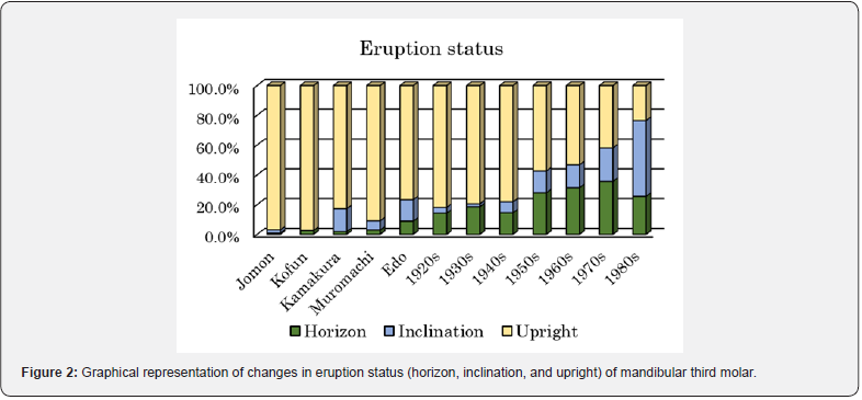
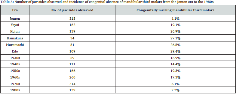
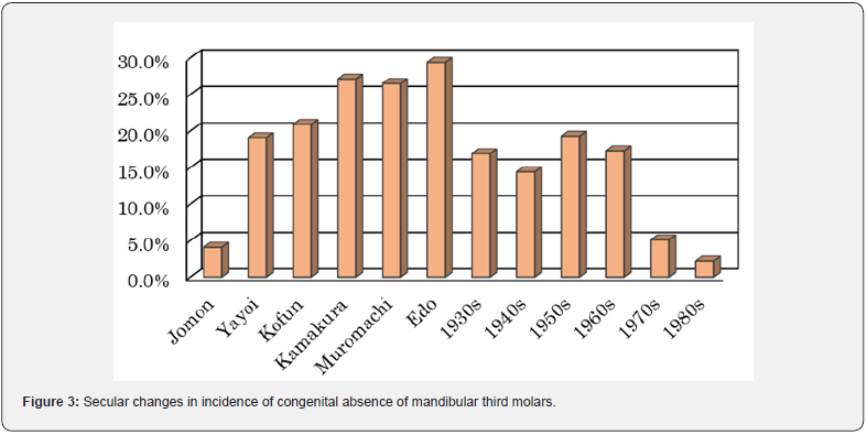
Table 3 shows the incidence of missing third molars from the Jomon to the Edo era and the incidence from the 1930s to the 1980s according to the year of birth in each decade. The incidence was low in the Jomon era (4.1%) and high in the Yayoi era (19.1%). There was a marked difference between the two eras. It then increased slightly in the Kofun era and fluctuated between 25% and 30% from the Kamakura to the Edo eras. In modern times, the incidence of congenital absence began to decrease dramatically in the 1930s (16.9%), a trend that continued, albeit gradually, in the 1940s. The rate of decline then accelerated significantly in the 1970s, reaching 5.1%, with a further decline to 2.2% in the 1980s (Figure 3).
Discussion
There have been many studies of third molars in the Japanese population. Koganei [14] studied the frequency of congenital absence of the third molar from the skull for human populations in and around Japan, and Saito, Ozaki [15] studied the incidence of eruption and absence of the third molar from radiographs of students at the University of Tokyo. Yamada [16] also studied eruption rates for each age group from 18 to 40 years. Osuga [17] studied the relationship between third molar eruption style (full impaction, half impaction and full eruption) and mandibular size using radiographic cephalograms of modern populations. In this paper, the author focused on the eruption status and nutritional conditions of the lower third molars and examined their chronological changes. In general, teeth erupt upright against the occlusal plane. However, mandibular third molars can erupt vertically, inclined, sometimes horizontally or even embedded.
As noted in Table 1, the eruption patterns of the third molars are highly dependent on the size of the M2-retromolar space: if the M2-retromolar gap is larger than the mesiodistal diameter of the lower second molar, they tend to erupt vertically; if it is smaller, they tend to erupt inclined or horizontally. And when the third molars are inclined or horizontal, eruption becomes extremely difficult. In palaeoanthropology, all three lower molars of a 40,000-year-old Neanderthal skull from the Amud Cave in Israel [18] erupted completely upright, and there is a fairly wide retromolar space behind the third molar.
Measuring the retromolar space is difficult even in excavated skulls and even more impossible in vivo. To determine the size of the retromolar space in the present study, a “Z” point was developed on the radiographs [12]. This point is slightly different from the Xi point (near the mandibular foramen) used in orthodontics [19]. In addition, an attempt was made to compare the relative size of the M2-retromolar space with the size of the second molar over time.
The relationship between eruption status and M2-retromolar clearance showed that as the eruption style of mandibular third molars changed from upright→inclined→horizontal, the size of the M2-retromolar space gradually decreased. In congenital absence, most of the M2-retromolar gap was less than 1/2 of the mesiodistal diameter of the second molar (Table 1). In modern populations, the eruption alignment of the mandibular third molars was largely dependent on the size of the M2-retromolar clearance. Similar results were obtained in a quantitative analysis of the M2-retromolar space [12].
In the Japanese archipelago, the Jomon era began about 16,000 years ago, at the end of the last ice age, and lasted until 3,000 years ago. The Jomon people were hunter-gatherers. As for the physical features of the Jomon, their heads were large in both length from front to back and width from left to right, and their faces were wide and short at the top and bottom, giving them a square appearance. Although their teeth are small [20], the heavy wear on their teeth probably indicates that they put considerable strain on their teeth when chewing their food.
Human remains from the Yayoi era (1,000 BC - 300 AD) are on average larger and taller than those from the Jomon era. The Yayoi were rice farmers. Their forearms and shins were relatively short in relation to their limbs, and their body shape was adapted to cold climates and was completely different in size, shape and form from that of the Jomon people. Their teeth were larger than those of the Jomon. This suggests that they were probably a group with different genes that migrated from the Korean peninsula to the Kyushu and Yamaguchi regions of Japan [21,22]. Even the size of the teeth is very different between the Jomon and Yayoi eras. According to Shinoda [23], who has studied the divergence of human populations based on mitochondrial DNA, there are clear differences in haplogroup frequencies between the Jomon and the migrating Yayoi, and it is recognised that they are populations with different lineages.
The Kofun era, which followed the Yayoi era, lasted from the middle of the 3rd century to the end of the 12th century at the beginning of the Kamakura era (1192-1333). According to Mizoguchi [24], changes in body measurements from the Kofun era to the present have been continuous, if at all, and there have been no sudden or disjointed changes such as the transition from the Jomon to the Yayoi era. In the Jomon and Kofun eras, more than 97% of the mandibular third molars erupted vertically. Inclined or horizontal third molars, which are common among modern peoples, are rarely seen. This suggests that the genetic effects of immigration and dietary changes had little effect on the direction of eruption of mandibular third molars. In terms of the difficulty of eruption, it was less than about 3% in the Jomon and Kofun eras, but rose to 17% in the Kamakura era, and then fluctuated in the range of 20% to 30% until the 1940s. Before 1945 (the end of World War II), rice was the staple food for ordinary people in Japan, supplemented by vegetables, potatoes and cereals. Until the early 1950s, during the post-war reconstruction period, people were poor due to food shortages and lived a simple life in the traditional Japanese style. However, the Korean War, which began in 1950, triggered a ‘bubble’ known as ‘Korean Special Demand’ and people experienced a period of instant relief from poverty.
In the 1950s, the difficulty of erupting the third molar in the lower jaw rose to 43%. Around 1955, after the end of the Korean War, the Japanese economy entered a period of rapid economic growth that lasted until the oil crisis of 1973, and from the late 1980s the country entered a period of economic prosperity known as the ‘bubble economy’. During this period of prosperity, which reflected a consumer economy, the dietary habits of the Japanese people changed dramatically and the market was flooded with high protein, high fat and high carbohydrate foods with high nutritional value. The incidence of third molar eruption problems also increased by almost 15% or more between the generations from the 1960s to the 1980s. From prehistoric times up to the 1950s, upright third molars were in the majority, but since the 1960s, eruption difficulties have been in the majority and most of the population now suffers from eruption problems.
On the other hand, the congenital absence of mandibular third molars was low in the Jomon era (4%), but increased rapidly in the Yayoi era (19%). It then gradually increased to 29% in the Edo era [25]. Although not shown in Table 3 because the year of birth is unknown, there was a radiographic survey in the 1930s. Ohashi and Matsumura [26] and Koganei [14] found the congenital absence of third molars to be relatively high at 46% and 39% respectively. The incidence of missing third molars increased in the 1950s, but fell dramatically in the 1970s and continued to fall to 2.2% in the 1980s. In the near future, most people in Japan may have all their lower third molars. The change in diet towards soft foods has led to a decrease in masticatory function, which in turn has led to an opening of the mandibular angle and changes in the eruption of the third molars. In addition, the increase in the number of mandibular third molars in recent years is thought to be due to a more nutritious diet. Looking at the relationship between third molar eruption status and congenital absence over time, eruption alignment is strongly influenced by diet, especially in the post-war period when the number of difficult eruption conditions (inclined + horizontal position) increased dramatically. Congenital defects of the mandibular third molars were strongly influenced by heredity from prehistoric times to the Edo era. However, in modern society, since the 1930s, they have been influenced by the environment, particularly diet, with the incidence of congenital defects decreasing as the diet becomes richer. The low rate of third molar agenesis in the Jomon era is thought to be due to the fact that people lived close to nature, hunting animals in the mountains and fields and gathering wild vegetables. In modern times, however, this balance has been altered and it is thought that richer diets and better nutrition have prevented the lack of third molars. The M2-retromolar gap strongly influences the eruption status of the mandibular third molar, but it is not involved in congenital absence.
Conclusion
The eruption status of the mandibular third molars depends on the size of the M2-retromolar space, and the smaller the amount of M2-retromolar space, the more the eruption alignment goes from upright to inclined to the horizon. In congenital absence, most of the M2-retromolar space is less than 1/2 of the mesiodistal diameter of the second molar. From the Jomon era until the late 1940s, most mandibular third molars erupted vertically, but after the end of the Second World War, nutrition improved rapidly and the majority of people began to have problems with eruption. The frequency of missing mandibular third molars was low in the Jomon era, but increased rapidly in the Yayoi era, followed by a gradual increase to a peak in the Edo era. It then declined sharply in the 1930s and even more sharply after the 1970s.
It is thought that the development of dietary habits had a positive influence on the environment for the development of third molars. In terms of the relationship between eruption status and congenital absence, eruption condition is less influenced by heredity, as little change in eruption status was found between the Jomon and Yayoi eras. Rather, the enriched diet and soft foods of the post-war period have a strong influence on the incidence of eruption problems.
Congenital defects of the mandibular third molars are strongly influenced by heredity because of the great changes in agenesis between the Jomon and Yayoi eras. It is also influenced by the environment and, particularly after the 1930s, the incidence of congenitally missing third molars decreased as lifestyles became more comfortable. The low rate of third molar agenesis in the Jomon era is thought to be due to the fact that their lives were adapted to nature. In modern times, however, this balance with nature has been compromised, and it is thought that richer diets and better nutrition have prevented the lack of third molars. The M2-retromolar clearance strongly influences the eruption status of the mandibular third molar, but it is not involved in congenital absence. The results for the M2-retromolar space calculated by the present non-metric method were in good agreement with the results of the metric method [12].
Acknowledgments
The materials of human bones excavated from archaeological sites of various eras (in the collection of the National Museum of Nature and Science) were provided by Dr. Naohiko Inoue and his research collaborators who kindly provided X-rays (intraoral radiographs). I hereby express my deepest gratitude to them. The author would like to express sincere gratitude to Dr. Yuji Mizoguchi, former Director of the Department of Anthropology, National Museum of Nature and Science, for his willingness to cooperate in the research of the materials. In addition, Dr Shinichi Muroi, Director of the Muroi Dental Clinic in Koriyama, Fukushima Prefecture, and Mr Shinji Okumura, X-ray technologist-in-chief at the School of Dentistry, Aichi-Gakuin University, cooperated in collecting data on modern people for this study. I would like to express my sincere gratitude to them.
References
- Garn SM, Lewis AB, Vicinus JH (1963) Third molar polymorphism and its significance to dental genetics. Journal of Dental Research Supplement 42(6): 1344-1363.
- Garn SM, Lewis AB, Kerewsky RS (1963) Third molar agenesis and size reduction of the remaining teeth. Nature 200: 488-489.
- Brothwell DR, Carbonell VM, Goose DH (1963) Congenital absent of teeth in human populations. In: Brothwell DR (ed.), Dental Anthropology. Pergamon Press, pp. 179-190.
- Byahatti SM, Nayak R, Jayade B (2011) Eruption status of third molars in South Indian city. Journal of Indian Academy of Oral Medicine and Radiology 23(3): 328-332.
- Sivaramakrishnan SM, Raman P (2015) Study on the prevalence of eruption status of third molars in South Indian population. Biology and Medicine (Aligarh) 7(4): 1-4.
- Ragini, Singh N, Goyal S, Padmanabhan P, Munjal P (2003) Prediction of third molar eruption. Journal of Indian Orthodontic Society 36: 103-112.
- Kumar PS, Dhupar V, Akkara F, Kumar GBA (2015) Eruption status of third molar and its possible influence on the location of mandibular angle fracture: A retrospective analysis. Journal of Maxillofacial and Oral Surgery 14(2): 243-246.
- Tuteja M, Bahirwani S, Balaji P (2012) An evaluation of third molar eruption for assessment of chronologic age: A panoramic study. Journal of Forensic Dental Sciences 4(1): 13-18.
- Shahid M, Joshi S, Alqhtani NR, AlSaidan M, Aldossari K, et al. (2017) Single nucleotide polymorphism (SNPs) in the genes associated with tooth agenesis. European Journal of Experimental Biology 7: 1-13.
- Yamada H, Hanamura H (1996) Secular change of third molar emergence times in a modern Japanese population ―For the past 80 years―.The Aichi-Gakuin Journal of Dental Science 34(4): 697-705.
- Yamada H (2015) A small Encyclopedia of Dentition―Dental Anthropology from the Viewpoint of Dentists—. Maruzen Pub. Co. Ltd, Tokyo, pp. 153.
- Yamada H (2023) Chronological changes in retromolar space viewed from radiographs in Japanese. International Journal of Surgery and Clinical Practice 5(2): 1-5.
- Inoue N, Itou G, Kamegai T (1987) Microevolution and Dental disease in Human Occlusion—Studies on tooth to denture base discrepancy—. Ishiyaku Co. Ltd., pp. 251.
- Koganei Y (1934) Human occlusal typologies and their phylogenetic significance. Acta anatomica Nipponica 7(3): 283-325.
- Saito H, Ozaki Y (1936) Die Statische Beobachtung über die Sogenannte Alveolar Phyorrhoe und den Weisheitzahn Durchbruch in Unseren Jugend. Japanese dental community 202: 1-17.
- Yamada H (1992) Third molar emergence in a modern Japanese population. Anthropological Science 100(4): 425-432.
- Osuga N, Kubota M, Miyazawa H (1999) The relationship between the eruption status of the mandibular third molars and the mandibular morphology in Japanese. The Japanese Journal of Pediatric Dentistry 37(1): 1-13.
- Suzuki H, Takai F (1999) The Amud Man and His Cave Site. In: Suzuki H and Takai F, (eds.), Therapeis Co., Ltd. Tokyo, pp. 443.
- Enlow DH (1975) Handbookof Facial Growth. WS Sanders company, Philadelphia, USA.
- Yamada H (2020) Tooth size in Japanese islands. In: Japanese Dentition. World Scientific Publishing Co. Pte. Ltd., Singapore, pp. 85-112.
- Kanaseki T (1976) Nippon Minzoku no Kigen (The Origin of the Japanese Race). Hosei Univ Press, Tokyo, Japan.
- Nakahashi T (2005) The Origin of Japanese. Kodansha Pub. Co. Ltd., Tokyo, pp. 270.
- Shinoda K (2009) Ancestors who became Japanese. NHK Books [1078], Nihon Hoso Shuppan Kyokai, Tokyo, pp. 219.
- Mizoguchi Y (2020) How Humans Originated in Africa and became Japanese. SB New Books, Tokyo, pp. 195.
- Yamada H, Kondo S, Hanamura H (2004) Secular change of third molar agenesis in the Japanese population. Anthropological Science (Japanese Series) 112: 75-84.
- Ohashi H, Matsumura W (1933) Incidence of wisdom tooth and its occurrence attitude based on blood type. Journal of the Japanese Dental Society 26(6): 413-468.


