




 CTOIJ Home




Introduction
Müllerian inclusions confined to lymph node emerges as a benign condition comprised of glandular inclusions layered by salpingeal or ovarian epithelium impregnating the lymphoid parenchyma. The inclusions appear to lack circumscribing foci of endometrial stroma or focal aggregates of hemosiderophages. Additionally designated as endosalpingiosis, the condition preponderantly arises within pelvic or para-aortic lymph nodes. Up to 41% of intra-abdominal lymph nodes of female subjects or ~20% of females demonstrating gynaecological malignancies display Müllerian inclusions permeating the para-aortic or pelvic lymph nodes. Lymph node inclusions are posited to arise due to transport of epithelial cells through the tubal ostium to assist implantation within enveloping peritoneum. Subsequently, phenomenon such as peeling and transport of inclusions via the lymphatics with consequent amalgamation within pelvic lymph node is encountered.
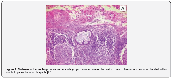
Additionally, postulates such as metaplastic origin from peritoneum, foci of endometriosis, vestiges of paramesonephricus ducts or distant metastases from ovarian serous borderline tumours may be implicated in the genesis of Müllerian inclusions within lymph node [1,2]. Clinically, the condition may be diagnosed as an incidental feature. Besides, lymph nodes pervaded with Müllerian inclusions appear enlarged [1,2]. Grossly, lymph nodes impregnated with Müllerian inclusions appear enlarged [2,3]. Upon microscopy, lymph node parenchyma is pervaded with glandular or cystic inclusions layered by cuboidal or columnar epithelial cells demonstrating a Müllerian or coelomic countenance. Frequently, inclusions appear embedded within the capsule [3,4] (Figures 1 & 2, Table 1).
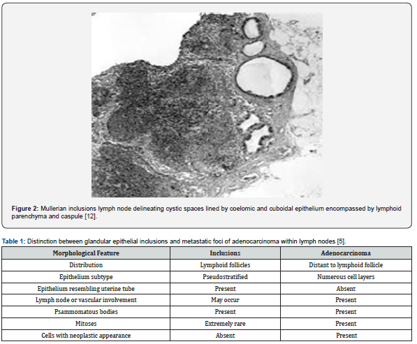
Psammoma bodies are commonly observed. The inclusions may depict focal squamous metaplasia. Morphological features as mitotic figures or mild cellular or nuclear atypia may be minimal to absence. Focal desmoplastic stroma or endometrial stroma encompassing the inclusions appear absent [3,4]. Müllerian inclusions confined to the lymph node appear immune reactive to keratin and mucin stains [6,7]. Müllerian inclusions confined to the lymph node necessitate segregation from conditions as distant metastasis from borderline or malignant ovarian tumours or various uterine carcinomas [8,9]. Müllerian inclusions confined to the lymph node configure as benign condition devoid of specific clinical significance [9,10].
References
- Quillen K, Williams HJ, Pfaendler KS (2025) Polypoid Müllerianosis Mimicking Primary Gynecologic Malignancy. OG Open 2(4): e103.
- Sirong Tao, Yan Zhang, Wei Wang, Ying He, Lili Jiang (2025) Müllerian papilloma: two case reports of malignant transformation and literature review. Front Oncol 15: 1573747.
- Saman Salih Fakhralddin, Sarwar Noori Mahmood, Deedar Kamaran Qader, Alaa Abbas Ali, Fahmi H, et al. (2021) Mullerianosis of the urinary bladder: a case report. Int J Surg Case Rep 83: 106040.
- Almatrafi MH, Alhazmi AM, Almosaieed BN (2020) Mullerianosis of the urinary bladder. Urol Case Rep 33: 101333.
- Spinardi J R, Goncalves I R D, La Falce T S et al. (2007) Benign inclusions in lymph nodes. Int J Morphol 25(3): 625-629.
- Rolando B Salada, Daniel Yong, Chi Shern Bernard Ho, Yew Lam Chong (2019) Müllerianosis: a case report. J Endourol Case Rep 5(3): 124-127.
- Yasushi Iida, Junya Tabata, Takashi Yorozu, Satomi Kitai, Kazu Ueda, et al. (2017) Polypoid endometriosis of the ovary and müllerianosis of pelvic lymph nodes mimicking an ovarian carcinoma with lymph node metastasis. Int Cancer Conf J 6(4): 145-148.
- Val-Bernal J-F, Mayorga M (2016) Incidental vaginal müllerianosis. Pathol Res Pract 212: 568-572.
- Winuthayanon W, Li S (2018) Fallopian tube/oviduct: structure and cell biology. In: Skinner MK, editor. Encyclopedia of reproduction. 2nd ed. Academic Press 2018: 282-290.
- Hirashima Y, Takekuma M, Takahashi N et al. (2019) Modified radical hysterectomy. In: Mikami M, editor. Surgery for Gynecologic Cancer. Comprehensive Gynecology and Obstetrics. Springer.