




 CTOIJ Home



Simple Summary
5-Aminolevulinic acid (5-ALA) fluorescence-guided surgical resection has become standard in glioblastoma treatment, utilizing the accumulation of fluorescent protoporphyrin IX (PpIX) from 5-ALA to optimize tumor resections. Beyond surgery, there’s a growing interest in 5-ALA photodynamic therapy (PDT), where PpIX is stimulated with a 620 nm light source. Recent research spans from laboratory studies exploring PDT effects on glioblastoma models to clinical trials investigating its application in patients. This review organizes current knowledge, offering fresh insights into the potential development of 5-ALA PDT as a promising therapeutic tool, with a focus on optimizing future clinical trials.
Abstract
The application of 5-Aminolevulinic acid (5-ALA) fluorescence-guided surgical resection has firmly established itself as a pivotal technique in the neurosurgical armamentarium for managing glioblastoma over the past decade. By leveraging the fluorescence induced by protoporphyrin IX accumulation, derived from 5-ALA and excited by 420 nm light, this method facilitates the identification of tumor-rich regions, thereby optimizing surgical tumor resections. Beyond its immediate surgical utility, the potential diagnostic and therapeutic ramifications of protoporphyrin IX accumulation are being explored. Of particular interest is the application of 5-ALA in photodynamic therapy (PDT), with a 620 nm light source stimulating protoporphyrin IX. While PDT has shown promise as a treatment modality in various tumors, recent attention has shifted towards its application in glioblastoma.
This comprehensive review systematically navigates through the evolving landscape of 5-ALA PDT research in glioblastoma, transcending from foundational “in vitro” laboratory studies elucidating photodynamic effects in diverse glioblastoma models to the forefront of clinical studies exploring the application of PDT in glioblastoma patients. In emphasizing the pivotal role of clinical trials, this review underscores the imperative for well-designed investigations to ascertain the therapeutic efficacy and safety profile of 5-ALA PDT in the context of glioblastoma treatment. By organizing and synthesizing existing knowledge, this review not only serves as a benchmark for current research but propels the field forward, guiding the design and execution of clinical trials that hold the key to unlocking the full potential of 5-ALA PDT in glioblastoma therapy
Keywords:Glioblastoma; Photodynamic Therapy; 5-Aminolevulinic Acid; Protoporphyrin; Fluorescence
Abbreviations: 5-ALA: 5-Aminolevulinic Acid; PDT: Photodynamic Therapy; FGS: Fluorescence-Guided Surgery; FECH: Ferrochelatase; ABC: ATP-Binding Cassette; MGd: Motexafin Gadolinium; NaBu: Na-Butyrate; HGF: Hepatocyte Growth Factor; FGFR: Fibroblastic Growth Factor Receptor; EGFR: Epidermal Growth Factor Receptor; βPDGFR: Β-Platelet-Derived Growth Factor Receptor; PFS: Progression-Free Survival; OS: Overall Survival; GSC: Glioma Stem Cells
Introduction
Glioblastoma (GB) treatment has represented, for the last decades, a considerable challenge in neuro-oncology. From 2005 to date, the standard GB treatment has not experienced significant variations. After surgery, Stupp et al. [1] proposed that Temozolomide-radiotherapy protocols represent the best alternative in GB patients, achieving mean overall survival rates of around 15 months [1]. This dramatic prognosis makes it necessary to investigate different therapeutic strategies to combat GB. Among these different strategies, electromagnetic fields, immunotherapy, angiogenesis modulation, and photodynamic therapy stand out because of increasing research and clinical studies reported in the literature.
Photodynamic therapy (PDT) is a minimally invasive therapeutic modality based on phototherapy that involves light and a photosensitizing chemical substance to elicit cell death [2]. PDT uses a primary or secondary (prodrug) photosensitizing agent, which accumulates specifically inside a target tissue, for example in the cancer cells. By exposing the tissue to light at a specific wavelength, PDT causes cellular damage and leads to targeted cell death. Nowadays, there is a broad spectrum of conditions in which PDT can be used, including controlling various dermatological conditions and as an adjuvant treatment to various neoplasms (e.g., digestive, pulmonary, and cerebral). In the last three decades, multiple drugs have been approved for PDT, including porfimer sodium, 5-aminolevulinic acid, temoporfin, norpseudoephedrine, and others.
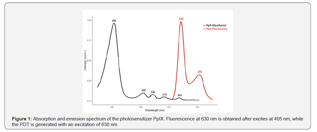
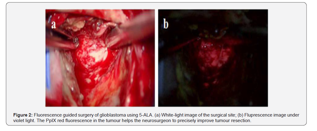
Among these drugs, 5-Aminolevulinic-Acid (5-ALA) highlights its increasingly important role in treating GB. 5-ALA is a prodrug, the first metabolite in the synthesis pathway of group heme, that generates, under certain conditions, protoporphyrin IX (PpIX) cell accumulation. Protoporphyrin IX is a photosensitizer that can produce fluorescence and photodynamic effects through specific light radiation wavelengths (Figure 1). In 1999, 5-ALA was approved in the USA by FDA to be used in the PDT of Actinic Keratosis. One year before, the first clinical use of 5-ALA in fluorescence-guided surgery (FGS) was reported by Stummer et al. [3] In 2006, this group led a multicenter phase III clinical trial consolidating 5-ALA FGS as a safe and effective method to detect high-grade glioma solid mass during neurosurgical procedures, thus improving resection rates and patient outcomes (Figure 2).
5-ALA-mediated PDT in GB is currently the subject of multiple studies focused on the ability of this agent to produce selective tumor cell death. Most published studies describe “in vitro” features of 5-ALA, pharmacodynamics, kinetics, Protoporphyrin IX cell metabolism, intracell accumulation, PDT light radiation doses, effect enhancers, and inhibitors. Translational studies focus on “in vivo” models demonstrating selective mediated 5-ALA PDT in glioblastoma. Finally, few clinical studies have been reported until now describing the use of PDT in GB patients. In the present manuscript, the authors aim to organize the literature published about 5-ALA-mediated PDT in GB.
In this comprehensive review, our primary objective is to meticulously analyze the existing body of literature on 5-ALAmediated PDT in the context of glioblastoma. As GB remains a formidable challenge in neuro-oncology, our focus extends beyond summarizing in vitro and in vivo studies to discern the intricacies of 5-ALA PDT. Emphasizing its crucial role in GB treatment, we aim to extract valuable insights from pharmacodynamics, kinetics, Protoporphyrin IX cell metabolism, intracellular accumulation, PDT light radiation doses, effect enhancers, and inhibitors. By synthesizing current knowledge, our intent is not only to present a comprehensive overview but to pave the way for future clinical trial designs, offering a strategic roadmap for optimizing 5-ALAmediated PDT in the treatment of GB patients.
Basic Research
Basic research in 5-ALA PDT has evolved in the last two decades, trying to solve different questions about its application in GB. Authors have published various manuscripts using multiple in vitro and in vivo glioma study models. A summary of in vitro and in vivo studies is provided in Tables 1 & 2, respectively. The topics covered stand out: optimal treatment scheme, the effectiveness of 5-ALA derivatives, 5-ALA mediated PDT enhancers and inhibitors, physiological and biomolecular PDT-mediated changes and cell death, and resistance to PDT.
Optimal Scheme of Treatment
Like with other therapeutical techniques, 5-ALA mediated PDT implies searching for an optimal application regimen to achieve the maximal effect on the tumoral cells with minimal impact on non-tumoral ones. Different studies have evaluated, in terms of light fluence and light fluence rate, to find the best in vitro and in vivo PDT light doses. Madsen et al. [4] were the first to investigate the effectiveness of different light exposure levels and their photodynamic therapy efficacy in the 2000s. After different light doses and light fluence rates, they concluded that 635 mm wavelength light administered at low fluences (less than 50 J/ cm2) with multifractionated (low fluence rate 10-25 mWcm2) was the most effective condition to induce cell death. In addition, the proliferation and growth of glioma spheroid cells decreased after this light stimulation [4-7].
In vivo assays have been reported that the low rate of light is the best condition to produce necrosis. In 2006, Angel-Petersen et al. [8] observed that only the treatment with ALA-PDT with light at low rates induces the necrosis of glioma cells in a rat glioma model. In addition, this treatment prolonged the survival of tumorbearing animals [8]. In contrast, in 2006, Hirschberg et al. [9] observed, through collagen matrix combined with tumor glioma spheroids, that the low fluence 6 J/cm2 inhibit the migration of cells but the cytotoxicity was obtained with high fluence 25 J/cm2 [9]. Similar results were obtained in their animal model. Tumor in rats treated with 125 mg/Kg of 5-ALA and 26 J of fluency increased survival, although only one case out of 17 experienced tumor regression. In this study, fractionated long-term treatment protocols were proposed to reduce the regression of tumor [10]. On the same way, Tetard et al. [11] group concluded that more fractionation schemes should be studied, since low fluence rates of 4.8mW were better tolerated, but fractionated high fluence treatment delivery caused more necrosis [11].
In 2015, Guo et al. [12] observed that using an organic lightemitting diode in mice with a low fluence rate (3 mW/cm2) and long duration (3.7 h), the mean survival increases from 26 to 40.5 [12]. Leroy et al. [13] observed that in human glioma engrafted in rat brains, the delivery of fractionated light may enhance treatment efficacy by reoxygenating tissues. They used optic fiber in the tumor and compared three groups: no light, two, and five fractions. They found that five fractions produced more apoptosis and more neovascularization. All treatments also had edema and macrophagic infiltration [13]. Finally, Vermandel et al., [13] in 2019, studied different treatment schemes in U87 nude rats. The five fractions and low fluence rate (5 mW) group showed the same cell response but had limited toxicity with less brain edema [14]. Probably, an interesting study was conducted by Lou et al. [15] in 2014. They described a high throughput system to test almost 1000 different PDT conditions in parallel. This platform can be helpful in the development of new photosensitizers and the optimization of current PDT protocols. Its application should be considered when looking for the best treatment regimen [15].
Effectiveness of 5-ALA Derivatives
In recent years, different molecules derived from 5-ALA have been studied to find a molecule with advantages over 5-ALA as a photosensitizer. In 2002, Hirschberg et al. [9] used 5-ALA (benzyl- and hexyl-) esters, generating the same standard 5-ALA PDT-mediated effect in glioma cells but with 10-20 times less concentration than the parent compound. Wu et al. [16] confirmed these findings regarding hexyl-5-ALA, demonstrating higher protoporphyrin IX cell accumulation and a better response to PDT. Finally, Berkovitch-Luriain et al. [17] 2012 used five different 5-ALA prodrugs, which differed in the specific chemical attachments to Alanine. It has been designed to optimize drug delivery, bioavailability, blood-brain barrier permeability, or lipophilicity depending on the desired therapeutic application. In the study, AlaAcBu, AlaAcPi, AlaFaBu, ALA, and AlaFaPi were tested to PDT and concluded that the PDT potency of these prodrugs was in the following order: AlaAcBu > AlaAcPi > AlaFaBu ≥ ALA > AlaFaPi. AlaAcBu is a prodrug that can be converted into Alanine for improved drug delivery and bioavailability.
AlaAcBu can be converted into Alanine through the cleavage of the ester bond between the Ac and Bu groups, enabling improved drug delivery and bioavailability, while AlaAcPi is a potential prodrug for neuroactive compounds with enhanced blood-brain barrier permeability, and AlaFaBu enhances drug delivery to fatty tissues and promotes lipophilicity. On the other hand, ALA is not a prodrug itself but is used in medicine for photodynamic therapy and disease diagnosis. Finally, AlaFaPi combines both fatty acid and phosphinate groups for enhanced bioavailability and brain distribution in the central nervous system. The superiority of AlaAcBu resided in the necessity of lower molar concentrations and light intensity to activate cell death following light exposure [17]. However, currently, any of them is being used as a photosensitizer, because more experiments are needed to confirm that some of them might be used in the clinical practices.
5-ALA-Mediated PDT Enhancers and Inhibitors
The effectivity of PDT may be influenced by different physiological conditions, such as temperature [18,19], or by the interaction with other drugs, such as levetiracetam [20]. One of the first modifiers of the PDT effect investigated was temperature in 2004 by Hirschberg et al. [19] During the study about the impact of hyperthermia (40-46 degrees C) and synergism with 5-ALAmediated PDT, they observed increased tumoral cell apoptosis in human and rat glioma spheroids when combining hyperthermia and PDT [19]. In contrast, another group, who studied the temperature as a mediator of PDT’s effectivity, found that hypothermia increased PpIX fluorescence, improving rat survival, and protecting typical brain structures [18]. Contrary to the first study, this group concluded that hypothermia might improve the patient’s outcome. Therefore, further experiments should be conducted to determine whether high or low temperatures can benefit the patient’s outcome and how these conditions might affect PDT therapy.
Oxygen concentrations are crucial to achieving PDT oxygenmediated toxicity. In 2014, Albert et al. [21] described that glioma cell lines cultured under atmospheric (pO2= 19%) and physiological (pO2= 9%) oxygen concentrations required different light doses to generate the PDT effect. They observed that, with physiological pO2, light doses required were 20% higher than with atmospheric O2 concentrations [21]. PDT is strictly dependent on the presence of oxygen, since it is indispensable to generate ROS and promote the cell death. The excitation of the PS with light results in the move of an electron to the first excited singlet state. The following intersystem crossing yields a triplet state. The triplet PS transfers energy to triplet oxygen, resulting in the generation of reactive singlet oxygen (1O2), which is the responsible to kill the cancer cells, damaging vascular structures as well as inducing immune responses [22].
Therefore, the levels of oxygen may be influencing the effectiveness of PDT. Ihata et al. [23] in 2022, used six human GSC lines, mesenchymal types HGG13, HGG30, HGG1123, and Proneural types HGG146, HGG157, and HGG528. Hypoxia-GSCs had lower intracellular PpⅨ accumulation than normoxia-GSCs due to increased gene expression of FECH, and their sensitivity to ALAPDT was reduced less, despite accumulating lower concentrations of PpⅨ. ALA-PDT is a potentially effective therapy for hypoxiatolerant GSCs that exist in hypoxia at 5% oxygen concentration [23]. Later, Ihata T et al. [24] studied the effect of hypoxia on 5-ALA mediated PDT with glioma stem cells. After analyzing two groups of GCSs cell lines: normoxia and hypoxia groups, authors conclude that hypoxia-GSCs had lower intracellular PpIX accumulation than normoxia-GSCs due to increased gene expression of FECH, and that their sensitivity to ALA-PDT was reduced less, despite accumulating lower concentrations of PpIX.
ALA-PDT is a potentially effective therapy for hypoxiatolerant GSCs that exist in hypoxia at 5% oxygen concentration [24]. Recently, Fahey et al. [25] in 2016 studied Oxid Nitric Synthase (NOS neural and induced counterpart) in glioma. They observed that extent of ALA/light-induced apoptosis increased substantially when an iNOS inhibitor or NO scavenger was present, implying that iNOS/NO was acting cytoprotectively. The use of iNOS inhibitors could enhance the effect of 5-ALA-mediated PDT [25]. Bazak et al. [26], in 2019, talked about NOs Bystander effect in cells non-targeted increasing growth and migration. If an actual tumor occurring in PDT setting and not suppressed (e.g., by iNOS activity or transcription inhibitors), then such effects could compromise treatment efficacy or even stimulate disease progression if PDT’s anti-tumor potency is not great enough [26].
Apart of the temperature or oxygen, there are some drugs and molecules that might be affect the PDT therapy by interacting directly or indirectly to 5-ALA or PpIX. The nuclear encoded mitochondrial enzyme ferrochelatase (FECH) catalyzes the insertion of ferrous iron into protoporphyrin IX in the last step of heme biosynthesis [27]. Genetic silencing of FECH has been shown to increase ALA-PpIX fluorescence and PDT response both in vitro [28] and in vivo [29] indicating that inhibition of PpIX bioconversion could be an effective strategy for enhancing ALA applications. Pharmacological inhibition of the conversion of PpIX to heme includes the use of FECH inhibitors [30] or, more commonly, iron chelators [31]. Blake et al. [32], in 2010, analyzed the role of iron chelator 1, 2-diethyl-3-hydroxy pyridine-4-one hydrochloride (CP94) and concluded its utility as an adjuvant to 5-ALA for photodiagnosis and PDT.
A year later, the same group compared CP94 with another iron chelator, dexrazoxane, concluding that CP94 was most favourable to PDT [32,33]. Later, Teng et al. [34], in 2011, reported that depletion of FECH by small interference RNA enhanced PpIX fluorescence and PDT after exposure to 5-ALA concomitant with increased intracellular PpIX accumulation in glioma cells [34]. These studies suggested that the inhibition of FECH increases ALA-PpIX fluorescence and enhances the response to PDT. Thus, targeting the bioconversion of PpIX could be a viable strategy for improving the application of ALA in PDT. Another interesting field was the interaction between PDT and different drugs typically administered in patients with glioma. In 2012, Hefti M. et al. [20] studied whether levetiracetam and phenytoin may reduce the cellular accumulation of PpIX in patients with high-grade gliomas. They observed that phenytoin decreases PpIX concentrations, but levetiracetam did not [20].
How phenytoin may interact with 5-ALA, PpIX, and PDT in glioblastoma is still not well-documented [20,21]. In 2016, JE Lawrence et al. [35] analyzed the role of dexamethasone, desipramine, phenytoin, valproic acid, and levetiracetam on the production and accumulation of PpIX. All these drugs, except levetiracetam, reduce the total amount of PpIX produced by GB cells (p < 0.05) [35,36]. Dexamethasone is a glucocorticoid that can suppress the immune system and inhibit inflammatory responses, affecting PDT by reducing the inflammatory response induced by this therapy. On the other hand, desipramine, phenytoin, and valproic acid, might indirectly influence PDT by modulating cellular signaling pathways related to ROS generation, metabolism, or scavenging. However, further research and clinical studies are required to provide a comprehensive understanding of these interactions [20,21]. Despite that, these findings highlight the potential impact of co-administered drugs on the effectiveness of PDT in glioma treatment [20,21,32,36-38].
A promising target to achieve better results with PDT is the family of ATP-binding cassette (ABC) transporters due to its essential role in transporting PpIX in glioma cells. In 2013, Sun W. et al. [37] explored the role of Gefitinib in glioma and malignant glioma cell lines. Decreasing levels of ABCB6 generated an increase in PpIX in these cells. The same year, G. Zhao et al. [38] studied the crucial role of ABCB6 in ALA metabolism and accumulation of PpIX in glioma. ABCB6 overexpression is a potential approach to enhance the collection of PpIX for optimizing the subjective discrimination of faint fluorescence and improving the efficacy of ALA-based photodynamic therapy [37,38]. In 2019, N Kawai et al. [39] also studied the importance of ABCG2. If this transporter had the highest levels, 5-ALA staining and PDT effect would be fewer. Inhibition of the ABCG2 transporter may enhance 5-ALA-mediated FGS and PDT. A year later, P Muller et al. [40] demonstrated that GB cells with high ABCG2 expression accumulate less photosensitizer and require higher light doses to be eliminated.
Inhibition of ABCG2, by KO143, during photosensitizer accumulation and irradiation promises to restore the full susceptibility of this crucial tumor cell population to photodynamic treatment [39,40]. Finally, Mansi et al. [41], in 2022, determined the activity of FECH and ABCG2 and correlated them with intracellular and extracellular PpIX levels and PDT response. They revealed ABCG2 as an essential biological determinant of PpIX fluorescence in glioma cells. They suggested ABCG2 inhibition with lapatinib as a promising therapeutic enhancement approach. Ferrochelatase inhibition and iron chelator were not effective [41]. On conclusion, the family of ATP-binding cassette (ABC) transporters, specifically ABCB6 and ABCG2, play a crucial role in the transport of PpIX in glioma cells. Studies have shown that decreasing levels of ABCB6 increase PpIX levels, while high levels of ABCG2 decrease 5-ALA staining and PDT effect. Inhibition of these transporters is a potential approach to enhance the collection of PpIX and improve the efficacy of ALA-based photodynamic therapy [23,26,27].
Among other 5-ALA mediated PDT modifiers, Madsen et al. [42] in 2009 studied the effect of radiosensitizer Motexafin Gadolinium (MGd). They reported that MGd could potentiate the cytotoxic and migration-inhibitory effects of ALA-PDT [42]. Also, in 2009, Flores-Ancona et al. [43] used previous Na-Butyrate (NaBu), increasing PDT-derived cell death by activating apoptosis genes. Later, in 2012, Bueno-Carrazco et al. [44] proved the synergistic effect of NaBu on cytotoxic damage induced by PDT. They studied NaBu-induced expression of caspase-3, caspase-9, and bcl-2 and increased bax in glioma cells. They concluded that genes and differentiation induced mainly by NaBu improve tumoral cell death after PDT. NaBu is a histone deacetylase inhibitor that can alter gene expression patterns and promote cell differentiation. Combining NaBu with PDT may modify the tumor microenvironment, sensitizing cancer cells to the cytotoxic effects of PDT [44,45]. Wang et al. [45], in 2013, reported the use of Trioxide of Arsenic (ATO).
We concluded that ATO was a potential optional approach in enhancing intracellular PpIX accumulation and improving the benefits of 5-ALA-induced FGR and PDT in glioma. ATO is known to inhibit DNA repair enzymes, such as topoisomerase II and telomerase, leading to DNA damage accumulation and cell death. Combining ATO with PDT may further impair DNA repair mechanisms, exacerbating the DNA damage caused by PDT and enhancing the therapeutic effects [45]. Finally, Chen et al. [45], in 2014, preconditioned with calcitriol, a form of vitamin D3, during 48h human glioblastoma cell and astrocytes. Calcitriol has been shown to inhibit proliferation and promote differentiation in cancer cells, potentially enhancing the effects of PDT. In this study, calcitrol induces increased levels of PpIX and fluorescence and better response to 5-ALA-mediated PDT [45]. Therefore, the use of modifiers such as motexafin gadolinium, Na-butyrate, trioxide of arsenic, and calcitriol have been explored and found to enhance PpIX accumulation and improve the benefits of 5-ALA-induced PDT in glioma cells.
These findings highlight the potential for combination therapies to optimize photodynamic treatment outcomes in glioma patients. Finally, in the most updated bibliography, there is a study published by Mandl GA et al. [46] in 2023 where U251 GB cell lines were treated by 5-ALA-PDT and NaLuF4: Pr (3+)-doped nanoradiosensitizer. This combination allowed to sensitize PpIX to induce radio-PDT and permitted radiation doseenhancement effect after analyzing the effect of the combination in cell death, viability, stress, senescence and proliferation. It represents a proof of concept for nanomedicine applied to PDT [46]. In 2020, Wang et al. [45] developed biocompatible periodic mesoporous organosilicon-coated Prussian blue nanoparticles (PB@PMOs). They were constructed to load a biosafe prodrug 5-ALA, pronouncedly converted to PpIX in malignant cells. PB@ PMO-5-ALA induces a higher accumulation of PpIX in glioma cells than free 5-ALA.
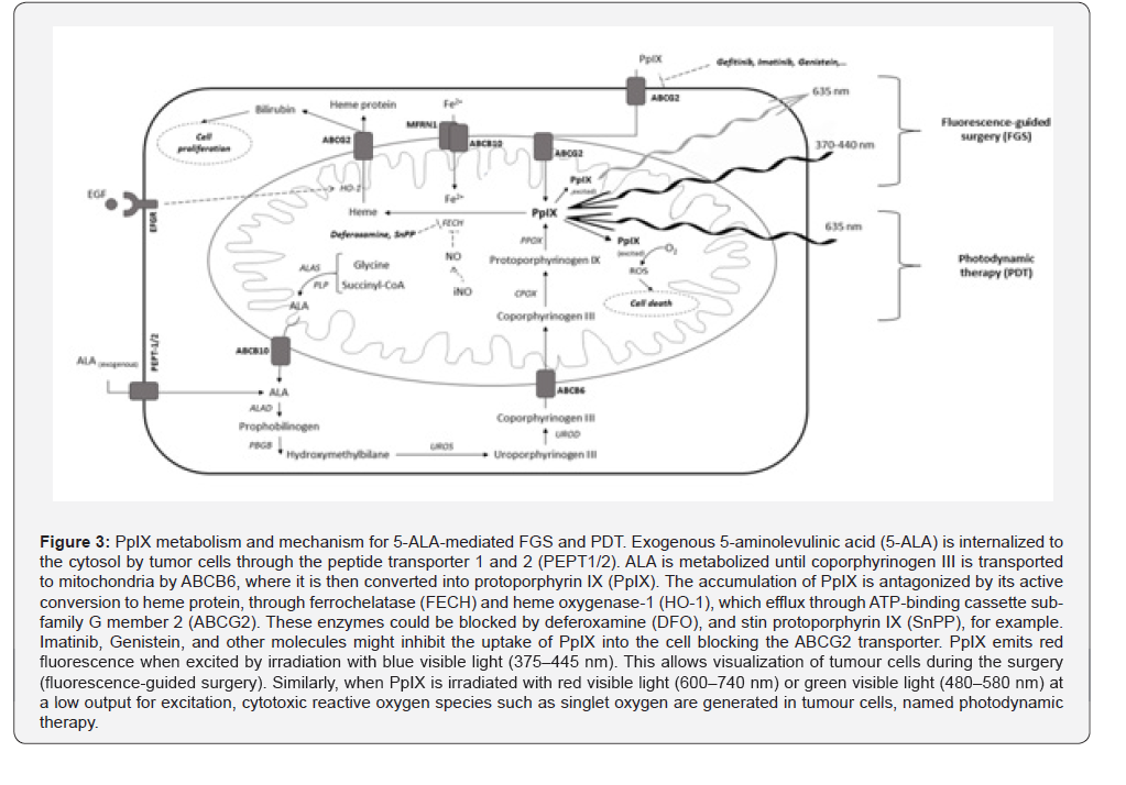
Meanwhile, the PB@PMOs, with a mean edge length of 81 nm and good biocompatibility, effectively decompose hydrogen peroxide to oxygen in a temperature-responsive manner. Oxygen supply further contributes to the promotion of 5-ALA-PDT. PpIX increases by 75% and suppresses tumor growth [47]. Finally, another group, in 2021, studied Near InfraRed-based chronic PDT. This was achieved in an untethered and non-invasive manner in a mouse xenograft GB model. It is postulated that such encapsulated UCNPs implants represent a translational shift for wireless deep-tissue phototherapy by enabling the sequestration of UCNPs without compromising wireless deep-tissue light delivery [48]. Overall, understanding the factors that can affect the efficacy of PDT, such as FECH inhibition, drug interactions, and design of nanoparticles that improve the accumulation of PpIX in glioblastoma cells can contribute to the development of more effective treatment strategies and enhance the clinical application of ALA in PDT. A summary of PpIX metabolism and mechanism for 5-ALA-mediated PDT is shown in Figure 3.
Physiological and Biomolecular PDT Mediated Changes and Cell Death
Photodynamic therapy produces different molecular changes that multiple studies have tried to elucidate, not without controversy. Once the photosensitizer is administered, it selectively accumulates in tumor cells. Then, light of a specific wavelength is applied to the area of interest, which activates the photosensitizer to produce ROS, such as singlet oxygen and oxygen free radicals. These reactive oxygen species cause damage to tumor cells by interacting with cellular structures and inducing oxidative damage. Cellular damage induced by PDT can trigger various pathways of cell death, including apoptosis, necrosis, autophagy, and necroptosis. Depending on the specific characteristics of the tumor and cellular environment, one or several of these pathways can be activated (Figure 4) [45-51].
There are several studies that confirm the activation of apoptosis after PDT in glioblastoma. Inoue et al. [51] showed in 2007 that glioma cell PDT induced apoptosis through mitochondrial cytochrome C Karmakar et al. [50] observed protease activation and survival factors suppression as a mechanism of apoptosis in glioma cells [50,51]. In the same way, Vogel et al [52], in 2013, observed that 5-ALA PDT promotes apoptosis of glioma stem cells by regulating hepatocyte growth factor (HGF) [52]. However, Zelenko et al. [53] observed differences between small and big glioma tumor spheroids. Smaller spheroids suffered higher cell death after 5-ALA PDT, mainly cell necrosis, especially with higher oxygen concentrations, while the big glioma tumor spheroids activate the apoptosis [53]. A year later, Kamoshima et al. [54] also described acute necrosis as a PDT effect in glioma cells spheroids [54]. In 2011, Coupienne et al. [55] studied the inhibition of nuclear factor kappa B (NFκβ) and demonstrated an increased sensitivity to PDT treatment due to a pronecrotic effect.
In a parallel study, the same author confirmed 5-ALA PDTinduced RIP-3-dependent necrosis [52-56]. Finally, Golla et al. [57], in 2020, showed that bcl-2/bcl-xl (antiapoptotic) inhibition (ABT-263) increases the response of glioblastoma cells toward photodynamic therapy. This effect can be partly attributed to cytotoxicity and is likely related to a pro-apoptotic shift because of an increased noxa/mcl-1 ratio. Thus, both apoptosis and necrosis might be activated after treating glioblatoma cells with PDT [48,52-54,58-60].
An interesting study was carried out by Uzdensky et al. [59] in 2012. This is one of the complete studies about 5-ALA PDTmediated cell changes. They determined more than 224 proteins expressed after sub-lethal 5ALA PDT. Sub-lethal PDT induces complex response of glioblastoma cells, including changes in activity and expression of proteins involved in adhesion-mediated signaling, signal transduction, cytoskeleton remodeling, cell cycle regulation, and anti-apoptotic processes.
Multiple reactions of various cellular subsystems, including adhesion, cytoskeleton, signal transduction, cell cycle, and apoptosis, are integrated into the general cell response to a sublethal impact [59]. Another study that observed morphological and migration changes in glioma cells after PDT was carried out by Etminnan et al. [58] in 2011. ALA/PDT altered invasiveness, possibly because of the cytoskeletal organization and matrix metalloproteinase expression. Morphology changes were related to the cytoskeleton [57]. Finally, as a miscellaneous study, Yamamoto et al. [60], in 2012, pre-treated glioma cells with 5-ALA and exposed them to ionizing irradiation. They observed that pre-treatment with 5-ALA not only photosensitizes but also radiosensitizers and increases the effect of ionic radiation [60].
Resistance to PDT
Photodynamic therapy has shown remarkable potential in glioblastoma treatment, but despite its initial success, resistance to PDT has emerged as a major obstacle. This resistance mechanism, still not fully understood, involves intricate cellular processes that allow cancer cells to evade the destructive effects of PDT. Unveiling the underlying mechanisms of resistance to PDT in glioblastoma is crucial for the development of novel strategies to overcome this resistance and enhance the effectiveness of this promising therapy. In 2021, Vilchez and colleagues directed their attention towards investigating resistance to PDT. In their research obtained resistant cells to the PDT that displayed reduced accumulation of photosensitizer, formed spheroids with an increased number of cells, exhibited enhanced tumorigenic capacity, and showcased elevated mRNA levels of fibroblastic growth factor receptor (FGFR), epidermal growth factor receptor (EGFR), and β-platelet-derived growth factor receptor (βPDGFR).
When compared to the original cells. These results suggested that βPDGF, FGFR and EGFR might be involved in the resistance to PDT [61]. In 2022, Mastrangelopoulou et al. [62] provided in vitro study 5-ALA mediated results with glioblastoma (T98G and U87) and breast cancer (MCF7, MDA-MB-231 and T47D) cell lines. They determined ABCG2, FECH and heme oxygenase (HO- 1) levels as predictive biomarkers for response to 5-ALA PDT. Glioblastoma cell lines demonstrated to be more resistant to PDT than breast cancer cells. Regarding to glioblastoma, inhibition of ABCG2 transporters was a common enhancer of PDT while FECH and HO-1 inhibition was only effective with T98G cell line. They concluded that inhibition of these specific molecular targets could overcome inherent resistance to 5-ALA-PDT [62].
5-ALA PDT and Glioma Stem Cells
In recent years, there has been a growing interest in studying glioma stem cells (GSC) and their role in tumor recurrence and resistance to treatment. GSC are a population of tumororiginating cells capable of self-renewal and differentiation, generating resistance and tumor recurrence after treatment. Therefore, several studies have focused on in vitro experiments to test the effective of treatment to promote GCS’ death. These studies explore different factors such as iron chelation, gene expression, and hypoxia that affect the accumulation of PpIX in GSC and their response to treatment. Additionally, researchers have investigated the impact of certain molecules and therapies on GSC viability, proliferation, and differentiation, to contribute to a better understanding of GSC biology and the development of potential therapies targeting these cells.
In 2016, Schimanski et al. [63] reported for the first time that glioblastoma stem-like cells accumulate PpIX when subjected to 5-aminolaevulinic acid and are sensitive to 5-aminolevulinic acid-based photodynamic therapy [63]. In 2017, Wang et al. [64] explained how iron chelation was necessary for PpIX production and fluorescence in GSC. They detected a side population of stem cells with low PpIX production after 5-ALA treatment, those present high levels of heme-oxygenase-1 compared with the GCS which accumlates high levels of PpIX. In these cells, the accumulation of PpIX might be increased by deferoxaminemediated iron chelation but not by the inhibition of ABCG2, which efflux PpIX from the cell [64].
Also, in 2017, Fujishiro et al. [65] studied how glioma stem cells expressed higher mRNA levels of PpIX biosynthesis enzymes and their transporters PEPT1/2 and ABCB6 when compared to the parental glioma cells. They revealed that upon incubation with ALA, glioma stem cells accumulate a higher level of PpIX. Finally, we showed that GSC was more sensitive to ALA-PDT than the original A172 cells and confirmed that all patient-derived glioma sphere lines also showed significantly increased sensitivity to ALA-PDT if cultivated under the pro-stem cell condition [65]. That is why most of the last studies about PDT efficacy and obtaining the optimal therapy of PDT are performed with GSC.
Normal tissue vs. glioma cell PDT effects
The interaction between normal brain tissue and tumors during 5-ALA PDT is a crucial factor in its application in clinical settings, that is why is important to study the PDT in a model that allow us to test the toxicity of treatment to glioblastoma cells and the healthy tissue and the possible changes in the microenvironment. To this aim there are in vivo and co-culture in vitro models. The first study analyzing this topic was conducted by Ito et al. [3], in 2005, who found that 5-ALA-mediated PDT induced edema. In the study, generating tumour in the brain of rats by C6 Glioma cells, observed that photo-irradiation therapy with 5-ALA induces edema, which is partly counteracted by steroid therapy. The possibility of steroid-resistant edema formation should be considered when planning human trials with this treatment modality. [3].
Later, Mathews et al. [66], in 2011, applied increasing laser light doses and monitored by MRI T2-weighted the edema. They described the vasogenic mechanism in response to corticoids [66]. These results suggested that MRI might be performed to evaluate the outcome of patients treated with PDT, detecting brain damage, such edema. Leroy et al. [13], in 2018, demonstrated the utility of MRI in assessing treatment outcomes after PDT in brain lesions. In this study, they determined that diffusion and perfusion MRI revealed histological lesions after PDT comparing the treated group with control brain images. Interstitial PDT (iPDT) induced specific lesions in the tumor tissue, which were observed with MRI and confirmed by histopathological analysis. Thus, MRI may provide a noninvasive and reliable tool to assess treatment outcomes after PDT. In addition, in other studies with animal model, disruption of BBB and edema were observed by MRI, after PDT [67]. In 2019, Zhang et al. [68] observed more pronounced PDT-induced BBB disruption in juvenile mice than adult mice, suggesting age differences in PDT-related BBB opening.
This might be an essential informative platform for a new application of PDT as a method for brain drug delivery, especially for the postsurgical treatment of malignant gliomas [68]. The BBB regulates the passage of substances between the blood vessels and the brain. One of the main challenges in GB treatment is the poor delivery of therapeutic agents to the tumor site, mainly due to the presence of the BBB. When the BBB is disrupted, it allows better penetration of drugs into the brain, increasing the efficacy of chemotherapy and targeted therapies. In addition, BBB acts as a physical barrier that limits the infiltration of immune cells into the brain. Thus, BBB-disruption might allow immune cell infiltration, including T cells, natural killer cells, and macrophages, which have an important role in anti-tumor immune responses and can contribute to the destruction of cancer cells. Overall, PDT might disrupt the BBB and can potentially enhance treatment outcomes for GB patients.
On the other hand, less microvessel density was found after 5-ALA PDT in a glioma rat model. In 2015, Yi et al. found that the treated group of animal model with 5-ALA PDT showed more necrosis and less microvessel density than non-treated group. These results suggest that the PDT are altering the brain tissue around glioblastoma. With the intention of creating more realistic vitro models, our group have recently published the application of 5-ALA-mediated PDT to brain organoids infiltrated by high-grade glioma stem cells tumorospheres. This model helps better understand the selective injury process (normal versus pathologic tissue) and create more realistic representation threedimensional model. In this study, we have demonstrated that this powerful experimental model permits study the effectiveness of therapeutic approaches, modeling the symbiosis between normal and malignant cells and providing a real insight into the tumor microenvironment interaction. According to our in vitro experiments, PDT seems to specifically target glioblastoma cells while sparing the surrounding tissue. In addition, this 3D model would allow to test different therapy strategies with multiple conditions and test the specificity to promote the tumour cell death without affect the normal cells [69].
Combined Therapies
Photodynamic therapy has emerged as a promising treatment option for GB due to its ability to selectively destroy tumor cells using light-activated photosensitizers. However, the effectiveness of standalone PDT in treating GB is limited. As a result, combination therapy approaches are being explored to enhance therapeutic outcome. Combination therapy refers to the simultaneous or sequential use of multiple treatment modalities to target different aspects of GB, aiming to achieve synergistic effects and improve patient outcomes. One such approach is the combination of PDT with other established treatments, such as surgery, chemotherapy, or immunotherapy. Combining PDT with surgery allows for the precise removal of visible tumor tissue, while PDT targets the remaining cancer cells that may not be visible or accessible to the surgeon.
This combination approach enhances the extent of tumor debulking and improves the overall efficacy of the treatment. Furthermore, PDT can also be combined with chemotherapy or targeted agents to tackle the underlying biological processes driving GB progression. The photosensitizers used in PDT can enhance drug uptake and sensitization of tumor cells to chemotherapy, potentially overcoming drug resistance and strengthening the treatment response. Moreover, recent advancements in immunotherapy have shown promise in treating GB. By combining PDT with immunotherapeutic strategies, such as immune checkpoint inhibitors or cancer vaccines, the immune system can be activated to recognize and eliminate GB cells more effectively.
The combination of PDT and boron neutron capture therapy (BNCT) using boronated porphyrins has shown promise in the treatment of gliomas. Boronated porphyrins have high tumor affinity, low cytotoxicity in dark conditions, and can easily be synthesized with high boron content. In the Ryo Hiramatsu et al. [70] study, the boronated porphyrin H2OCP was evaluated for its applicability in both PDT and BNCT. The results showed that H2OCP accumulated within cells more effectively than other boron delivery agents currently used in clinical BNCT studies. Additionally, H2OCP was found to be a promising photosensitizer for PDT and could potentially replace or be used in combination with existing boron delivery agents for BNCT. These findings suggest that H2OCP could be a novel and effective dual sensitizing agent for both PDT and BNCT. [70].
Yonsei et al. [71] published this year an interesting study about combined effects of Focused Ultrasound and Photodynamic treatment for GB. They used C6 Glioma Rat Model to compare photodynamic, sonodynamic and sonophotodynamic therapy. They observed that photodynamic therapy alone was able to inhibit GB growth while ultrasound could not generate this effect. Regarding the combination of both therapies, authors described an increase in oxidative stress parameters compared with PDT alone [71]. Another recent study has developed a new therapy called X-ray-mediated photodynamic therapy (X-PDT) using radioluminescent nanoparticles and an endogenous photosensitizer PpIX. The nanoparticles NaLuF4:Pr3+ were tested at different concentrations and found to emit strong light that overlaps with the PpIX to perform photodynamic therapy
This combination improved treatment outcomes for cancer cells. Additionally, the nanoparticles enhanced the effectiveness of radiation therapy. The researchers tested the nanoparticles alone and in combination with PpIX and observed their effects on cell viability, death, stress, senescence, and proliferation. Overall, this study demonstrates the potential of using these nanoparticles in nanomedicine for the treatment of glioblastoma [46]. In summary, combination therapy with PDT offers a multifaceted approach for the treatment of glioblastoma. By combining the selective tumor cell killing of PDT with surgery, chemotherapy, or immunotherapy, it holds the potential to improve treatment outcomes, enhance patient survival rates, and ultimately contribute to a more comprehensive management of this challenging brain cancer.
Clinical Studies
According to the experimental results obtained in both vitro and in vivo studies, photodynamic therapy has demonstrated potential as an adjunctive treatment alongside standard therapies, including surgery, chemotherapy, and radiotherapy, for glioblastoma. However, it is important to note that patient-based experimentation with ALA PDT remains limited, with only a few studies conducted, most of which are Phase I-III trials involving a small number of patients. A summary of clinical studies is provided in Table 3. There are ongoing studies investigating the efficacy and safety of PDT in glioblastoma patients, such as the INDOMETAFLU study (NCT02748188), which aims to evaluate the use of PDT combined with metronomic fluorouracil chemotherapy in recurrent glioblastoma, and the trial NCT02422979 (phase II/ III study), which is investigating the addition of PDT to standard treatment for newly diagnosed glioblastoma.
Metronomic fluorouracil chemotherapy is a treatment approach that involves the continuous administration of lowdose fluorouracil (a chemotherapy drug) over an extended period. Unlike the traditional chemotherapy regimen where high doses are given in cycles with rest periods, metronomic therapy provides a consistent and relatively lower dose of the drug without extended breaks. This treatment strategy targets blood vessels that support tumor growth rather than directly attacking the cancer cells themselves. By inhibiting the formation of new blood vessels, metronomic chemotherapy aims to starve the tumor of its blood supply, thereby slowing down its growth and spread. Metronomic fluorouracil chemotherapy is considered a relatively low-toxicity treatment option and can be administered orally as a pill or intravenously. It is used in the treatment of a variety of cancers, including colorectal, breast, and gastric cancers. In some cases, it may be utilized as an adjuvant therapy after surgery or in combination with other anti-cancer agents.
The first clinical trial about 5-ALA PDT in glioblastoma was in 2007, where Beck et al. [72] published two series of patients with recurrent small, circumscribed GB who underwent interstitial 5-ALA PDT (up to six cylindrical light diffusers placed by stereotaxic technique and planned previously with 3D light and temperature distribution). The total light fluence applied was from 4,320 to 11,520 J with a laser beam of 633 nm wavelength. No side effects were reported, and the survival median of these patients was 15 months. These authors studied PpIX concentration in GB vs. cortex, finding a 100-fold increased PpIX in glioma. Photobleaching was also studied and increased with high fluence doses. Therefore, as was described in the experimental models, glioblastoma cells accumulate PpIX, those might be used as a photosensitizer to PDT [72,73].
A year later, Ejamel et al. [74] reported a Phase III randomized clinical trial where 27 patients were recruited. Thirteen were exposed to a combination of Photophine and 5-ALA and underwent repetitive PDT after surgical FGS resection. Postsurgical cavity balloons were used to administer light planned fluence in the treatment group. These patients were compared with a control group determining 52.8 vs. 24.6 weeks survival, respectively, with 20 more average KPS scores. The time of progression was 8.6 vs. 4.8 months. These results suggested that this approach of PDT imrove the clinical outcome [74]. In 2008, Stummer et al. [75] report a case of a patient who had previously been treated for glioblastoma in the left frontal lobe who developed a new tumor in the left insula, which was resistant to standard treatments. After 5-ALA and 633 nm laser application, the tumor disappeared, within 24 hours. Although some contrast enhancement was observed at 72 hours, it resolved over subsequent months.
Additionally, the patient’s edema completely resolved, and they have remained free of recurrence for an impressive 56 months after treatment. These results demonstrate a long-lasting and significant response to this innovative therapy. Further research and clinical trials are needed to confirm these findings and determine the optimal use of ALA in the treatment of glioblastoma multiforme [75]. In 2011, Hennig et al. [76] described a method to calculate the consumption of photosensitizer (bleaching) during interstitial PDT. This method allowed for individualized exposition time during PDT application [76]. Building on this research, Johansson et al. [77] conducted a 2013 study concerning PDT and photobleaching where five patients with non-resectable GB were treated with interstitial PDT mediated by 5-ALA. The study found that high intra-tumoral PpIX concentrations, characterized by strong fluorescence intensity and complete photobleaching after PDT, were associated with favorable outcomes.
Real-time monitoring of PpIX fluorescence intensity and photobleaching was feasible and safe and might be employed for early treatment prognosis of PDT. In GB, intra-tumoral PpIX concentrations exhibited pronounced inter- and intra-tumoral variations, which are directly correlated with levels of fluorescence intensity [76,77]. In 2019, Dupont et al. [78] reported a preliminary protocol to carry out a Phase I clinical trial based on 5-ALA. In this trial, PDT was delivered immediately after resection surgery, and the feasibility and safety of the procedure was measured. In 2021, Vermandel et al. [79] described preliminary results from the same study, the INDIGO trial. Ten patients were enrolled with newly diagnosed GB, who underwent intraoperative PDT. The authors did not report unacceptable or unexpected toxicities or serious adverse effects and the median of progression-free survival (PFS) and overall survival (OS) of treated patients, were 17.1 and 23.1 months, respectively. They conclude that PDT added to standard GB treatment is safe and reliable and could help localize the tumor after maximal surgical resection [78,79].
Schiemann et al. [80] 2020 studied 5-ALA PDT in 20 patients with recurrent GB. Patients underwent PDT for 60 min (635 nm, 200 mw/cm diffuser). One surgical site infection after treatment was noted at six months as the only adverse event. MRI revealed cytotoxic edema around the cavity in 80% of patients. The edema was selective for infiltrated tissue and non-resected tumor. The median of PFS was six months (95% CI 4.8-7.2 months). These promising results suggest that combining FGS and PDT in the recurrent tumor could improve PFS in these patients though more studies are needed [80]. In 2021, Kustov et al. [81] published six clinical studies applying a new two-channel video system device for FGS and PDT. The radiation source of this device was in the red range of the spectrum, which allowed for increasing the depth of probing into biological tissues and slowing the photobleaching effect [81].
Regarding the largest series of patients treated with 5-ALA mediated PDT, Leroy et al., [82] in 2021, described a series of de novo and recurrent 251 GB patients who underwent interstitial 5-ALA PDT. Up to 6 optical fibers were introduced inside the tumor, delivering 200 mW/cm at 630 nm. Overall mortality was 1%. Transient and persistent morbidity were both 5%. No permanent deficit occurred. The tumor response rate after PDT was 92% (IQR, 67; 99). Regarding GB, progression-free-survival was respectively 14.5 months for de novo lesions and 14 months for recurrent lesions, while overall survival was 19 months and eight months, respectively. 13% were considered long-term survivors (> 2 years) after PDT [82]. Liedtke et al. [83], also in 2021, described 44 patients retrospectively evaluated after being treated with interstitial PDT. The study included 37 GB and seven anaplastic astrocytomas (WHO grade III).
Thirty (68.2%) tumors were O-6-methylguanine-DNA methyltransferase (MGMT)-methylated, 29 (65.9%)-isocitrate dehydrogenase (IDH)-wildtype. Twenty-six (59.1%) patients were treated for their first, 9 (20.5%)-for their second, and 9 (20.5%)-for the third or further recurrence. Severe neurologic deterioration lasted for more than six weeks in one patient only. The median TTF was 7.1 months, and the median PRS was 13.0 months. The 2- and 5-year PRS rates were 25.0% and 4.5%, respectively. Finally, in 2023, Quach et al. selected 16 with newly diagnosed, small-sized, not safely resectable supratentorial GB who underwent 5-ALA iPDT as upfront eradicating local therapy followed by standard chemoradiation.
iPDT was defined as a safe and feasible treatment concept and might be associated with long-term PFS in a subgroup of GB patients when comparing retrospectively with a n=110 patient cohort of GB treated with optimal standard of care (surgery plus chemoradiation) [83]. The same group, published later, a longterm follow-up of the same patients. They described that, mainly patients with MGMT metilation, showed significant prolonged PFS and OS. They also observed MRI changes (iPDT remmant) in relation to the therapy as an important finding to be considered during this patient radiological assessment [84]. In 2022, our group published a retrospective study that attempted to identify if 5-ALA mediated photodynamic therapeutic effect after gross total glioblastoma resection has inadvertently occurred due to the exposition of protoporphyrin IX charged peripheral tumoral cells to operative room light sources. Thirty-three patients intervened from glioblastoma between 2015 and 2020 were included. Two groups were created regarding the location of recurrence (group A: up to 1 centimeter from the surgical cavity, and group B: beyond 1 centimeter from the surgical cavity). The strict control of possible confounding variables was achieved.
In both univariate and multivariate analysis, in patients who received 5-ALA, the recurrence was less frequently localized within the first centimeter from the surgical cavity. RR=0,655 (95% CI 0,442-0,970) p=0,045 for the univariate analysis, and RR=0,730 (95% CI 0,340-0,980) p=0,017 for the multivariate analysis. These results suggested that there is a statistically significant decrease in the frequency of recurrence within the first centimeter from the surgical cavity in patients who received 5-ALA. The strict control of possible confounding variables suggests that this finding is reliable and not likely influenced by other factors [85]. Finally, also in 2023, Kozlikina EI et al. [86] described for the first time the combined use of 5-ALA and Chlorin e6 as theranostic tools in a case of recurrent GB with encouraging results [86]. In Conclusions, 5-ALA mediated PDT shows promise in the treatment of GB, with potential benefits in terms of PFS and OS. It appears to be safe and reliable, and when combined with other treatment modalities, it may further improve outcomes. However, more studies are needed to fully understand its effectiveness, optimize treatment protocols, and identify patient selection criteria.
Future Directions
The present review has treated different aspects of using 5-ALA-mediated PDT in treating high-grade gliomas, from basic research to clinical implantation. Despite the favorable evolution of this therapeutic adjuvant up to the present, further investigation is still necessary regarding this promising therapeutic tool.
Optimized and more accurate “in vitro models” will be necessary to get more information about therapy effectiveness and selective targeted results. In vitro and in vivo studies are mandatory to improve translational laboratory knowledge to the clinical setting assessing safety and biological effectiveness. Glioma stem cells (GSC) play a crucial role in tumor recurrence and treatment resistance, making them an important focus for research in the field.
Recent studies have brought attention to the sensitivity of GSC to 5-ALA-based PDT, suggesting the potential for targeted therapeutic interventions. Notably, GSC exhibit increased sensitivity to 5-ALA PDT, with factors such as iron chelation influencing the production of protoporphyrin IX, a key element in PDT. Therefore, a comprehensive understanding of these cellular responses is paramount in the development of effective treatments that specifically target GSC. It is imperative to delve deeper into the mechanisms underlying GSC sensitivity to 5-ALA PDT and explore innovative approaches to combat glioma recurrence and treatment resistance. Studies suggest that a wavelength of 635mm, low light fluences (less than 50J/cm2), and a low fluence rate (10-25mW/cm2) are effective in inducing cell death without causing adverse effects. Recognize the variability in responses. While low fluence rates may inhibit cell migration, cytotoxicity may be achieved with higher fluence rates.
Thus, Clinical trials should consider optimizing light fluence and fluence rate, and adopt findings from in vivo studies, emphasizing the use of low light rates to induce necrosis in glioma cells, potentially prolonging survival. Fractionate longterm treatment protocols should be explored to minimize tumor regression and should be considered incorporating innovative approaches, such as using organic light-emitting diodes and fractionated light delivery, to enhance treatment efficacy. These approaches aim to increase survival rates and minimize treatmentrelated toxicities. In addition, to explor new techonologies and new approaches, it might be usefull utilize high-throughput systems, as suggested by Lou et al. to test a wide range of PDT conditions in parallel. This can aid in the development of new photosensitizers and optimization of PDT protocols. In addition, to investigate the long-term efficacy and safety of PDT, the continuous monitoring and assessment of treatment response might be crucial for establishing the sustained benefits of the therapy.
Regarding to the effectiveness of 5-ALA derivatives as photosensitizers studied since nowadays, Benzyl- and hexyl- 5-ALA esters showed similar PDT effects in glioma cells with significantly lower concentrations compared to 5-ALA and prodrugs like AlaAcBu demonstrated enhanced PDT potency, requiring lower concentrations and light intensity for cell death. Despite promising results, none of the derivatives are currently employed as photosensitizers in clinical practice. Thus, more experiments are deemed necessary to validate their clinical applicability. In addition, there are several physiological conditions that might influence the effectiveness of PDT, and these conditions should be studied. As mentioned earlier, studies have demonstrated conflicting effects of hyperthermia and hypothermia on PDT effectiveness. However, additional research is necessary to determine the optimal temperature conditions for achieving positive patient outcomes. Furthermore, oxygen concentrations also play a role in PDT effectiveness, with lower levels necessitating higher light doses.
This is due to the reduced sensitivity of GSCs in hypoxic conditions to ALA-PDT, despite lower PpIX accumulation. Certain drugs, such as levetiracetam and phenytoin, have been found to affect the cellular accumulation of PpIX in glioma cells. The impact of dexamethasone, desipramine, and valproic acid on PpIX production suggests potential implications for the effectiveness of photodynamic therapy. Inhibition of ferrochelatase (FECH) has been shown to increase ALA-PpIX fluorescence, offering a potential strategy for enhancing the application of ALA. The transporters ABCB6 and ABCG2 play a crucial role in PpIX transport in glioma cells, and inhibiting these transporters hold promise as an approach to improve PDT efficacy. Additionally, Motexafin Gadolinium, Na-Butyrate, Trioxide of Arsenic, and calcitriol have been investigated for their ability to enhance PpIX accumulation, suggesting the potential for combination therapies to optimize PDT outcomes in glioma patients.
Recent studies have also explored combining 5-ALA-PDT with nanoradiosensitizers, periodic mesoporous organosiliconcoated nanoparticles, and Near Infra-, red-based chronic PDT, showcasing innovative approaches for enhancing efficacy. While numerous studies demonstrate the potential of 5-ALA derivatives and modifiers to enhance PDT efficacy, many challenges remain in translating these findings into clinical practice. The complex interactions between physiological conditions, drugs, enzymes, and transporters necessitate comprehensive studies to understand their collective impact on PDT effectiveness. Continued investigations that concentrate on identifying optimal conditions, developing personalized treatment approaches, and exploring innovative combinations will be instrumental in refining 5-ALA-mediated PDT for the treatment of gliomas. Clinical literature quality must be improved through multicentric randomized clinical trials to consolidate the knowledge of the efficacy and effectiveness of 5-ALA-mediated PDT to promote this therapy as a useful weapon to fight against the GB.
Overall, the future directions for 5-ALA mediated PDT in glioma treatment should focus on optimizing treatment parameters, exploring combination therapies, understanding molecular changes and cell death pathways, evaluating microenvironment interactions, and conducting long-term follow-up studies. Personalized treatment strategies based on patient characteristics, such as GSC presence and oxygen levels, should be considered to tailor treatment regimens. Collaboration between research institutions, pharmaceutical companies, and regulatory bodies is essential for translating preclinical findings into well-designed and rigorously conducted human clinical trials. By addressing these future directions, the efficacy and safety of 5-ALA mediated PDT in glioma treatment can be further improved, ultimately leading to better outcomes for patients.
Taking everything into account, we propose several points to improve the furute clinical trials and propose different topics to study:
1. Derivative Evaluation:
• Conduct human clinical trials to assess the safety
and efficacy of 5-ALA derivatives, especially those identified as
potentially advantageous in preclinical studies.
• Evaluate the optimal dosage and administration regimen
for derivative-based PDT in human subjects.
2. Temperature and Oxygen Conditions:
• Investigate the impact of temperature variations on PDT
effectiveness in glioma patients. Determine whether hyperthermia
or hypothermia enhances treatment outcomes.
• Examine the influence of oxygen concentrations on PDT
efficacy in human glioma patients. Tailor treatment protocols
based on physiological oxygen levels to optimize therapeutic
effects.
3. Drug Interactions
• Explore the interactions between 5-ALA and commonly
administered drugs in glioma patients, such as levetiracetam,
phenytoin, dexamethasone, desipramine, and valproic acid.
• Assess the impact of these drug interactions on PpIX
production and accumulation, aiming to optimize PDT outcomes
while considering the standard treatment regimens for glioma
patients.
4. Pathways of Cell Death:
• Conduct clinical trials to further investigate and
understand the various pathways of cell death induced by PDT
in glioblastoma, including apoptosis, necrosis, autophagy, and
necroptosis.
• Explore the specific characteristics of tumors and the
cellular environment that determine the activation of one or a
combination of these pathways.
• Initiate a clinical trial to explore the pathway-specific
targeting of cell death induced by PDT. Assess the activation of
apoptosis, necrosis, autophagy, and necroptosis in response to
PDT, and correlate these pathways with treatment response.
5. Molecular Changes and Proteomic Studies:
• Initiate clinical studies to delve into the molecular
changes induced by PDT in glioblastoma cells. Employ proteomic
analyses to identify key proteins and pathways affected by PDT.
• Evaluate the impact of sub-lethal PDT on glioblastoma
cells, as demonstrated by Uzdensky et al. [58], to understand the
complex responses involving adhesion-mediated signaling, signal
transduction, cytoskeleton remodeling, cell cycle regulation, and
anti-apoptotic processes.
6. Microenvironment Interaction Studies:
• Initiate a clinical trial using in vivo and co-culture in
vitro models to study the interaction between normal brain tissue
and tumors during PDT. Evaluate the impact on the blood-brain
barrier, edema formation, and the selective injury process in both
normal and pathologic tissues.
• Conduct clinical trials to study the interactions between
normal brain tissue and tumors during 5-ALA PDT. Utilize in vivo
and co-culture in vitro models to assess the toxicity of PDT to
glioblastoma cells and healthy tissue.
7. Combination Therapies:
• Conduct clinical trials to evaluate the inhibition of
ferrochelatase (FECH) as a strategy to increase ALA-PpIX
fluorescence in glioma patients.
• Investigate the efficacy and safety of targeting ATPbinding
cassette (ABC) transporters, specifically ABCB6 and
ABCG2, in enhancing PpIX collection and improving ALA-based
PDT outcomes.
• Investigate combination therapies involving PDT with
molecular targets identified in resistance studies. Evaluate the
effectiveness of inhibiting specific proteins such as ABCG2, FECH,
and HO-1 to overcome inherent resistance to 5-ALA-PDT.
• Explore the potential of combining PDT with other
therapeutic modalities, such as targeted agents or chemotherapy,
to enhance treatment responses and overcome resistance
mechanisms.
• Explore combination therapies with modifiers identified
in preclinical studies, including Motexafin Gadolinium, Na-
Butyrate, Trioxide of Arsenic, and calcitriol.
• Assess the safety and efficacy of these combination
therapies in human subjects, aiming to enhance PpIX accumulation
and improve overall treatment outcomes.
• Investigate the application of nanoradiosensitizers in
combination with 5-ALA-PDT, as demonstrated in recent studies.
Assess the safety, feasibility, and potential synergistic effects of
this approach in human glioma patients.
• Explore a clinical trial specifically focused on optimizing
the combination of PDT with other modalities (surgery,
chemotherapy, immunotherapy). Assess the sequencing, timing,
and dosages to achieve synergistic effects and improve treatment
outcomes.
8. Biomarker Validation for Resistance:
• Validate the identified biomarkers (e.g., FGFR, EGFR, βPDGFR) associated with resistance to PDT in glioblastoma through a prospective clinical trial. Evaluate their predictive value and potential for guiding personalized treatment strategies.
9. Patient Stratification:
• Implement patient stratification based on individual
characteristics, including GSC presence, oxygen levels, and drug
sensitivities, to tailor treatment regimens for optimized outcomes.
• Stratify patients based on molecular markers (e.g.,
MGMT methylation) and conduct a clinical trial to evaluate
the differential responses to PDT. Investigate whether certain
subgroups benefit more from PDT and explore the underlying
mechanisms.
10. Long-Term Efficacy and Safety:
• Evaluate the long-term efficacy and safety of 5-ALA
mediated PDT in human glioma patients.
• Monitor treatment responses, recurrence rates, and
potential adverse effects over an extended period.
• Conduct long-term follow-up studies to assess the
sustained effectiveness and safety of 5-ALA PDT in glioblastoma
patients.
• Investigate the factors influencing long-term
progression-free survival (PFS) and overall survival (OS).
• Investigate the dose-response relationship in
glioblastoma patients undergoing PDT.
• Evaluate the correlation between varying light doses
and the extent of tumor debulking, progression-free survival, and
overall survival.
• Design a long-term follow-up clinical trial to assess
treatment response durability.
• Evaluate the sustained effectiveness of PDT by
monitoring recurrence patterns, progression-free survival beyond
2 years, and overall survival outcomes.
11. Optimization of Derivative Delivery:
• Explore optimal methods for delivering 5-ALA
derivatives to glioma patients, considering factors such as
bioavailability, blood-brain barrier permeability, and lipophilicity.
• Investigate prodrugs identified in preclinical studies,
such as AlaAcBu, to determine their feasibility and effectiveness
in human subjects.
• Explore the effects of PDT on the blood-brain barrier
and its potential to disrupt the BBB, allowing better penetration
of therapeutic agents into the brain and improving treatment
outcomes.
12. Theranostic Approaches:
• Explore the use of theranostic tools, as demonstrated by
Kozlikina EI et al. [86], combining 5-ALA and other agents for both
diagnostic and therapeutic purposes.
• Investigate the potential of theranostic approaches
to enhance treatment monitoring, localization, and overall
effectiveness in glioblastoma.
• Implement a clinical trial to validate the feasibility and
effectiveness of theranostic approaches, such as combining 5-ALA
with other agents for diagnostic and therapeutic purposes, in a
larger patient cohort.
13. Collaborative Platforms:
• Encourage collaboration between research institutions, pharmaceutical companies, and regulatory bodies to facilitate the translation of promising preclinical findings into well-designed and rigorously conducted human clinical trials.
14. Optimization of PDT Parameters:
• Conduct a prospective clinical trial to systematically optimize PDT parameters such as light dose, wavelength, and photosensitizer concentration based on tumor characteristics. Assess the impact of optimized parameters on treatment outcomes in glioblastoma patients.
15. Clinical Studies and Patient-Based Experimentation:
• Expand patient-based experimentation with 5-ALA PDT
through well-designed clinical trials, including Phase I-III trials
with larger patient cohorts.
• Evaluate ongoing studies, such as the INDOMETAFLU
study (NCT02748188) and trial NCT02422979, to gather more
evidence on the efficacy and safety of PDT in recurrent and newly
diagnosed glioblastoma.
16. International Collaborations:
• Foster international collaborations to pool data, share
insights, and accelerate the translation of research findings into
clinical practice.
• Establish standardized protocols and methodologies to
facilitate multi-center studies and ensure the reproducibility of
results across different research settings.
• Collaborate with international research centers to
establish standardized protocols for 5-ALA PDT in glioblastoma.
Implement multi-center clinical trials to validate findings
across diverse patient populations and treatment settings.
These specific recommendations aim to address key gaps in the
current understanding of 5-ALA PDT in glioblastoma and provide
actionable insights for future clinical research.
Conclusion
5-ALA-mediated PDT as an adjuvant for the treatment of highgrade gliomas, primarily glioblastoma, has been the subject of multiple studies focusing on the ability of this agent to produce selective tumor cell death. Most of the studies published are centered on “in vitro” features of 5-ALA, such as pharmacokinetics and pharmacodynamics, protoporphyrin IX cell metabolism and intracellular accumulation, PDT light radiation doses, possible effective treatment enhancers, and inhibitors. A lower number of translational studies have focused on “in vivo” models demonstrating selective mediated 5-ALA PDT in GB. Finally, few clinical studies are present in the literature describing the efficacy of the 5-ALA-mediated PDT in treating GB. In the present study, the authors aimed to organize published literature about this promising therapeutic tool.
Patents
This section is not mandatory but may be added if there are patents resulting from the work reported in this manuscript. Supplementary Materials: The following supporting information can be downloaded at: www.mdpi.com/xxx/s1, Figure 1: PpIX metabolism and mechanism for 5-ALA-mediated FGS and PDT; Table 1: Summary of in vitro studies with 5-ALA to treat glioblastoma through PDT; Table 2: Summary of in vivo studies with 5-ALA to treat glioblastoma through PDT; Table 3: Summary of clinical studies of 5-ALA-PDT.


Data Availability Statement
Not applicable.
Conflict of Interest
The authors declare no conflict of interest.
References
- R Stupp, WP Mason, MJ van den Bent, M Weller, B Fisher, et al. (2005) Radiotherapy, and G National Cancer Institute of Canada Clinical Trials, Radiotherapy plus concomitant and adjuvant temozolomide for glioblastoma. N Engl J Med 352(10): 987-996.
- K Mahmoudi, KL Garvey, A Bouras, G Cramer, H Stepp, et al. (2019) Hadjipanayis, 5-aminolevulinic acid photodynamic therapy for the treatment of high-grade gliomas. J Neurooncol 141(3): 595-607.
- S Ito, W Rachinger, H Stepp, HJ Reulen, W Stummer (2005) Oedema formation in experimental photo-irradiation therapy of brain tumours using 5-ALA. Acta Neurochir (Wien) 147(1): 57-65.
- SJ Madsen, CH Sun, BJ Tromberg, VP Wallace, H Hirschber (2000) Photodynamic therapy of human glioma spheroids using 5-aminolevulinic acid. Photochem Photobiol 72(1): 128-134.
- SJ Madsen, CH Sun, BJ Tromberg, H Hirschberg (2001) Development of a novel indwelling balloon applicator for optimizing light delivery in photodynamic therapy. Lasers Surg Med 29: 406-412.
- SJ Madsen, CH Sun, BJ Tromberg, H Hirschberg (2003) Repetitive 5-aminolevulinic acid-mediated photodynamic therapy on human glioma spheroids. J Neurooncol 62: 243-250.
- MS Mathews, E Angell-Petersen, R Sanchez, CH Sun, V Vo, et al. (2009) The effects of ultra-low fluence rate single and repetitive photodynamic therapy on glioma spheroids. Lasers Surg Med 41(8): 578-584.
- E Angell-Petersen, S Spetalen, SJ Madsen, CH Sun, Q Peng, et al. (2006) Hirschberg, Influence of light fluence rate on the effects of photodynamic therapy in an orthotopic rat glioma model. J Neurosurg 104(1): 109-117.
- H Hirschberg, CH Sun, T Krasieva, SJ Madsen (2006) Effects of ALA-mediated photodynamic therapy on the invasiveness of human glioma cells. Lasers Surg Med 38(10): 939-945.
- H Hirschberg, S Spetalen, S Carper, P Hole, T Tillung, et al. (2006) Minimally invasive photodynamic therapy (PDT) for ablation of experimental rat glioma. Minim Invasive Neurosurg 49: 135-142.
- MC Tetard, M Vermandel, HA Leroy, B Leroux, CA Maurage, et al. (2016) Interstitial 5-ALA photodynamic therapy and glioblastoma: Preclinical model development and preliminary results. Photodiagnosis Photodyn Ther 13: 218-224.
- HW Guo, LT Lin, PH Chen, MH Ho, WT Huang, et al. (2015) Low-fluence rate, long duration photodynamic therapy in glioma mouse model using organic light emitting diode (OLED). Photodiagnosis Photodyn Ther 12(3): 504-510.
- HA Leroy, M Vermandel, B Leroux, A Duhamel, JP Lejeune, et al. (2018) MRI assessment of treatment delivery for interstitial photodynamic therapy of high-grade glioma in a preclinical model. Lasers Surg Med 50(5): 460-468.
- M Vermandel, M Quidet, AS Vignion-Dewalle, HA Leroy, B Leroux, et al. (2019) Comparison of different treatment schemes in 5-ALA interstitial photodynamic therapy for high-grade glioma in a preclinical model: An MRI study. Photodiagnosis Photodyn Ther 25: 166-176.
- X Lou, G Kim, HK Yoon, YE Lee, R Kopelman, et al. (2014) A high-throughput photodynamic therapy screening platform with on-chip control of multiple microenvironmental factors. Lab Chip 14(5): 892-901.
- C Wang, X Chen, J Wu, H Liu, Z Ji, et al. (2013) Low-dose arsenic trioxide enhances 5-aminolevulinic acid-induced PpIX accumulation and efficacy of photodynamic therapy in human glioma. J Photochem Photobiol B 127: 61-67.
- G Berkovitch-Luria, M Weitman, A Nudelman, A Rephaeli, Z Malik (2012) Multifunctional 5-aminolevulinic acid prodrugs activating diverse cell-death pathways. Invest New Drugs 30(3): 1028-1038.
- CJ Fisher, C Niu, W Foltz, Y Chen, E Sidorova-Darmos, Jet al. (2017) ALA-PpIX mediated photodynamic therapy of malignant gliomas augmented by hypothermia. PLoS One 12(7): e0181654.
- H Hirschberg, CH Sun, BJ Tromberg, AT Yeh, SJ Madsen (2004) Enhanced cytotoxic effects of 5-aminolevulinic acid-mediated photodynamic therapy by concurrent hyperthermia in glioma spheroids. J Neurooncol 70: 289-299.
- M Hefti, I Albert, V Luginbuehl (2012) Phenytoin reduces 5-aminolevulinic acid-induced protoporphyrin IX accumulation in malignant glioma cells. J Neurooncol 108(3): 443-450.
- I Albert, M Hefti, V Luginbuehl (2014) Physiological oxygen concentration alters glioma cell malignancy and responsiveness to photodynamic therapy in vitro. Neurol Res 36(11): 1001-1010.
- DE Dolmans, D Fukumura, RK Jain (2003) Photodynamic therapy for cancer. Nat Rev Cancer 3(5): 380-387.
- T Ihata, N Nonoguchi, T Fujishiro, N Omura, S Kawabata, et al. (2022) The effect of hypoxia on photodynamic therapy with 5-aminolevulinic acid in malignant gliomas. Photodiagnosis Photodyn Ther 40: 103056.
- T Ihata, N Nonoguchi, T Fujishiro, N Omura, S Kawabata, et al. (2022) The effect of hypoxia on photodynamic therapy with 5-aminolevulinic acid in malignant gliomas. Photodiagnosis Photodyn Ther 40: 103056.
- JM Fahey, JV Emmer, W Korytowski, N Hogg, AW Girotti (2016) Antagonistic Effects of Endogenous Nitric Oxide in a Glioblastoma Photodynamic Therapy Model. Photochem Photobiol 92(6): 842-853.
- J Bazak, W Korytowski, AW Girotti (2019) Bystander Effects of Nitric Oxide in Cellular Models of Anti-Tumor Photodynamic Therapy. Cancers (Basel) 11(11): 1674.
- H Fukuhara, K Inoue, A Kurabayashi, M Furihata, H Fujita, et al. (2013) The inhibition of ferrochelatase enhances 5-aminolevulinic acid-based photodynamic action for prostate cancer. Photodiagnosis Photodyn Ther 10(4): 399-409.
- W Kemmner, K Wan, S Ruttinger, B Ebert, R Macdonald, et al. (2008) Silencing of human ferrochelatase causes abundant protoporphyrin-IX accumulation in colon cancer. FASEB J 22(2): 500-509.
- K Wan, B Ebert, J Voigt, Q Wang, Y Dai, et al. (2012) In vivo tumor imaging using a novel RNAi-based detection mechanism. Nanomedicine 8(4): 393-398.
- SI Beale, T Foley (1982) Induction of delta-Aminolevulinic Acid Synthase Activity and Inhibition of Heme Synthesis in Euglena gracilis by N-Methyl Mesoporphyrin IX. Plant Physiol 69(6): 1331-1333.
- A Curnow, A Pye (2007) Biochemical manipulation via iron chelation to enhance porphyrin production from porphyrin precursors. J Environ Pathol Toxicol Oncol 26(2): 89-103.
- E Blake, A Curnow (2010) The hydroxypyridinone iron chelator CP94 can enhance PpIX-induced PDT of cultured human glioma cells. Photochem Photobiol 86(5): 1154-1160.
- E Blake, J Allen, A Curnow (2011) An in vitro comparison of the effects of the iron-chelating agents, CP94 and dexrazoxane, on protoporphyrin IX accumulation for photodynamic therapy and/or fluorescence guided resection. Photochem Photobiol 87(6): 1419-1426.
- L Teng, M Nakada, SG Zhao, Y Endo, N Furuyama, et al. (2011) Silencing of ferrochelatase enhances 5-aminolevulinic acid-based fluorescence and photodynamic therapy efficacy. Br J Cancer 104 (5): 798-807.
- JE Lawrence, CJ Steele, RA Rovin, RJ Belton, RJ Winn (2016) Dexamethasone alone and in combination with desipramine, phenytoin, valproic acid or levetiracetam interferes with 5-ALA-mediated PpIX production and cellular retention in glioblastoma cells. J Neurooncol 127(1): 15-21.
- L Shi, A Buchner, H Pohla, T Pongratz, A Rühm, et al. (2019) Methadone enhances the effectiveness of 5-aminolevulinic acid-based photodynamic therapy for squamous cell carcinoma and glioblastoma in vitro. J Biophotonics 12(10): e201800468.
- W Sun, Y Kajimoto, H Inoue, S Miyatake, T Ishikawa, et al. 2013) Gefitinib enhances the efficacy of photodynamic therapy using 5-aminolevulinic acid in malignant brain tumor cells. Photodiagnosis Photodyn Ther 10(2013): 42-50.
- SG Zhao, XF Chen, LG Wang, G Yang, DY Han, et al. (2013) Increased expression of ABCB6 enhances protoporphyrin IX accumulation and photodynamic effect in human glioma. Ann Surg Oncol 20(13): 4379-4388.
- N Kawai, Y Hirohashi, Y Ebihara, T Saito, A Murai, et al. (2019) ABCG2 expression is related to low 5-ALA photodynamic diagnosis (PDD) efficacy and cancer stem cell phenotype, and suppression of ABCG2 improves the efficacy of PDD. PLoS One 14(5): e0216503.
- P Müller, SA Abdel Gaber, W Zimmermann, R Wittig, H Stepp (2020) ABCG2 influence on the efficiency of photodynamic therapy in glioblastoma cells. J Photochem Photobiol B 210: 111963.
- M Mansi, R Howley, S Chandratre, B Chen (2022) Inhibition of ABCG2 transporter by lapatinib enhances 5-aminolevulinic acid-mediated protoporphyrin IX fluorescence and photodynamic therapy response in human glioma cell lines. Biochem Pharmacol 200: 115031.
- SJ Madsen, MS Mathews, E Angell-Petersen, CH Sun, V Vo, et al. (2009) Motexafin gadolinium enhances the efficacy of aminolevulinic acid mediated-photodynamic therapy in human glioma spheroids. J Neurooncol 91(2): 141-149.
- RM Flores-Ancona, FY García-Gómez, AM Jiménez-Betanzos, M Solis-Paredes, V Castro-Leyva, et al. (2009) Effects of sodium butyrate on cell death induced by photodynamic therapy in U373-MG and D54-MG astrocytoma cell lines. Photochem Photobiol 85 (5): 1182-1188.
- J Bueno-Carrazco, V Castro-Leyva, F García-Gomez, M Solís-Paredes, E Ramon-Gallegos, et al. (2012) Sodium butyrate increases the effect of the photodynamic therapy: a mechanism that involves modulation of gene expression and differentiation in astrocytoma cells. Childs Nerv Syst 28: 1723-1730.
- X Chen, C Wang, L Teng, Y Liu, X Chen, et al. (2014) Calcitriol enhances 5-aminolevulinic acid-induced fluorescence and the effect of photodynamic therapy in human glioma. Acta Oncol 53(2014): 405-413.
- GA Mandl, F Vettier, G Tessitore, SL Maurizio, K Bietar, et al. (2023) Combining Pr(3+)-Doped Nanoradiosensitizers and Endogenous Protoporphyrin IX for X-ray-Mediated Photodynamic Therapy of Glioblastoma Cells. ACS Appl Bio Mater 6(6): 2370-2383.
- X Wang, Y Tian, X Liao, Y Tang, Q Ni, et al. (2020) Enhancing selective photosensitizer accumulation and oxygen supply for high-efficacy photodynamic therapy toward glioma by 5-aminolevulinic acid loaded nanoplatform. J Colloid Interface Sci 565: 483-493.
- DBL Teh, A Bansal, C Chai, TB Toh, RAJ Tucker, et al. (2020) A Flexi-PEGDA Upconversion Implant for Wireless Brain Photodynamic Therapy. Adv Mater 32(29): e2001459.
- SK Bisland, EA Goebel, NS Hassanali, C Johnson, BC Wilson (2007) Increased expression of mitochondrial benzodiazepine receptors following low-level light treatment facilitates enhanced protoporphyrin IX production in glioma-derived cells in vitro. Lasers Surg Med 39(8): 678-684.
- S Karmakar, NL Banik, SJ Patel, SK Ray (2007) 5-Aminolevulinic acid-based photodynamic therapy suppressed survival factors and activated proteases for apoptosis in human glioblastoma U87MG cells. Neurosci Lett 415(3): 242-247.
- H Inoue, Y Kajimoto, MA Shibata, N Miyoshi, N Ogawa, et al. (2007) Massive apoptotic cell death of human glioma cells via a mitochondrial pathway following 5-aminolevulinic acid-mediated photodynamic therapy. J Neurooncol 83(3): 223-231.
- S Vogel, C Peters, N Etminan, V Börger, A Schimanski, et al. (2013) Migration of mesenchymal stem cells towards glioblastoma cells depends on hepatocyte-growth factor and is enhanced by aminolaevulinic acid-mediated photodynamic treatment. Biochem Biophys Res Commun 431(3): 428-432.
- P Zelenkov, R Baumgartner, K Bise, M Heide, R Meier, et al. (2007) Acute morphological sequelae of photodynamic therapy with 5-aminolevulinic acid in the C6 spheroid model. J Neurooncol 82(1): 49-60.
- Y Kamoshima, S Terasaka, Y Iwasaki (2008) Photodynamic therapy mediated with 5-aminolevulinic acid for C6 glioma spheroids. Hokkaido Igaku Zasshi 83(2008): 167-173.
- I Coupienne, S Bontems, M Dewaele, N Rubio, Y Habraken, et al. (2011) NF-kappaB inhibition improves the sensitivity of human glioblastoma cells to 5-aminolevulinic acid-based photodynamic therapy. Biochem Pharmacol 81(5): 606-616.
- I Coupienne, G Fettweis, N Rubio, P Agostinis, J Piette (2011) 5-ALA-PDT induces RIP3-dependent necrosis in glioblastoma. Photochem Photobiol Sci 10 (12): 1868-1878.
- C Golla, M Bilal, A Dwucet, N Bader, J Anthonymuthu, et al. (2021) Photodynamic Therapy Combined with Bcl-2/Bcl-xL Inhibition Increases the Noxa/Mcl-1 Ratio Independent of Usp9X and Synergistically Enhances Apoptosis in Glioblastoma. Cancers (Basel) 13(16): 4123.
- N Etminan, C Peters, J Ficnar, S Anlasik, E Bünemann, PJ et al. (2011) Modulation of migratory activity and invasiveness of human glioma spheroids following 5-aminolevulinic acid-based photodynamic treatment. Laboratory investigation. J Neurosurg 115(2): 281-288.
- A Uzdensky, B Kristiansen, J Moan, A Juzeniene (2012) Dynamics of signaling, cytoskeleton and cell cycle regulation proteins in glioblastoma cells after sub-lethal photodynamic treatment: antibody microarray study. Biochim Biophys Acta 1820(7): 795-803.
- J Yamamoto, S Ogura, T Tanaka, T Kitagawa, Y Nakano, et al. (2012) Radiosensitizing effect of 5-aminolevulinic acid-induced protoporphyrin IX in glioma cells in vitro. Oncol Rep 27(6): 1748-1752.
- ML Vilchez, LB Rodríguez, RE Palacios, CG Prucca, MD Caverzán, et al. (2021) Isolation and initial characterization of human glioblastoma cells resistant to photodynamic therapy. Photodiagnosis Photodyn Ther 33: 102097.
- M Mastrangelopoulou, M Grigalavicius, TH Raabe, E Skarpen, P Juzenas, (2022) Predictive biomarkers for 5-ALA-PDT can lead to personalized treatments and overcome tumor-specific resistances. Cancer Rep (Hoboken) 5: e1278.
- A Schimanski, L Ebbert, MC Sabel, G Finocchiaro, K Lamszus, et al. (2016) Human glioblastoma stem-like cells accumulate protoporphyrin IX when subjected to exogenous 5-aminolaevulinic acid, rendering them sensitive to photodynamic treatment. J Photochem Photobiol B 16: 203-210.
- W Wang, K Tabu, Y Hagiya, Y Sugiyama, Y Kokubu, et al. (2017) Enhancement of 5-aminolevulinic acid-based fluorescence detection of side population-defined glioma stem cells by iron chelation. Sci Rep 7: 42070.
- T Fujishiro, N Nonoguchi, M Pavliukov, N Ohmura, S Kawabata, et al. (2018) 5-Aminolevulinic acid-mediated photodynamic therapy can target human glioma stem-like cells refractory to antineoplastic agents. Photodiagnosis Photodyn Ther 24: 58-68.
- MS Mathews, D Chighvinadze, HM Gach, FA Uzal, SJ Madsen, et al. (2011) Cerebral edema following photodynamic therapy using endogenous and exogenous photosensitizers in normal brain. Lasers Surg Med 43(9): 892-900.
- S Kimura, T Kuroiwa, N Ikeda, N Nonoguchi, S Kawabata, et al. (2018) Assessment of safety of 5-aminolevulinic acid-mediated photodynamic therapy in rat brain. Photodiagnosis Photodyn Ther 21: 367-374.
- C Zhang, W Feng, Y Li, J Kürths, T Yu, et al. (2019) Age differences in photodynamic therapy-mediated opening of the blood-brain barrier through the optical clearing skull window in mice. Lasers Surg Med 51(7): 625-633.
- L Pedrosa, C Bedia, D Diao, A Mosteiro, A Ferres, et al. (2023) Preclinical Studies with Glioblastoma Brain Organoid Co-Cultures Show Efficient 5-ALA Photodynamic Therapy. Cells 12(8): 1125.
- R Hiramatsu, S Kawabata, S Miyatake, T Kuroiwa, MW Easson, et al. (2011) Application of a novel boronated porphyrin (H(2) OCP) as a dual sensitizer for both PDT and BNCT. Lasers Surg Med 43(1): 52-58.
- J Park, C Kong, J Shin, JY Park, YC Na, et al. (2023) Combined Effects of Focused Ultrasound and Photodynamic Treatment for Malignant Brain Tumors Using C6 Glioma Rat Model. Yonsei Med J 64(4): 233-242.
- TJ Beck, FW Kreth, W Beyer, JH Mehrkens, A Obermeier, et al. (2007) Interstitial photodynamic therapy of nonresectable malignant glioma recurrences using 5-aminolevulinic acid induced protoporphyrin IX. Lasers Surg Med 39(5): 386-393.
- H Stepp, T Beck, T Pongratz, T Meinel, FW Kreth, et al. (2007) ALA and malignant glioma: fluorescence-guided resection and photodynamic treatment. J Environ Pathol Toxicol Oncol 26(2): 157-164.
- MS Eljamel, C Goodman, H Moseley (2008) ALA and Photofrin fluorescence-guided resection and repetitive PDT in glioblastoma multiforme: a single centre Phase III randomised controlled trial. Lasers Med Sci 23(4): 361-367.
- W Stummer, T Beck, W Beyer, JH Mehrkens, A Obermeier, et al. (2008) Long-sustaining response in a patient with non-resectable, distant recurrence of glioblastoma multiforme treated by interstitial photodynamic therapy using 5-ALA: case report. J Neurooncol 87(1): 103-109.
- G Hennig, H Stepp, A Johansson (2011_) Photobleaching-based method to individualize irradiation time during interstitial 5-aminolevulinic acid photodynamic therapy. Photodiagnosis Photodyn Ther 8(3): 275-281.
- A Johansson, F Faber, G Kniebühler, H Stepp, R Sroka, et al. (2013) Protoporphyrin IX fluorescence and photobleaching during interstitial photodynamic therapy of malignant gliomas for early treatment prognosis. Lasers Surg Med 45(4): 225-234.
- C Dupont, M Vermandel, HA Leroy, M Quidet, F Lecomte, et al. (2019) INtraoperative photoDYnamic Therapy for GliOblastomas (INDYGO): Study Protocol for a Phase I Clinical Trial. Neurosurgery 84(6): E414-e419.
- M Vermandel, C Dupont, F Lecomte, HA Leroy, C Tuleasca, et al. (2021) Standardized intraoperative 5-ALA photodynamic therapy for newly diagnosed glioblastoma patients: a preliminary analysis of the INDYGO clinical trial. J Neurooncol 152(3): 501-514.
- S Schiemann, M Müther, L Stögbauer, S Zimmer, B Brokinkel, et al. (2020) Combination of ALA-induced fluorescence-guided resection and intraoperative open photodynamic therapy for recurrent glioblastoma: case series on a promising dual strategy for local tumor control. J Neurosurg 134(2): 426-436.
- DM Kustov, EI Kozlikina, KT Efendiev, MV Loshchenov, PV Grachev, et al. (2021) Laser-induced fluorescent visualization and photodynamic therapy in surgical treatment of glial brain tumors. Biomed Opt Express 12(3): 1761-1773.
- HA Leroy, L Guérin, F Lecomte, G Baert, AS Vignion, et al. (2021) Is interstitial photodynamic therapy for brain tumors ready for clinical practice? A systematic review. Photodiagnosis Photodyn Ther 36: 102492.
- S Lietke, M Schmutzer, C Schwartz, J Weller, S Siller, et al. (2021) Interstitial Photodynamic Therapy Using 5-ALA for Malignant Glioma Recurrences. Cancers (Basel) 13(8): 1767.
- M Foglar, M Aumiller, K Bochmann, A Buchner, M El Fahim, et al. (2023) Interstitial Photodynamic Therapy of Glioblastomas: A Long-Term Follow-up Analysis of Survival and Volumetric MRI Data. Cancers (Basel) 15(9): 2603.
- A Ferres, A Di Somma, A Mosteiro, TE Topczewski, P Roldan, et al. (2022) Photodynamic therapy in glioblastoma: Detection of intraoperative inadvertent 5-ALA mediated photodynamic therapeutical effect after gross total resection. Front Oncol 12: 1080685.
- EI Kozlikina, IS Trifonov, MV Sinkin, VV Krylov, VB Loschenov (2022) The Combined Use of 5-ALA and Chlorin e6 Photosensitizers for Fluorescence-Guided Resection and Photodynamic Therapy under Neurophysiological Control for Recurrent Glioblastoma in the Functional Motor Area after Ineffective Use of 5-ALA: Preliminary Results. Bioengineering (Basel) 9(3): 104.

























