




 CTOIJ Home



Abstract
Purpose: India has developed its first ring gantry-mounted Linear Accelerator (LA) with a stereotactic imaging system, capable of delivering IGRT-based treatment modalities such as 3D CRT, IMRT, IGRT, SBRT, and VMAT. Some important commissioning and beam characterization results are presented, demonstrating that this is one of the most advanced systems, suitable for confident clinical use with all modern features.
Methods: The Siddharth-II Superia, an advanced linear accelerator with a large 150 cm diameter O-ring gantry, has been developed to deliver image-guided radiation therapy techniques such as 3D Conformal Radiation Therapy (3DCRT), Intensity-Modulated Radiation Therapy (IMRT), Volumetric Modulated Arc Therapy (VMAT), and Stereotactic Body Radiation Therapy (SBRT). This system, a product of Panacea Medical Technologies, India, is now fully installed and commissioned two years back at our hospital. The Siddharth-II Superia features a 6 MV linear accelerator capable of both small and large field treatments, utilizing two flattening filters. It is equipped with a tertiary multileaf collimator (MLC) comprising 92 leaves (32 leaves of 5 mm width and 14 leaves of 10 mm width), enabling a maximum field size of 300 × 300 mm. The O-ring gantry design allows for a full 360° rotation around the horizontal axis. The Siddharth-II Superia features an in-gantry stereotactic imaging system. This system includes dual orthogonal kV imaging arms, precisely mounted at 45° angles from the MV beam on the O-ring, enabling the acquisition of both orthogonal X-ray images and kV cone-beam CT imaging. For precise patient positioning, the system incorporates a robotic couch with six degrees of freedom, conveniently controlled via a wireless hand pendant. Additionally, a built-in beam stopper, rated at 3 TVT, is integrated into the system. Extensive mechanical and radiation measurements, along with thorough testing of the imaging systems, have been completed and are detailed in this report.
Results: The commissioning of this indigenously developed large-bore, ring gantry linear accelerator is now complete. Panacea Medical Technologies has already secured FDA approval for the system. Furthermore, comprehensive testing was conducted to obtain both type approval and clinical clearance from India’s Atomic Energy Regulatory Board (AERB) for patient treatment. Over the past 24 months, this system has successfully treated more than 400 patients. We found good agreement between the mechanical and radiation measurements and the TG-142 standard data. Treatment planning system calculations, quality assurance measurements, and patient-specific quality assurance (QA) were all satisfactory. For patient-specific QA, the 2D Array-based measurements demonstrated a gamma (γ) passing rate of over 95% (with criteria of 3% dose difference / 3 mm distance to agreement).
Conclusion: Based on the commissioning results, the system’s performance is deemed fully adequate for delivering all major treatment techniques, including 3DCRT, IMRT, VMAT, and SBRT. Its large gantry size, minimal imaging time, and non-coplanar beams in O-gantry collectively offer significant clinical advantages.
Keywords:Siddharth-II Superia; Ring Gantry-Mounted Stereotactic Imaging System; IGRT; VMAT; Commissioning; Beam Characterization; FDA Approval; AERB; Advanced Radiotherapy Techniques
Abbreviations: QA: Quality Assurance; RA: Rapid Arc; LA: Linear Accelerator; 3DCRT: 3D Conformal Radiation Therapy; IMRT: Intensity-Modulated Radiation Therapy; VMAT: Volumetric Modulated Arc Therapy; SBRT: Stereotactic Body Radiation Therapy; MLC: Multileaf Collimator; AERB: Atomic Energy Regulatory Board; QA: Quality Assurance; FF: Flattening Filter; FFF: Flattening Filter-Free; CBCT: Cone-Beam CT; DRRs: Digitally Reconstructed Radiographs; AAPM: American Association of Physicists in Medicine; ODI: Optical Distance Indicator; SSD: Source-to-Surface Distance; PDD: Percent Depth Dose
Introduction
This study details the commissioning experience of India’s first indigenously manufactured, O-ring gantry-mounted, IGRT- capable VMAT delivery system. This innovative system offers several significant advantages over conventional linear accelerators [1-3]. For both type approval and clinical clearance from the local regulatory authority, the Atomic Energy Regulatory Board (AERB), the vendor, Panacea Medical Technologies of Bengaluru, India, collaborated with our institution, Karkinos Healthcare in Imphal, India. This installation of the Siddharth-II Superia represents a pioneering achievement, being the first of its kind both in India and globally. The primary objective of this work is to demonstrate the system’s performance and unique capabilities. This is achieved through the comprehensive commissioning process and the execution of all tests mandated by the AERB for both type approval and clinical testing, ultimately proving this system to be one of the leading ring-gantry based linear accelerators for radiation treatment.
Background
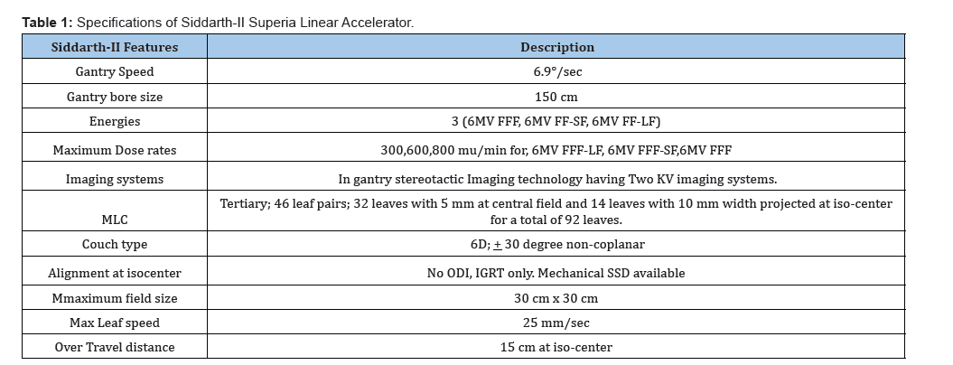
The Siddharth-II Superia linear accelerator can deliver various IGRT-based treatment modalities, including 3DCRT, IMRT, VMAT, and SBRT. It features a tertiary multi-leaf collimator (MLC) that supports both dynamic and sliding window techniques. Table 1 summarizes the main characteristics of this advanced treatment system. The machine features an enclosed, ring-mounted gantry equipped with two 6 MV flattening filter-free (FFF) photon beams and a single 6 MV beam with a flattening filter (FF). For imaging purposes, the system includes two kV imaging arms that facilitate simultaneous orthogonal image acquisition. Additionally, either kV imaging unit can be utilized for cone-beam CT (CBCT) scans.
The MLC leaves of this system operate at a maximum speed of 2.5 cm/s, which is comparable to conventional C-arm linear accelerators. The Siddharth-II Superia comes equipped with a built-in beam stopper, strategically positioned opposite the MV beam, offering inherent shielding equivalent to 3 TVT. For imaging, the system incorporates in-gantry stereotactic technology that enables the acquisition of both kV images and cone-beam CT (CBCT). While MV imaging and portal dosimetry are not available, the system fully supports both kV CBCT and kV orthogonal projections for each imaging method, users have the flexibility to choose between Cone-Beam CT (CBCT) and digitally reconstructed radiographs (DRRs). Notably, this system boasts the largest gantry in the industry, with a 150 cm bore size. Furthermore, it offers non-coplanar beam delivery, an advanced capability typically absents in other ring gantry-based linear accelerators.
As a standard procedure with the Siddharth-II Superia, initial patient positioning-or the alignment of physics QA equipment-is performed outside the gantry bore using laser guidance. The treatment couch then moves the patient into the bore to achieve the precise treatment position. Given the absence of a light field, optical distance indicator (ODI), or mechanical distance measurement device, the system relies on MV imaging for highly precise verification and fine-tuning of patient alignment. This study highlights critical tests performed to obtain type approval and clinical licensing for the Siddharth-II Superia. The guidelines and protocols from the American Association of Physicists in Medicine (AAPM) and the International Atomic Energy Agency (IAEA) [4- 13] were followed for this commissioning.
Materials and Methods:
Several authors have previously documented quality assurance, beam data validation, and performance tests for various linear accelerators [14-16]. The Siddharth-II [17,18], however, stands out as India’s first advanced linear accelerator equipped with Image-Guided Radiation Therapy (IGRT) capabilities [19,20]. It boasts unique features such as a large bore size, a tertiary MLC design, in-gantry stereotactic imaging, and a robotic couch with six degrees of freedom. These advanced features facilitated robust testing for obtaining both type approval and a clinical license. Critical mechanical and radiation tests were conducted, including assessments of beam profiles, percent-depth dose, Dmax, and radiation isocenter. The results were then rigorously compared against vendor-supplied beam measurements and established protocols from both the AAPM and IAEA, thereby evaluating the system’s overall performance and safety.
Gantry - Radiation Iso-centre:
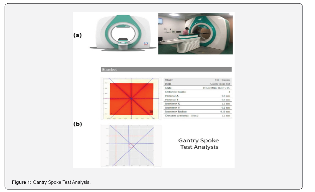
The Siddharth-II Superia linear accelerator, lacking a light field, requires alternative methods for verifying geometric accuracy. To address this, the gantry angle’s accuracy was assessed using the spoke test. For this test, a Gafchromic film, marked with a reference line, was precisely positioned at the isocentre. The irradiation field size was configured to 0.2 cm×30 cm. Spoke test exposures were conducted at gantry angles of 0∘,60∘,90∘,150∘,210∘, and 300∘. Following irradiation, the film was digitized. Image analysis software was then used to measure the angles formed by the radiation “spokes” relative to the reference line. The primary objective of this test was to evaluate the accuracy of the gantry angle as displayed by the system’s digital readout.
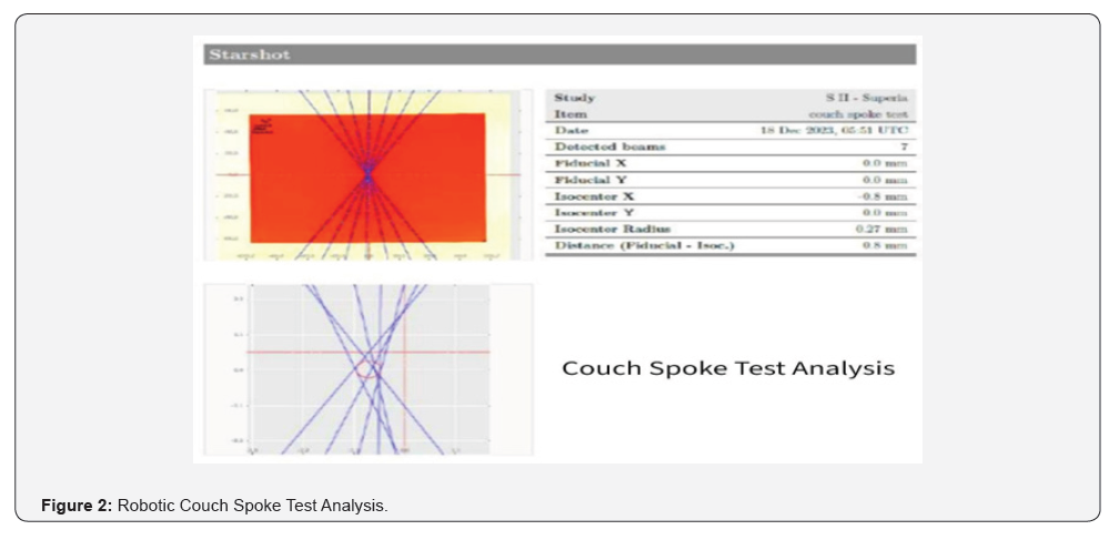
Table- Radiation Iso-Centre: In this linear accelerator, the treatment couch (Table) can rotate within a range of −20∘ to +20∘. Much like the gantry spoke test, the accuracy of the couch angle was evaluated by irradiating a Gafchromic film placed at the isocentre, which had a reference line marked on it. The irradiation field size was set to 0.2 cm×30 cm. Tests were performed at couch angles of −20∘, 0∘, and 20∘. The irradiated film was then digitized. The angles formed by the radiation “spokes” relative to the reference line were measured using image analysis software. This test’s purpose was to verify the accuracy of the couch rotation as indicated by the system’s digital table angle display.
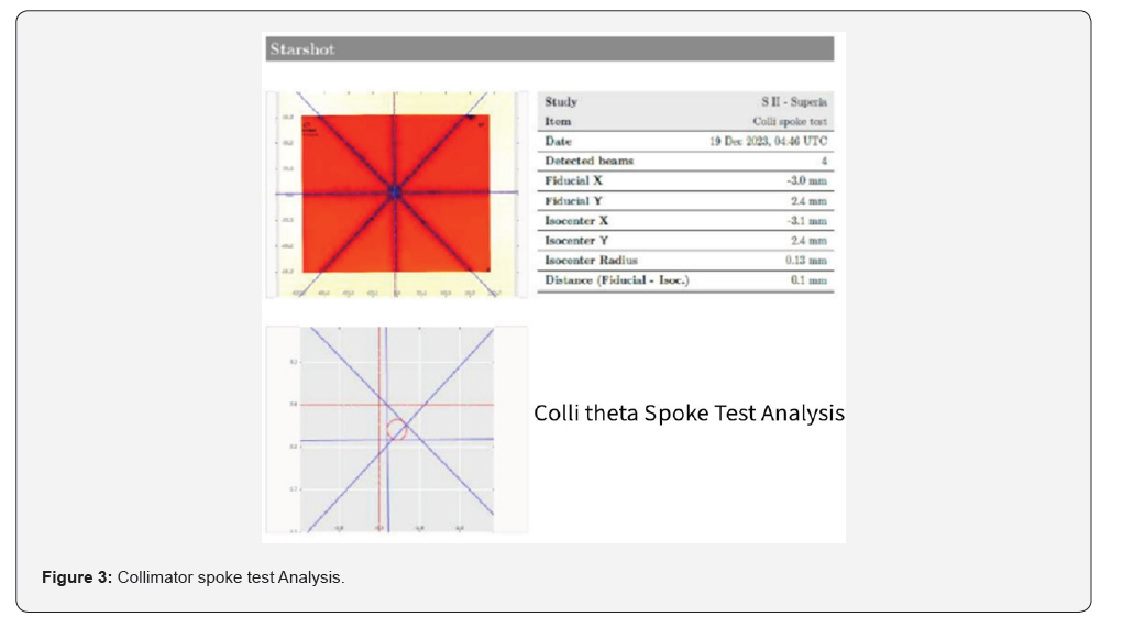
Collimator- Radiation Iso-center: Just like with the gantry angle, we used the spoke test to verify the accuracy of the collimator angle in the Siddharth-II Superia linear accelerator. For this test, a Gafchromic film with a reference line was placed at the isocentre to help with angle measurements. The irradiation field size was set to 0.2 cm×30 cm. We performed spoke test exposures at collimator angles of 0∘,30∘,60∘,90∘,300∘, and 330∘. After irradiation, the film was digitized. Then, we used image analysis software to measure the angles between the radiation “spokes” and the reference line. The goal of this test was to ensure the accuracy of the collimator’s rotation as shown on the system’s digital display.
Collimator- Radiation Iso-center: Just like with the gantry angle, we used the spoke test to verify the accuracy of the collimator angle in the Siddharth-II Superia linear accelerator. For this test, a Gafchromic film with a reference line was placed at the isocentre to help with angle measurements. The irradiation field size was set to 0.2 cm×30 cm. We performed spoke test exposures at collimator angles of 0∘,30∘,60∘,90∘,300∘, and 330∘. After irradiation, the film was digitized. Then, we used image analysis software to measure the angles between the radiation “spokes” and the reference line. The goal of this test was to ensure the accuracy of the collimator’s rotation as shown on the system’s digital display.
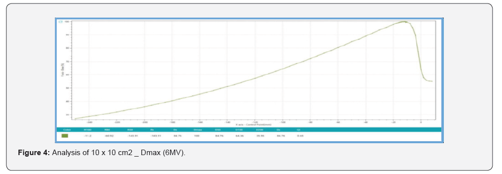
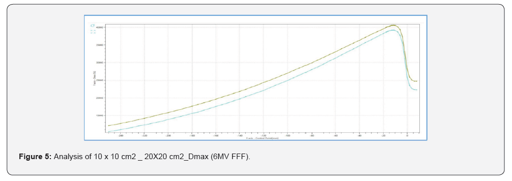
Dmax for 6X and 6XFFF (Small filter) and 6XFFF (Large filter): The depth of maximum dose (Dmax) was determined through profile analysis using the SmartScan radiation field analyzer (IBA). The measurements were acquired for a 10 × 10 cm² field size at a source-to-surface distance (SSD) of 100 cm.
Percentage Depth Dose Tests: The percent depth dose (PDD) curves for 6 MV flattening filter-free (FFF) photon beams were measured for various field sizes using CC13 ionization chambers and diode detectors (both from IBA) in the Smart SCAN water tank (IBA). The treatment bore diameter of 150 cm allows the use of standard scanning water tanks for measurements on the Siddharth-II Superia. However, since the water tank cannot be aligned directly with lasers, light fields, or mechanical measurement devices at the isocenter, initial alignment is performed at an external setup position using laser guidance. The tank is then shifted into the isocenter, where fine alignment is achieved with the help of image guidance. When setting the source-to-surface distance (SSD) to 100 cm, the air–water interface can be accurately visualized by acquiring images with the gantry positioned at either 90° or 270° in service mode. The 100 cm SSD is established by adjusting the treatment table height until the air–water interface aligns with the crosshair overlay corresponding to the isocenter in the service mode images. The acquired scanning data were analyzed using OmniPro Accept software (IBA). Percent depth dose (PDD) curves for field sizes ranging from 5 × 5 cm² to 30 × 30 cm² were evaluated and compared against AAPM TG-51 guidelines and Atomic Energy Regulatory Board (AERB) recommendations.
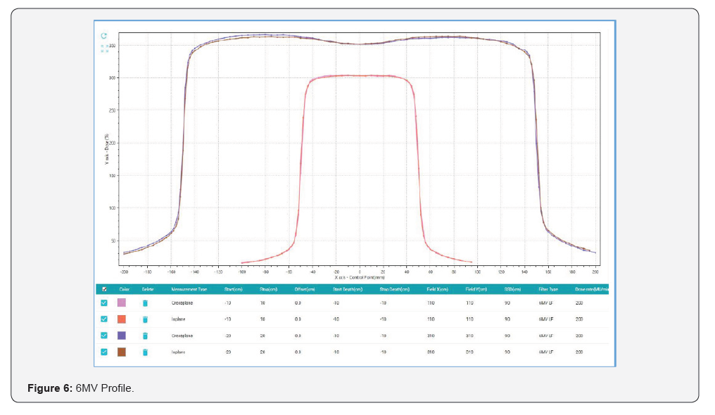
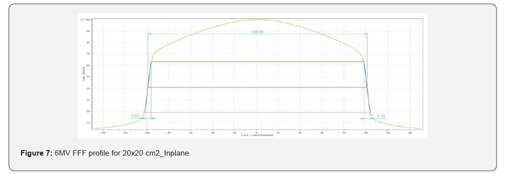
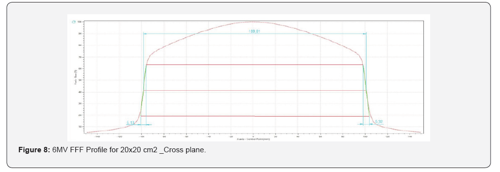
Profiles: Beam profiles were measured at multiple depths along both the in-plane and cross-plane directions at an SSD of 90 cm, for a range of field sizes: 2 × 2, 4 × 4, 6 × 6, 8 × 8, 10 × 10, 20 × 20, and 30 × 30 cm². Measurements were performed at five depths: 1.3 cm (dmax), 5 cm, 10 cm, 20 cm, and 30 cm. Additionally, beam profiles were acquired along the diagonal direction for the largest field size (30 × 30 cm²) at the same SSD of 90 cm. CC13 ionization chambers were used as the primary detectors for these measurements. The acquired beam profile data were initially smoothed, corrected for any central axis discrepancies, and symmetrized. The profiles were then re-normalized to 100% based on the central axis values. The resulting data were evaluated following AAPM and AERB protocols to ensure compliance with quality standards.
Output Measurements: Output factors were measured with the ionization chamber positioned at a depth of 10.0 cm and an SSD of 90 cm within a 30 × 30 × 30 cm³ water phantom. Measurements were taken for various field sizes and normalized to the output of the 10 × 10 cm² reference field to calculate relative output factors. A CC13 ionization chamber was used for field sizes smaller than 6 × 6 cm², while a 0.65 cc ion chamber was employed for field sizes larger than 6 × 6 cm².
Quality Index: The quality index (TPR 20/10) was measured for all three available photon beams using a 10 × 10 cm² field size.
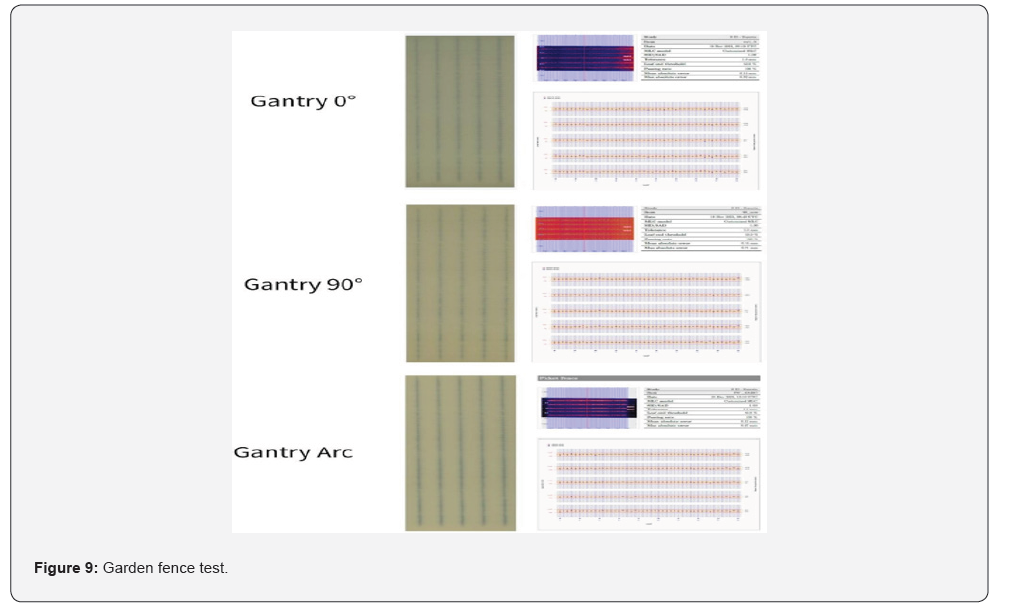
Leaf positioning accuracy induced error (Garden fence test): Leaf positioning accuracy e.g. Garden fence test at different MLC speeds and dose rate for 6 MV photon beam performed. Gantry set at 0 degrees and the Gafchromic film kept at 10 cm SSD. The plan delivered with the planned MU. Two induced leaf errors programmed.
Monitor chamber constancy during Arc mode of the gantry: 0.6 CC thimble chamber with build-up cap used for this study. Dose measurements were performed for 10x10 cm2 field size. Dose deviation from gantry 00 is derived with the following formula. Deviation in %= (Average of Measured Dose in dynamic beam/Average of Measured Dose in static beam)-1} * 100.
Accuracy of dose with variation of dose rate and gantry speed: 0.6 CC thimble chamber with build-up cap used for this study. Dose measurements were performed for 10x10 cm2 field size. Accuracy of dose with variation of dose rate and gantry speed is derived with the following formula for COV (And derived the Deviation From 0° Gantry Angle.
Treatment Plan validation: As part of the comprehensive dosimetric validation of the Siddharth-II Superia, multiple tests were conducted to verify that the beam model calculations accurately corresponded with commissioning measurements. Treatment plans were generated using both IMRT and Rapid Arc (RA) delivery techniques in the Ray Station treatment planning system, incorporating the complete Siddharth-II Superia beam data. Patient- specific quality assurance (QA) measurements were performed using the IBA iMatrixx detector. Dose distributions calculated by the treatment planning system were compared with the delivered dose planes. Gamma analysis was applied to all measurements using a 3% dose difference and 3 mm distance-to-agreement criteria to assess the accuracy of dose delivery.
Results
The results section summarizes the outcomes of the commissioning process, indicating successful completion of tests for FDA approval and obtaining clinical approval from AERB. Quantitative data on system characteristics demonstrating the system’s suitability for delivering various radiotherapy techniques including VMAT.


Gantry: Radiation iso-centrer: The diameter of the circle formed by the radiation lines at the center of the gantry angle spoke shot on the EBT3 film was measured to be within 0.3 mm, which is well below the tolerance limit of 2 mm. The accuracy of the gantry angle, evaluated relative to the reference line, ranged from 0.1° to 0.6° (Figure 1).
Table: Robotic Couch Radiation Isocentre: Measured rotational errors for the robotic treatment coach were consistently less than 0.2°, falling well within the clinical tolerance of ±1.0° for couch angle accuracy as given in Figure 2. This confirms the precision of patient positioning and target alignment during treatment delivery.
Collimator: For the collimator angle indicator accuracy test (Figure 3), the measured collimator angles were 0.13° and 89.86°, differing from the digital settings by only 0.13° and 0.14°, respectively.
Dmax for 6MV FFF and 6MV FF (Small filter) and 6MV FF (Large filter):
The Dose maximum (Dmax) observed for 6MV SF, 6MV FFF are 11.2 mm and 11 mm respectively for 10x10 cm2 field size. The Dmax for 6MV FFF for 20x20 cm2 field size found to be 11.5mm which are shown in Tables 2 & 3 and Figures 4 & 5.
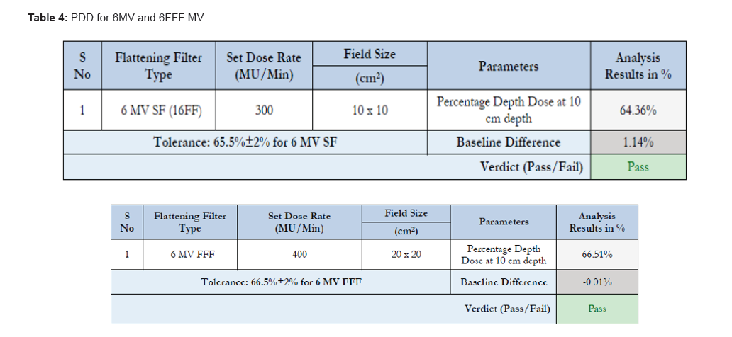
Percentage Depth Dose Tests: For all field sizes measured (2 X 2 cm2 to 30 X 30 cm2), PDD curves matched the Baseline values and are within 1.0% (step size = 1mm). For field sizes greater than 10 x 10 cm2, this maximum difference was 0.6%. The maximum dose difference observed is as follows for various field sizes (cm2) and is featured in Table 4.
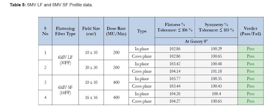
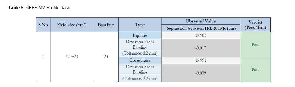
Profiles: The 6MV LF, SF and FFF profile data (Tables 5 to 6) showed that the profiles from this linear accelerator were well within the accepted limits. Profiles from this delivery system were shown in Figures 6 to 8.
Output Measurements: Output factors that were measured with the ionization chamber positioned at a depth of 10.0 cm and an SSD of 90 cm within a 30 × 30 × 30 cm³ water phantom resulted in good agreement within 1%–2% of any standard linear accelerator.
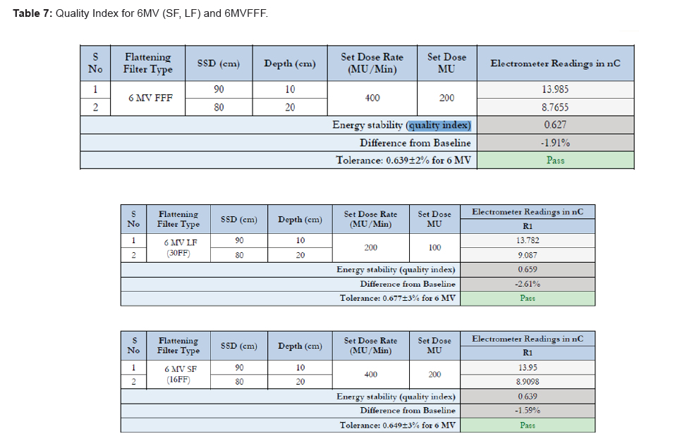
Quality Index: Quality index measured was 0.659 for 6MV LF, 0.639 for 6MV SF and 0.632 for 6MV FFF (Table 7).
Leaf positioning accuracy induced error (Garden fence test): Two induced errors are found at leaf positioning in Red colour (Figure 9), and remaining leafs are within the limit at different MLC speed and dose rate for 6MV photon beam.
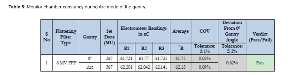
Monitor chamber constancy during Arc mode of the gantry: The Monitor chamber showed consistency with arc and showed less than 0.62% deviation compared to 0-degree gantry angle (Table 8).
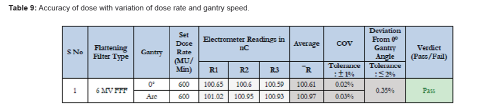
Accuracy of dose with variation of dose rate and gantry speed: The system shows less than 0.35% deviation dose with varying dose rate and gantry speed (Table 9).
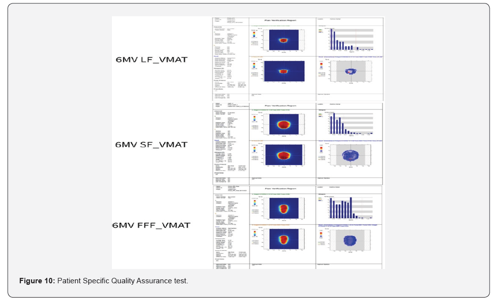
Treatment Plan validation: All IMRT and VMAT treatments (more than 300 cases) underwent patient specific QA with Imatrixx. The Ray Station treatment planning system does planes and I’MatriXX dose planes compared and calculated for gamma analysis. One of the patient analyses shown in Figure 10.
Conclusion
In conclusion, Siddharth-II Superia, which is India’s first ring gantry-mounted Linear Accelerator with a stereotactic imaging system, capable of delivering IGRT-based treatment modalities such as 3D CRT, IMRT, IGRT, SBRT, and VMAT, emerges as a one of the best treatment solutions for advanced radiotherapy treatments in India and abroad. The commissioning and beam characterization results affirm its efficacy, reliability, and patient- friendliness, demonstrating that this is one of the most advanced systems linear accelerators, suitable for confident clinical use with all modern features.
References
- Shiba S, Trauernicht C, Robar JL (2025) Treatment plan quality for stereotactic treatment of multiple cranial metastases: Comparison of C-arm and O-ring treatment platforms. Medical Dosimetry 50(1): 47-56.
- Guang-Yu Wang, Qi-Zhen Zhu, He-Ling Zhu, Ling-Juan Jiang, Nan Zhao, et al. (2022) Clinical performance evaluation of O-Ring Halcyon Linac: A real-world study. World J Clin Cases 10(22): 7728-7737.
- Chaofeng ShiYanjie JiangXi Wang, Yifu Chen (2021) Optimization design of O-ring linear accelerator gantry and mechanical isocenter detection method. Review of Scientific Instruments 92(3): 033308.
- Ezzell GA, Burmeister JW, Dogan N, Thomas J LoSasso, James G Mechalakos, et al. (2009) IMRT commissioning: multiple institution planning and dosimetry comparisons, a report from AAPM task group 119. Med Phys 36(11): 5359-5373.
- Agency Iae (2004) Commissioning and quality assurance of computerized planning systems for radiation treatment of cancer. Vienna: International Atomic Energy Agency.
- Das IJ, Cheng CW, Watts RJ, Anders Ahnesjö, John Gibbons, et al. (2008) Accelerator beam data commissioning equipment and procedures: report of the TG-106 of the therapy physics committee of the AAPM. Med Phys 35(9): 4186-4215.
- Bissonnette JP, Balter PA, Dong L, Dong, Katja M Langen, et al. (2012) Quality assurance for image guided radiation therapy utilizing CT-based technologies: a report of the AAPM TG-179. Med Phys 39(4): 1946-1963.
- Smilowitz JB, Das IJ, Feygelman V, Benedick A Fraass, Stephen F Kry, et al. (2015) Medical physics practice guideline 5.a.: commissioning and QA of treatment planning dose calculations - megavoltage photon and electron beams. J Appl Clin Med Phys 16(5): 14-34.
- Fontenot JD, Alkhatib H, Garrett JA, Andrew R Jensen, Steven P McCullough, et al. (2014) Medical physics practice guideline 2.a: commissioning and quality assurance of X-ray-based image-guided radiotherapy systems. J Appl Clin Med Phys 15(1): 4528.
- McEwen M, DeWerd L, Ibbott G, David Followill, David WO Rogers, et al. (2014) Addendum to the AAPM's TG- 51 protocol for clinical reference dosimetry of high-energy photon beams. Med Phys 41(4): 041501.
- Klein EE, Hanley J, Bayouth J, Fang-Fang Yin, William Simon, et al. (2009) Task group 142 report: quality assurance of medical acceleratorsa). Med Phys 36(9): 4197-4212.
- Almond PR, Biggs PJ, Coursey BM, WF Hanson, MS Huq, et al. (1999) AAPM's TG-51 protocol for clinical reference dosimetry of high-energy photon and electron beams. Med Phys 26(9): 1847-1870.
- Smith K, Balter P, Duhon J, Gerald A White, David L Vassy, et al. (2017) Medical physics practice guideline 8.a.: linear accelerator performance tests. J Appl Clin Med Phys 18: 23-39.
- Netherton T, Li Y, Gao S, Klopp A, Balter P, et al. (2019) Experience in commissioning the Halcyon linac. Medical Physics 46(10): 4304-4313.
- Chang Z, Wu Q, Adamson J, Lei Ren, James Bowsher, et al. (2012) Commissioning and dosimetric characteristics of TrueBeam system: composite data of three TrueBeam machines. Med Phys 39(11): 6981-7018.
- Glide-Hurst C, Bellon M, Foster R, C Altunbas, M Speiser, et al. (2013) Commissioning of the Varian TrueBeam linear accelerator: a multi-institutional study. Med Phys 40(3): 031719.
- Muralidhar KR, Venkataraman R, Subrahmanyam GV (2024) Siddharth II Superia: The First Indian Made Ring Gantry‑Mounted IGRT Capable VMAT Delivery System Commissioning and Beam Characterization. Abstract. Paper 8784, 66th AAPM annual Meeting & Exhibition, Los Angeles, California 52: 1252.
- Ravichandran R, Mondal T, Mani T, Kuppa AR, Govindappa M, et al. (2025) New-generation Indian Linear Accelerator: Validation of Specifications and Comparative Analysis. Journal of Medical Physics 50(02): 279-287.
- Report No. 180 - Image Guidance Doses Delivered During Radiotherapy: Quantification, Management, and Reduction (2018). American association of physcists in medicine.
- SP McCullough, H Alkhatib, KJ Antes, S Castillo, JD Fontenot, et al. (2021) AAPM Medical Physics Practice Guideline 2.b.: Commissioning and quality assurance of X-ray-based image-guided radiotherapy systems. J Appl Clin Med Phys 22(9): 73-81.

























