




 CTOIJ Home



Abstract
Human endogenous retroviruses (HERVs) constitute approximately 8% of the human genome. Their aberrant activation in cancer cells is strongly implicated in oncogenesis and malignancy progression. Current therapeutic interventions primarily focus on targeting HERV envelope (env) proteins through traditional immunotherapy. However, these methods have yielded suboptimal clinical outcomes. This study aims to evaluate the role of altered plasma membrane dynamics in propagating HERV-mediated pathogenicity. It seeks to establish a novel conceptual framework for inhibiting cancer progression by targeting the structural biophysics and biochemistry of the host cell membrane. We synthesized literature on tumour cell membranes’ lipidomic alterations and viral protein spreading mechanics. We analysed how modified membrane fluidity and lipid raft clustering in cancer cells facilitate the endogenous dissemination of oncogenic viral proteins. Building upon established membrane-targeted, resistance-avoiding oncology therapies, we modelled how pharmacological reconfiguration of plasma membrane structure can disrupt HERV envelope protein trafficking and subsequent cellular signalling. This conceptualised approach could offer a promising alternative paradigm for targeted oncological interventions.
Keywords:HERV; Cancer; Membrane targeted therapy; Transmembrane; Cholesterol
Abbreviations: HERVs: Human Endogenous Retroviruses; LTR: Long-Terminal Repeat; TM: Transmembrane; SU: Surface Envelope; PS: Phosphatidylserine; SFAs: Saturated Fatty Acids; UFAs: Unsaturated Fatty Acids; ERVs: Endogenous Retroviruses; VLPs: Virus-Like-Particles; VLV: Virus-Like-Vaccine; PE: phosphatidylethanolamine; FASN: Fatty Acid Synthase; HTAs: Host-Targeting Antivirals; PA: Palmitic Acid; PPTI: Palmitoyl-Protein Thioesterase Inhibitor; PUFAs: Polyunsaturated Fatty Acids
Introduction
Human endogenous retroviruses (HERVs) originate from ancestral exogenous retroviral infections, where the proviral element became stably integrated into the host genome, now comprising approximately 8% of the human genome, and was subsequently vertically transmitted through germline cells to progeny generations [1]. Although most HERVs sequences have accumulated mutations rendering them incapable of replication or retrotransposition, a significant portion retains the ability to be transcribed, particularly under certain pathological or epigenetically dysregulated conditions [2]. Many recent scientific reports confirm its activation and is responsible for origin and growth of numerous types of cancer [3].
Hence, during recent times the HERV Env genome protein has received special attention among the researchers for its significant clinical utility as diagnostic biomarkers and therapeutic targets across major malignancies [4-6,2,7]. Although the major therapeutic attempts to neutralize its effects on cancer growth use immunotherapy, other alternative therapeutic measures such as preventing its initial colonization onto the host cell membrane have not been attempted. Since the Env genome protein of the HERV depends on the ideal cell membrane chemical architecture for its initial endogenous colonization and subsequent safety that are provided by the cancer host, it is presumed that reversing such facilities could prevent the virus genome from initial colonization attempt.
It is well established that the cancer cells alter or modify their membrane chemical architecture as a part of their survival mechanism and thus differ in many ways from the normal cells in their plasma membrane chemistry [8]. Therefore, modulating such modified architecture of the Cancer cell membrane by some therapeutic means, not only prevents the Virus genome from initial spreading and getting shelter, but also do not harm the normal cells. Further, the difference in lipid composition between the cellular membranes of healthy cells and tumour cells allows for the development of novel therapies based on targeting membrane lipids in cancer cells to increase sensitivity to chemotherapeutic agents and consequently defeat multidrug resistance [8].
The study of Tan et al, [9], Kaynak et al [10], and Paulraj et al [11] pave way for targeting such modified chemical architecture of the cancer cells as one of the novel methods in cancer therapy. The significant aspect of cancer therapy via membrane disruption is that it will not lead to any resistance which normally happen in conventional therapies [12]. Keeping advantage of this approach, it is attempted here to inhibit HERV spreading and Downstream Pathogenicity through structural reconfiguration of the Cancer Cell Plasma Membrane using various therapeutic means. This necessitates the background study of the chemical architectures of HIRV genome as well as plasma membrane of cancer cells.
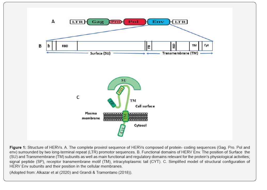
Chemical Configuration of HERV Genome:
Alcazar et al. [13] and Grandi & Tramontano [14] have given a detailed account of HERV genome configuration and figuratively explained the general structure of HERV DNA sequences (Figure 1A, B and C). The complete proviral sequence of HERVs is composed of four protein-coding sequences (Gag, Pro, Pol and Env) surrounded by two long-terminal repeat (LTR) promoter sequences which can act as promoters to drive the transcription of HERVs in cancer. The env gene encodes envelope proteins including transmembrane (TM) and surface envelope (SU) proteins responsible for fusion and receptor recognition. (Refer Figures: 1A, B and C for detailed configuration of the entire genome and how the HERV env genome parts inserted onto the host cell membrane).
Structure of the Altered Chemical Configuration of Cancer Cell Plasma Membrane:
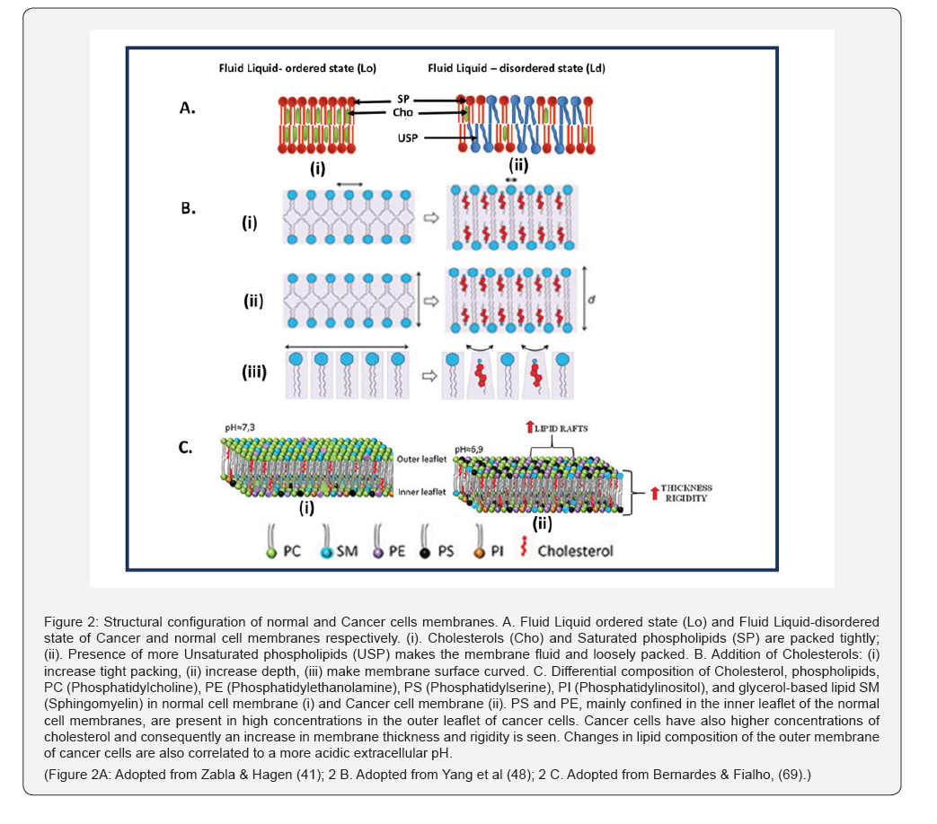
The physical properties of the bilayer cellular membrane depend on the density and location of phospholipids and cholesterols inside the bilayer membrane. The liquid ordered phase (Lo) typically harbours high density packing of saturated phospholipids and cholesterol has relatively rigid nature while under liquid -disordered phase (Ld) due less density of both saturated phospholipids and cholesterol and addition of more unsaturated phospholipids, loose packing occurs resulting in less rigid nature (Figure 2). These two stages, Lo and Ld decide differential functional rolls of the cells [15]. Cell membrane of normal cells remains in a Ld state, while the cancer cells are generally in Lo state because of their above said lipids and cholesterol composition. Here, membrane fluidity is influenced by the presence of unsaturated lipid content [15].
Influence of Modified Structural Changes in Cancer Cell Membrane on the Successful Colonization of HERV Env:
It an established fact that the physic- chemical nature of the cancer cell membrane differs from normal cell membrane in many ways which make the cancer cell membrane behave differently from normal cells thereby the cancels could express pathological characters including encouraging the HERV to colonize. In this respect it is important to find out the various molecular level structural modifications in the cancer cell membrane that enable the easy access and dissemination of HERV. Modulating the cancer cell’s plasma membrane can prevent HERV env attachment by physically altering the “docking sites” and the biochemical environment the protein requires to anchor itself. Because HERV Env proteins often use specific cell-surface receptors to mediate membrane fusion or anchor themselves, changing the membrane’s physical properties can disrupt this process.
Key Membrane Modulation Strategies
The key membrane modulation strategies include; a. cholesterol density, b. phospholipid composition, c. Fatty Acids composition and d. Transmembrane domine protein.
Cholesterol density
A growing body of evidence supports the idea that cholesterolrich regions serve as platforms for the entry of many enveloped viruses [16]. Cancer cell membranes are often enriched with cholesterol and lipid rafts, [17-19]. Cholesterol intercalates between the saturated hydrocarbon chains of sphingolipids, increasing the lateral order and thickness of the bilayer. This creates a “liquid-ordered” (Lo) phase that is more rigid and less fluid than the surrounding membrane (Figure 2). This provides a stable physical base for viral proteins [20].ug exposure or unnecessary interruption of anticancer therapy.
Here is how cholesterol’s unique properties facilitate the fusion process
Membrane fusion is not a straight path; it requires the two flat membranes to bend into a high-energy, hourglass-shaped intermediate called a fusion stalk. The Shape Factor of the cholesterol facilitates the membrane fusion process. Cholesterol is a “cone-shaped” lipid (small headgroup, bulky ring structure). When it clusters in the outer leaflet of the cancer cell membrane, it naturally pushes the lipid heads apart and induces negative curvature. Cholesterol’s curvature-promoting ability helps create concave “pits” or micro-domains. The HERV TM subunits migrate into these curved areas because they fit better there than on flat surfaces. Without cholesterol, the membrane is too flat and the energy required for the viral protein to bend it would be too high (Figure 2).
Phospholipid composition
One of the characteristics of Cancer cell membranes is the expression of more PE and PS phospholipids at the outer phase of the outer membrane [9]. Such symmetry is another advantageous feature for the easy entry of the HERV. Fusion-active tumors show an intense concentration of Phosphatidylserine (PS) and PE at these junctions (Figure 2). The “point of contact” is a chemical battlefield where the energy required to bend the membrane is determined by the specific shape and flexibility of the lipids present. Because the HERV Env protein needs to create extreme curves to fuse cells, it only succeeds if the local lipid composition at that exact spot is “cooperative.”
Lipids are not all shaped like cylinders; their geometric “packing parameter” dictates which way the membrane wants to bend. Lipids with small heads and bulky tails (like PE) act like wedges (Figure 2). At the point of contact, they force the membrane to curve inward toward the tails. This is essential for forming the “fusion stalk.” If these are dominant at the contact point, they will physically resist the HERV protein’s attempt to form a fusion stalk, effectively “jamming” the process. The point of contact is also influenced by the “stickiness” of the lipid heads. The contact point is rich in negatively charged lipids, PS [10]. So, they can “pull” the positively charged parts of the HERV Env protein closer, increasing the force applied to the membrane and forcing a deeper curve.
PS is detected through two fundamentally distinct modes of interaction: direct molecular recognition by PS-binding proteins (e.g., annexins and related PS-binding domains) and electrostatic/ biophysical recognition of PS-rich membrane surfaces as negatively charged platforms [10,21]. In the latter mode, proteins respond not to individual PS molecules but to collective membrane properties—most notably charge density and curvature-which can strongly shape binding and downstream activity [21]. For two membranes to fuse, the water layer between them must be pushed out. Lipids like PE are “dehydrating”—they don’t hold onto water tightly. If the contact point is rich in PE, the membranes can get closer together, allowing the curvature to become much tighter.
Fatty acids composition
It was found that the saturated fatty acid makes the model membrane more rigid, while the presence of unsaturated fatty acid increases its fluidity [22]. In the context of HERV-mediated fusion in cancer cells, the ratio of Saturated Fatty Acids (SFAs) to Unsaturated Fatty Acids (UFAs) dictates the “stiffness” and “organization” of the membrane platforms where these viral proteins operate. UFAs (like oleate or linoleate) have “kinks” in their chemical chains that prevent tight packing, making the membrane more fluid and disordered [23]. Cancer cells often ramp up de novo lipogenesis (fatty acid synthesis), leading to a high concentration of SFAs (like palmitate and stearate).
Saturated fatty acid chains are straight and pack together tightly (Figure 2). This tight packing is what allows cholesterol to wedge in and form the rigid, stable lipid rafts. Without a high SFA content and with a presence of high UFA, lipid rafts would be too “leaky” or fluid (Figure 2). The SFAs provide the structural “walls” that keep HERV Env proteins and their receptors (like ASCT2) trapped in the same small area, ensuring they find each other to initiate fusion. Many viral envelope proteins, including those from HERVs, undergo a process called palmitoylation, where a saturated fatty acid (palmitate) is chemically attached to the protein [24].
This acts as a “zip code” that anchors the HERV protein specifically into the SFA-rich lipid rafts. Thus, the structural modifications with high density cholesterols and SFAs, and with hyper expression of PS & PE at the outer phase of the cell membrane facilitate perfect accommodation and subsequent dissemination for the HERV env antigen. Targeting the cell membrane by reducing the cholesterol and SFA contents, enhancing the USFA composition and by deactivating the actions of PS & PE are the therapeutic strategies proposed for inhibiting role of HERV in cancer growth and spreading.
Discussion
Under normal physiological conditions, HERVs remain transcriptionally silent; however, as shown by multiple studies, that aberrant level of HERV env genome protein has been linked to numerous types of cancers [3]. It is established that this viral protein acts as a critical molecular switch for promoting growth factors like beta-catenin, ERK, Akt and Notch1 [25,26] and down regulation of this prompted a decrease in cell proliferation and a concomitant reduction of RAS, p-ERK, and p-AKT expression [27]. Antibodies against HERV-K-originated protein products have been detected at high titres in the sea of patients with various tumours [28]. Remarkably, a bioinformatic analysis of different datasets revealed that increased expression of HERV in cancer patients was linked to reduced survival [29,30]. Analyzing differential HERV expression and its correlation with clinicopathological patient characteristics could be valuable for identifying novel biomarkers and exploring potential therapeutic targets [31]. Thus, Endogenous retroviruses (ERVs) are emerging as promising therapeutic targets in cancer [2,14].
Immunotherapy is the main therapeutic measure as this virus has immunosuppressive capacity which aid cancer to escape immunosurveillance. In that regard, virus-like-vaccine (VLV) technology, combining adenoviral vectors and virus-like-particles (VLPs), can be ideal to target ERVs and elicit B-cell responses, as well as CD8+ and CD4+ T-cells responses [7]. Costa et al [32] in their latest review highlighted the latest research advancements and potential treatment strategies including usage of antiviral drugs to combat HERV-induced diseases.
However, no studies have been attempted to prevent this virus from endogenous spreading in the cancer cells microenvironment and to stop its cancer promoting action at initial stage itself. As discussed in this paper, cancer cells have developed some adaptive features in their membrane which facilitate the virus to invade into the cells. If we can deactivate these adaptive features from their functioning or existence, it could be possible to stop the virus from their colonization. In this respect the membrane biology of cancer cells has been explained in detail above. As much as those adaptive features of the cancer cells are their specific characteristics and not expressed by normal cells, targeting such characters not only makes them modify their role but also will not affect the normal cells. Such adaptive features are generally considered as hyper expressions [11] and targeting such characters like cholesterol with statins and anti-cancer drugs synergistically helps to overcome anti-cancer drug resistance [33].
The following are the hyper expressed characters in the cell membrane which facilitate the virus to invade the cancer cells: i. accumulation of cholesterol, ii. Hyper expression of PE and PS at the outer phase of the outer membrane and iii. Over accumulation of SFA. Targeting these characters may be considered as a doubleedged weapon as, such target results in affecting some essential molecular functions and affect the invasion of the virus into the cancer cells. For example, overexpressed cholesterol in cancer cells serves as a critical “building block” for rapid proliferation and a master regulator of oncogenic signalling pathways [34- 36]. High levels of Saturated Fatty Acids (SFAs) primarily support the rapid demands of tumour growth and survival by acting as building blocks for new membranes, energy sources, and signalling molecules [37-39].
And high expression of phosphatidylethanolamine (PE) and phosphatidylserine (PS) on the outer cell membrane—a loss of the normal lipid asymmetry found in healthy cells—primarily serves as a mechanism for immune evasion, angiogenesis, and metastatic progression [9,10,40]. Thus, structural reconfigurations of the said cell membrane components result in multiple anti-cancer effects including inhibition of pro-cancer effects of HERV. Given that fundamental differences exist between the cellular membranes of healthy cells and tumour cells, novel therapies based on targeting membrane lipids in cancer cells is a promising approach that deserves attention in the field of anticancer drug development [9].
Targeting membrane lipids of cancer cells
The selective targeting of membrane lipids represents a promising therapeutic strategy in oncology [41]. Tan et al. [9] detailed various mechanisms for targeting phosphatidylethanolamine (PE), a phospholipid central to diverse pathophysiological processes [42]. The development of PE-specific probes derived from duramycin and cinnamycin has facilitated recent breakthroughs in understanding PE’s biological functions in vivo. Research indicates that PE-binding cinnamycin induces significant membrane reorganization, ultimately triggering cell death [43].
Similarly, recent studies suggest that the upregulation of surface phosphatidylserine (PS) exposure-induced by chemotherapy, radiation, or external electric fields-serves as a novel approach to sensitize malignant cells to PS-targeting agents [10]. Cancer cells exhibiting elevated surface PS demonstrate increased sensitivity to specialized treatments, such as SapCDOPS (saposin C embedded in dioleoyl phosphatidylserine nanovesicles) [44-46]. Several clinical trials are currently evaluating the efficacy of SapC-DOPS and bavituximab to improve patient outcomes [10].
In a comprehensive review, Yoo and Kim [40] highlighted emerging PS-targeting platforms, including monoclonal antibodies, engineered PS-binding proteins, immunomodulatory adjuvants, and cellular therapies. Furthermore, PS-targeted nanoparticles that leverage electrostatic recognition of PS-rich membranes have been developed to enable selective tumour accumulation and the precise delivery of therapeutic payloads [47]. Given that both PE and PS are characteristically overexpressed on the outer leaflet of cancer cell membranes, it is hypothesized that targeting these lipids may not only induce direct cytotoxicity but also inhibit the invasion of Human Endogenous Retroviruses as these phospholipids help and facilitate successful invasion and spread of HERV env as already discussed above.
Targeting membrane cholesterol
Targeting membrane cholesterol represents a strategic therapeutic approach in oncology, as rapidly dividing cancer cells exhibit an increased demand for cholesterol to support membrane synthesis. Moreover, cholesterol can organize membrane fusion proteins in spatial arrangements that support the ensuing fusion process of viruses [48]. Clinical and preclinical data suggest that this requirement can be managed by reducing low-density lipoprotein cholesterol (LDL-C) [49,50]. Depleting cellular cholesterol levels significantly diminishes cholesterol content within membrane lipid rafts, thereby altering membrane fluidity and integrity. Modulation of membrane fluidity to revert MDR resistance and increase sensitivity to chemotherapeutics is the most promising strategy regarding lipid modulation [41].
Statins remain the primary pharmacological intervention for lipid reduction, with global usage estimated at 145.8 million individuals as of 2018 [51]. Recent evidence underscores the multifaceted role of statins in cancer; for instance, Simvastatin has been shown to reduce cellular cholesterol and disrupt lipid raft architecture [52,53,33]. Beyond pharmacotherapy, dietary interventions have demonstrated significant efficacy in cholesterol management [54]. Schoeneck et al. [55] provided an extensive classification of food supplements based on their lipidlowering potential, while Jenkins et al. [56] showed that a “dietary portfolio”—comprising plant sterols, viscous fibers, soy protein, and nuts—can reduce LDL-C by approximately 30%, a result comparable to low-dose statin therapy.
Furthermore, Jacobo-Velázquez [57] highlighted the additive and synergistic potential of combining functional foods with statins, introducing a cholesterol-lowering capacity index to evaluate these interactions. Collectively, these therapeutic strategies limit cholesterol bioavailability for malignant cells, consequently modifying lipid raft dynamics and compromising membrane stability. Such interventions not only inhibit the role of cholesterol in tumorigenesis and progression [58]. but may also reduce the susceptibility of cancer cells to HERV endogenous spreading.
Modifying SFA and USFA Concentration in the Cell Membrane:
It has been already discussed that the cancer cells prefer more concentration of SF in the cell membrane and less amount of USF as, such condition enhance the rigidity of the cell membrane which in turn facilitates the colonization of the HERV and provide the resistance to anticancer drugs. By altering this configuration by reducing the SFA and increasing the USFA in the cell membrane, it is hypothesised that the invasion of HERV could be curtailed. Mammalian cells can endogenously produce most fatty acids; however, they lack the desaturase enzymes necessary to produce n-6 or n-3 fatty acids de novo [59]. Therefore, these lipids must be obtained via the diet and are considered essential. These fatty acids absorbed and incorporated in cells from dietary sources. At the same time, cancer cells are capable of de novo synthesis of SFA using Fatty Acid Synthase (FASN) and incorporate in the cell membrane apart from using it for other malignancies [60].
Researchers are investigating host-targeting antivirals (HTAs) that block Fatty Acid Synthase (FASN) to “starve” viruses of the lipids they need to replicate. Fhu & Ali [60] have discussed several pivotal roles of FASN in lipid metabolism and considered it as an attractive target in the clinic with several new inhibitors currently being tested. The role of PUFA as antiviral agent has been demonstrated by scientists. The PUFAs being lipophilic molecules, they could interfere with the viral envelope itself, changing its dynamics and inactivate viruses by disrupting their envelopes [61]. Mechanistic experiments showed that EPA disrupted the membrane integrity of viral particles, leading to the release of viral RNA, together with the interruption of ZIKV from binding, adsorption and entry, and ultimately the inhibition of viral proliferation. Furthermore, EPA exerted antiviral effects in a dose-dependent manner [62].
Thus, it is presumed that enrichment of cancer cell membranes with omega 3 PUFA and depriving SFA may have profound effect in curtailing the endogenous spread of HERV evn. Palmitoylation is a process where a 16-carbon saturated fatty acid, palmitate, is chemically attached to a protein. Palmitoylation adds a “greasy tail” to viral proteins, making them highly lipophilic. For a virus, this process is a critical “host-jacking” manoeuvre [63]. Because viruses have tiny genomes, they rely on the host cell’s enzymes (DHHC-palmitoyltransferases) to tag viral proteins with palmitate. By anchoring these proteins to the membrane, the virus ensures all the “parts” (envelope proteins, spikes, etc.) are in the same physical location so they can bundle together and bud off as a new virus particle.
The palmitate tail helps brace the spike protein against the viral envelope. Without this anchor, the spike might not have the mechanical stability required to “harpoon” and fuse with a new host cell [64]. Thus, Palmitoylation is an essential process for the virus to survive and propagate. By stopping this process, one could prevent the survival of the virus. De-palmitoylation is a chemical process used by researchers using Palmitoyl-Protein Thioesterase Inhibitor (PPTI) enzyme which removes palmitate from the host defence protein. Ahmed et al. [65] have found out that palmitoyltransferase inhibitors like two bis-piperazine backbonebased DHHC9 inhibit SARS-CoV-2 spike protein palmitoylation and the resulting progeny virion particles released are defective in fusion and infection.
Moreover, palmitates are reported to poses pro-cancer effects such as a palm oil diet, or palmitic acid (PA) treatment, induces a prometastatic memory in human oral squamous cell carcinoma and melanoma cells [66]. FASN is the primary enzyme responsible for the de novo synthesis of palmitate. FASN inhibition presents a promising therapeutic strategy for treating a variety of cancers [67]. It is pertinent to point out here is that next-generation FASN inhibitors, including TVB-3166 and TVB-2640 (Denifanstat), have shown tremendous antitumor potential in preclinical breast and CRC models, enabling patients to actually benefit from FASN inhibition [68].
Therefore, targeting palmitate using FASN inhibitors has double anticancer effects – one by the way of targeting viral protein invasion and another by directly affecting pro-cancer pathways. Apart from using FASN inhibitors and Palmitoyl- Protein Thioesterase Inhibitor (PPTI) enzyme, naturalistically, reducing saturated fats (like palmitic acid) while boosting Polyunsaturated Fatty Acids (PUFAs) would act as additional anticancer measures. Different classes of drugs are arising with the purpose of modulating the biochemical and biophysical features of cancer cells acting directly at the membrane and interacting with lipids and membrane-resident proteins or at the enzymes responsible for sensing and synthesis of membrane lipids [69].
Conclusion
Evidence suggests that Human Endogenous Retrovirus (HERV) envelope proteins serve as significant pro-oncogenic drivers, contributing to tumour progression and metastatic spread across diverse tissues. As such, HERVs represent promising targets for innovative anti-cancer strategies, including targeted inhibitors and immunotherapeutic interventions [14]. Crucially, because HERV activity and invasive capacity are contingent upon the specific structural configuration of the cancer cell plasma membrane, the pharmacological modulation of membrane lipids may offer a compelling therapeutic avenue. By reducing cholesterol and saturated fatty acid (SFA) levels and targeting the hyperexpression of phosphatidylserine (PS) and phosphatidylcholine (PC), it may be possible to disrupt the permissive environment required for virion-mediated invasion.
Evidence suggests that Human Endogenous Retrovirus (HERV) envelope proteins serve as significant pro-oncogenic drivers, contributing to tumour progression and metastatic spread across diverse tissues. As such, HERVs represent promising targets for innovative anti-cancer strategies, including targeted inhibitors and immunotherapeutic interventions [14]. Crucially, because HERV activity and invasive capacity are contingent upon the specific structural configuration of the cancer cell plasma membrane, the pharmacological modulation of membrane lipids may offer a compelling therapeutic avenue. By reducing cholesterol and saturated fatty acid (SFA) levels and targeting the hyperexpression of phosphatidylserine (PS) and phosphatidylcholine (PC), it may be possible to disrupt the permissive environment required for virion-mediated invasion.
Author Contribution
Conceptualization, RK and AA; writing-original draft preparation, writing-review and editing, PS All authors have read and agreed to the published version of the manuscript.
References
- Jakobsson J, Vincendeau M (2022) Snapshot: human endogenous retroviruses. Cell 185(21): 400-400.e401.
- Jansz N, Faulkner GJ (2021) Endogenous retroviruses in the origins and treatment of cancer. Genome Biol 22(1): 147.
- Stricker E, Peckham-Gregory EC, Scheurer ME (2023) Cancer HERVdb: human endogenous retrovirus (HERV) expression database for human cancer accelerates studies of the retrovirome and predictions for HERV-based therapies. J Virol 97(7): e00059-23.
- Ramalingam K, Paulraj S, Karnan P (2026) The cellular expression dynamics of endogenous gp70 TAA antigens in cancer cells and their biomarkers and immunopotential for clinical relevance of drug combinations. Int J Cancer Res Ther 10(1): 01-03.
- Zhang J, Sun D, Zhan Y, Gao Q, C Bao, et al. (2025) The role of HERV envelope protein in ovarian cancer. Front Cell Dev Biol 13: 1618542.
- Ma W, Ji C, Abudushataer A, Liu N, Tao Xu, Kunlun Zhao, et al. (2025) The expression, regulation, and function of human endogenous retroviruses in genitourinary cancers. Cell Death Discov 11(1): 553.
- Vergara Bermejo A, Ragonnaud E, Daradoumis J, Holst P (2020) Cancer associated endogenous retroviruses: ideal immune targets for adenovirus-based immunotherapy. Int J Mol Sci 21(14): 4843.
- Preta G (2020) New insights into targeting membrane lipids for cancer therapy. Front Cell Dev Biol 8: 571237.
- Tan LT, Chan KG, Pusparajah P, Lee WL, LH Chuah, et al. (2017) Targeting membrane lipid a potential cancer cure? Front Pharmacol 8: 12.
- Kaynak A, Davis HW, Kogan AB, Lee JH, Daria A Narmoneva, et al. (2022) Phosphatidylserine: the unique dual-role biomarker for cancer imaging and therapy. Cancers 14(10): 2536.
- Paulraj S, Ramalingam K, Arulvasu A (2024) Targeting the Weakness of Hyper Expression of Phosphatidylethanolamine (PE) on the Cancer Cell Membrane Surface using Cyclotide Peptides, A Potential Pathway in the Cancer Cure. Canc Therapy Oncol Int J 28(1): 556226.
- Lin L, Chi J, Yan Y, Luo R, X Feng, Y Zheng, et al. (2021) Membrane-disruptive peptides/peptidomimetics-based therapeutics: Promising systems to combat bacteria and cancer in the drug-resistant era. Acta Pharm Sin B 11(9): 2609-2644.
- Alcazer V, Bonaventura P, Depil S (2020) Human Endogenous Retroviruses (HERVs): Shaping the Innate Immune Response in Cancers. Cancers 12(3): 610.
- Grandi N, Tramontano E (2018) HERV Envelope Proteins: Physiological Role and Pathogenic Potential in Cancer and Autoimmunity. Front Microbiol 9: 462.
- Gracià RS, Bezlyepkina N, Knorr RL, Lipowsky R, R Dimova (2010) Effect of cholesterol on the rigidity of saturated and unsaturated membranes: fluctuation and electrodeformation analysis of giant vesicles. Soft Matter 6(7): 1472.
- Manes S, del Real G, Martinez AC (2003) Pathogens: raft hijackers. Nat Rev Immunol 3(7): 557-568.
- Li YC, Park MJ, Ye SK, Kim CW, YNKim (2006) Elevated Levels of Cholesterol-Rich Lipid Rafts in Cancer Cells Are Correlated with Apoptosis Sensitivity Induced by Cholesterol-Depleting Agents. Am J Pathol 168(4): 1107-1118.
- Hryniewicz-Jankowska A, Augoff K, Sikorski AF (2019) The role of cholesterol and cholesterol-driven membrane raft domains in prostate cancer. Exp Biol Med 244(13): 1053-1061.
- Maja M, Tyteca D (2022) Alteration of cholesterol distribution at the plasma membrane of cancer cells: From evidence to pathophysiological implication and promising therapy strategy. Front Physiol 13: 999883.
- Pluhackova K, Gahbauer S, Kranz F, Wassenaar TA, RA Böckmann (2016) Dynamic Cholesterol-Conditioned Dimerization of the G Protein Coupled Chemokine Receptor Type 4. PLoS Comput Biol 12(11): e1005169.
- Bevers EM, Williamson PL (2016) Getting to the Outer Leaflet: Physiology of Phosphatidylserine Exposure at the Plasma Membrane. Physiol Rev 96(2): 605-645.
- Hac-Wydro K, Wydro P (2007) The influence of fatty acids on model cholesterol/phospholipid membranes. Chem Phys Lipids 150(1): 66-81.
- Bruno MJ, Koeppe RE 2nd, Andersen OS (2007) Docosahexaenoic acid alters bilayer elastic properties. Proc Natl Acad Sci USA 104(23): 9638-9643.
- Chai K, Wang Z, Xu Y, Zhang J, J Tan, et al. (2021) Palmitoylation of the Bovine Foamy Virus Envelope Glycoprotein Is Required for Viral Replication. Viruses 13(1): 31.
- Chen T, Meng Z, Gan Y, Wang X, F Xu, et al. (2013) The viral oncogene Np9 acts as a critical molecular switch for co-activating beta-catenin, ERK, Akt and Notch1 and promoting the growth of human leukemia stem/progenitor cells. Leukemia 27(7): 1469–1478.
- Lemaitre C, Tsang J, Bireau C, Heidmann T, M Dewannieux, et al. (2017) A human endogenous retrovirus-derived gene that can contribute to oncogenesis by activating the ERK pathway and inducing migration and invasion. PLoS Pathog 13(6): e1006451.
- Gao Y, Yu XF, Chen T (2021) Human endogenous retroviruses in cancer: Expression, regulation and function. Oncol Lett 21(2): 121.
- Cherkasova EA, Chen L, Childs RW (2024) Mechanistic regulation of HERV activation in tumors and implications for translational research in oncology. Front Cell Infect Microbiol 14: 1358470.
- Büscher K, Trefzer U, Hofmann M, Sterry W, Reinhard Kurth, et al. (2005) Expression of Human Endogenous Retrovirus K in Melanomas and Melanoma Cell Lines. Cancer Res 65(10): 4172-4180.
- Shah AH, Govindarajan V, Doucet-O’Hare TT, Rivas S, Leo Ampie, et al. (2022) Differential expression of an endogenous retroviral element [HERV-K(HML-6)] is associated with reduced survival in glioblastoma patients. Sci Rep 12(1): 6902.
- Dolci M, Civettini I, Bagnoli PF, Toumi W, et al. (2025) Expression of human endogenous retrovirus env-gene product is a hallmark of sidedness in operable colorectal cancer. Oncology 1-16.
- Costa B, Vale N (2023) Exploring HERV-K (HML-2) influence in cancer and prospects for therapeutic interventions. Int J Mol Sci 24(19): 14631.
- Tilija Pun N, Jeong CH (2021) Statin as a Potential Chemotherapeutic Agent: Current Updates as a Monotherapy, Combination Therapy, and Treatment for Anti-Cancer Drug Resistance. Pharmaceuticals (Basel) 14(5): 470.
- Li YC, Park MJ, Ye SK, Kim CW, YN Kim (2006) Elevated levels of cholesterol-rich lipid rafts in cancer cells are correlated with apoptosis sensitivity induced by cholesterol-depleting agents. Am J Pathol 168(4): 1107-1118.
- Halimi H, Farjadian S (2022) Cholesterol: An important actor on the cancer immune scene. Front Immunol 13: 1057546.
- Wu Y, Song W, Su M, He J, R Hu, et al. (2025) The Role of Cholesterol Metabolism and Its Regulation in Tumor Development. Cancer Med 14(1): e70783.
- Du A, Wang Z, Huang T, S Xue, C Jiang, et al. (2023) Fatty acids in cancer: metabolic functions and potential treatment. MedComm – Oncology 2(1): e25.
- He JW, Huang ZX, Su YF, Mo YQ (2025) Dietary saturated fatty acids and prostate cancer: insights into NF-κB pathway and lipid metabolism mechanisms. Discov Oncol 16(1): 1166.
- Sun J, Liu J, Chen F, Wang X, G Wu (2025) Exploring the significance of fatty acid metabolism reprogramming in the pathogenesis of cancer and anticancer therapy. Ann Med 57(1): 2445774.
- Yoo M, Kim KH (2026) Phosphatidylserine Externalization in Cancer: Biology, Immune Suppression, and Emerging Theragnostic Strategies. Int J Mol Sci 27(2): 697.
- Zalba S, Ten Hagen TL (2017) Cell membrane modulation as adjuvant in cancer therapy. Cancer Treat Rev 52: 48-57.
- Calzada E, Onguka O, Claypool SM (2016) Chapter Two-Phosphatidylethanolamine Metabolism in Health and Disease. Int Rev Cell Mol Biol 321: 29-88.
- Makino A, Baba T, Fujimoto K, Iwamoto K, Yoshiaki Yano, et al. (2003) Cinnamycin (Ro 09-0198) promotes cell binding and toxicity by inducing transbilayer lipid movement. J Biol Chem 278(5): 3204-3209.
- N’Guessan KF, Davis HW, Chu Z, Vallabhapurapu SD, CS Lewis, et al. (2020) Enhanced Efficacy of Combination of Gemcitabine and Phosphatidylserine-Targeted Nanovesicles against Pancreatic Cancer. Mol Ther 28(8): 1876-1886.
- Davis HW, Vallabhapurapu SD, Chu Z, Vallabhapurapu SL, Robert S Franco, et al. (2019) Enhanced phosphatidylserine-selective cancer therapy with irradiation and SapC-DOPS nanovesicles. Oncotarget 10(8): 856-868.
- Davis HW, Kaynak A, Vallabhapurapu SD, Qi X (2021) Targeting of elevated cell surface phosphatidylserine with saposin C-dioleoylphosphatidylserine nanodrug as individual or combination therapy for pancreatic cancer. World J Gastrointest Oncol 13(6): 550-559.
- De M, Ghosh S, Sen T, Shadab M, Indranil Banerjee, et al. (2018) A Novel Therapeutic Strategy for Cancer Using Phosphatidylserine Targeting Stearylamine-Bearing Cationic Liposomes. Mol Ther Nucleic Acids 10: 9-27.
- Yang ST, Kreutzberger AJB, Lee J, Kiessling V, LK Tamm, et al. (2016) The role of cholesterol in membrane fusion. Chem Phys Lipids 199: 136-143.
- Nielsen SF, Nordestgaard BG, Bojesen SE (2012) Statin use and reduced cancer-related mortality. N Engl J Med 367(19): 1792-1802.
- Gobel A, Rauner M, Hofbauer LC, Rachner TD (2020) Cholesterol and beyond - the role of the mevalonate pathway in cancer biology. Biochim Biophys Acta Rev Cancer 1873(2): 188351.
- Blais JE, Wei Y, Yap KKW, Alwafi H, TT Ma, et al. (2021) Trends in lipid-modifying agent use in 83 countries. Atherosclerosis 328: 44-51.
- Menter DG, Ramsauer VP, Harirforoosh S, Chakraborty K, Peiying Yang, et al. (2011) Differential effects of pravastatin and simvastatin on the growth of tumor cells from different organ sites. PLoS ONE 6(12): e28813.
- Zhuang L, Kim J, Adam RM, Solomon KR, MRFreeman (2005) Cholesterol targeting alters lipid raft composition and cell survival in prostate cancer cells and xenografts. J Clin Invest 115(4): 959-968.
- Ge Q, Yan Y, Luo Y, Teng T, Caixia Cao, et al. (2024) Dietary supplements: clinical cholesterol-lowering efficacy and potential mechanisms of action. Int J Food Sci Nutr 75(4): 349-368.
- Schoeneck M, Iggman D (2021) The effects of foods on LDL cholesterol levels: A systematic review of the accumulated evidence from systematic reviews and meta-analyses of randomized controlled trials. Nutr Metab Cardiovasc Dis 31(5): 1325-1338.
- Jenkins DJ, Nguyen TH, Kendall CW, Faulkner DA, B Bashyam, et al. (2008) The effect of strawberries in a cholesterol-lowering dietary portfolio. Metabolism 57(12): 1636-1644.
- Jacobo-Velázquez DA (2025) Functional Foods for Cholesterol Management: A Review of the Mechanisms, Efficacy, and a Novel Cholesterol-Lowering Capacity Index. Nutrients 17(11): 2648.
- Xiao M, Xu J, Wang W, Zhang B, Jiang Li, et al. (2023) Functional significance of cholesterol metabolism in cancer: from threat to treatment. Exp Mol Med 55(10): 1982-1995.
- Chapkin R (2007) Reappraisal of the Essential Fatty Acids. In Fatty Acids in Foods and Their Health Implications, (3rd edn), CRC Press, Boca Raton, FL, USA pp: 675-691.
- Fhu CW, Ali A (2020) Fatty Acid Synthase: An Emerging Target in Cancer. Molecules 25(17): 3935.
- Goc A, Niedzwiecki A, Rath M (2021) Polyunsaturated ω-3 fatty acids inhibit ACE2-controlled SARS-CoV-2 binding and cellular entry. Sci Rep 11(1): 5207.
- Feng Y, Qiu S, Zou S, Li R, H Chen, et al. (2025) Antiviral activity of eicosapentaenoic acid against zika virus and other enveloped viruses. Front Pharmacol 16: 1564504.
- Chlanda P, Mekhedov E, Waters H, Sodt A, Cindi Schwartz, et al. (2017) Palmitoylation Contributes to Membrane Curvature in Influenza A Virus Assembly and Hemagglutinin-Mediated Membrane Fusion. J Virol 91(14): e00947-17.
- Rousso I, Mixon MB, Chen BK, Kim PS (2000) Palmitoylation of the HIV-1 envelope glycoprotein is critical for viral infectivity. Proc Natl Acad Sci USA 97(25): 13523-13525.
- Ramadan AA, Mayilsamy K, McGill AR, Ghosh A, et al. (2021) Inhibition of SARS-CoV-2 spike protein palmitoylation reduces virus infectivity.
- Alkan HF, Altea-Manzano P, Fendt SM (2022) Palmitic acid: Enabling the tumor’s nerves. Cell Metab 34(1): 7-9.
- Richard Ventura, Kasia Mordec, Joanna Waszczuk, Zhaoti Wang, Julie Lai, et al. (2015) Inhibition of de novo Palmitate Synthesis by Fatty Acid Synthase Induces Apoptosis in Tumor Cells by Remodeling Cell Membranes, Inhibiting Signaling Pathways, and Reprogramming Gene Expression. EBioMedicine 2(8): 808-824.
- Menendez JA, Lupu R (2017) Fatty acid synthase (FASN) as a therapeutic target in breast cancer. Expert Opin Ther Targets 21(11): 1001-1016.
- Bernardes N, Fialho AM (2018) Perturbing the Dynamics and Organization of Cell Membrane Components: A New Paradigm for Cancer-Targeted Therapies. Int J Mol Sci 19(12): 3871.

























