




 CTOIJ Home



Abstract
Cowden syndrome (CS) is a rare autosomal dominant disorder predisposing to various extracutaneous benign and malignant tumors due to germline mutations in the PTEN gene. CS is characterized by highly variable expressivity and age-dependent penetrance. Mucocutaneous manifestations often represent important clinical clues to the diagnosis of CS and may precede the development of tumors in internal organs. We report on a boy in whom the presence of multiple collagenomas served as a clinical clue to the diagnosis of CS at the age of 4 years. Such early diagnosis enables the establishment of a screening programme years before malignant tumors develop.
Keywords:Cowden Syndrome; Collagenoma; Connective Tissue Nevi; Germline Mutation, Hamartoma, Macrocephaly; PTEN Phosphatase
Abbreviations:CS: Cowden Syndrome; PTEN: Phosphatase and Tensin Homolog; NCCN: National Comprehensive Cancer Network; BOS: Buschke–Ollendorff Syndrome; TS: Tuberous Sclerosis; MEN1: Multiple Endocrine Neoplasia Type 1; PHTS: PTEN Hamartoma Tumor Syndrome
Introduction
Cowden syndrome (CS) is an autosomal-dominant genetic disorder caused by germline phosphatase and tensin homolog (PTEN) mutations on chromosome 10, characterized by multiple hamartomatous lesions in the skin and various organs. PTEN is a phosphatase that acts as a tumor suppressor, inhibiting cell proliferation in various tissues. In CS, loss-of-function mutations in PTEN gene lead to overactivation of the PI3K/AKT/mTOR pathway, resulting in increased proliferation and survival of tissue cells. Mucocutaneous benign lesions most commonly develop much earlier compared with malignant tumors, making cutaneous lesions to important marker lesions for CS.
The most common skin lesions observed are trichilemmomas (6-38%), mucocutaneous neuromas (5–10%), oral papillomas (15–85%) and acral keratoses (10-82%) [1]. The most common extracutaneous manifestations include macrocephaly (80-100%) [2,3], gastrointestinal polyps (80-90%), and solid tumors such as breast (85% of female patients, with only anecdotal cases in male patients [4,5], thyroid (35%), endometrial cancer (28%) and renal cell carcinoma (34%) [6]. Early diagnosis is crucial for managing symptoms effectively, initiating cancer surveillance, improving outcomes and offering genetic counseling to affected families.
Diagnosis of CS is based on established clinical criteria, most commonly the revised criteria proposed by Pilarski et al. and incorporated into current National Comprehensive Cancer Network (NCCN) guidelines (Table 1) and is confirmed by PTEN genetic testing [7]. Differential diagnoses include Buschke– Ollendorff syndrome (BOS), isolated familial collagenomas [8], sporadic eruptive collagenomas [9], tuberous sclerosis (TS) and multiple endocrine neoplasia type 1 (MEN1) [10], as detailed in Table 2. Early diagnosis of CS, however, can be particularly challenging in case of a negative family history, when a de-novo mutation must be suspected. In this situation, the recognition of suggestive skin lesions can be paramount as shown in our case.
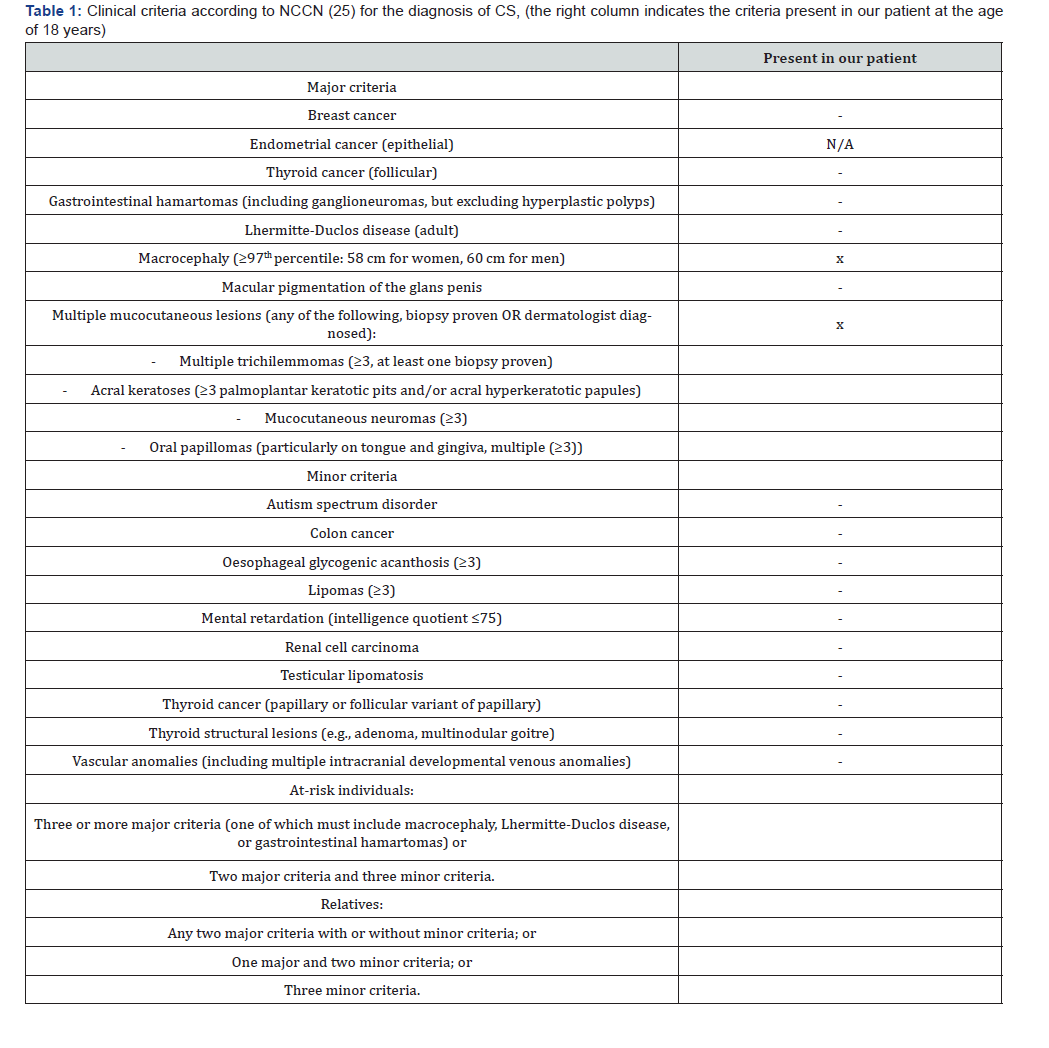
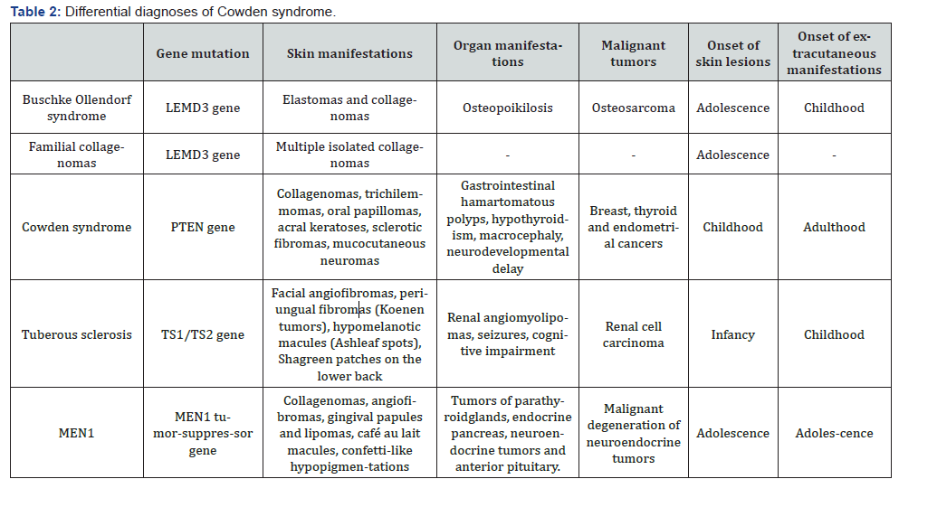
Table Abbreviations: NCCN: National Comprehensive Cancer Network; CS: Cowden Syndrome; MEN1: Multiple endocrine neoplasia.
Case Report
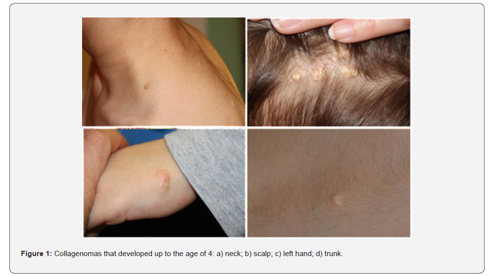
A 4-year-old boy was referred from his pediatrician due to multiple, firm, dome-shaped papules located on the neck, scalp, back, armpits, and left hand, each measuring approximately 0.5 to 1 cm in diameter (Figure 1). These lesions were asymptomatic but had increased in number since early childhood. The child had remarkable macrocephaly without symptoms. Initially, the cutaneous lesions were suspected to be connective tissue nevi, leading to a provisional diagnosis of BOS. However, genetic testing for the LEMD3 mutation, associated with this condition, was negative. The patient’s family history was unremarkable, with no reported cases of CS, associated cancers, or related syndromes. A larger lesion located on the left hand was surgically removed after a mechanical trauma.
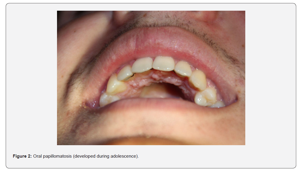
The resulting histopathology revealed tightly packed collagen bundles in a storiform pattern [11], consistent with a sclerotic fibroma- a specific subtype of collagenomas. This finding together with the important macrocephaly raised suspicion of CS. Subsequent gene testing by PCR and Next-Generation Sequencing confirmed a heterozygous germline mutation (c.289C>T) exon 5 of the PTEN gene, establishing the diagnosis of CS. Thereafter, the patient underwent baseline evaluation in accordance with current NCCN guidelines and expert recommendations, including thyroid ultrasound. Additional investigations, such as abdominal ultrasound and skeletal imaging, were performed to exclude differential diagnoses but revealed no pathological findings [12]. As the child grew older, oral papillomatosis (Figure 2) developed which is a typical feature of CS.
Results & Discussion
Collagenomas are rare benign connective tissue nevi first described in 1972 by Weary et al. [13]. They usually present as asymptomatic, localized, flesh-colored, waxy papules or nodules and can occur at any site; however, the head and neck are most commonly affected [14]. Histopathologically, a sharply defined hypocellular dermal nodule with hyalinized dense collagen arranged in a wavy or plywood pattern can be observed [11]. Immunohistochemistry is positive for CD34, vimentin, mucin, and also shows sporadic positivity for factor XIIIa; however, embryonic membrane antigen, S-100, and neuron-specific enolase are negative [15]. Although the collagenomas in our patient were initially misinterpreted as connective tissue nevi associated with BOS, their confirmation by histopathology raised the suspicion of CS which was confirmed by gene testing.
This highlights the importance of collagenomas as early indicators of CS as previously reported [14-16]. This is particularly true in case of multiple collagenomas, as single lesions can also occur sporadically [14,15]. Thus, the occurrence of multiple collagenomas in a patient with macrocephaly, potentially intellectual disability, overgrowth, or tumors early in life should raise suspicion of CS [14-16]. Apart from CS, collagenomas have also been described in other genodermatoses, such as BOS, familial collagenomas, MEN1, TS, sporadic eruptive collagenomas, as well as in non-CS conditions within the umbrella term PTEN hamartoma tumor syndrome (PHTS), including Bannayan-Riley- Ruvalcaba syndrome and Proteus syndrome [6].
Other important cutaneous indicators of CS are trichilemmomas, benign tumors derived from the outer root sheath epithelium of hair follicles, most commonly presenting as small, flesh-colored papules on the facial skin, especially around the mouth, nose, and ears. The presence of multiple facial trichilemmomas is even considered pathognomonic for CS, usually preceding the development of internal tumors [1,17]. Histologically, trichilemmomas show lobular proliferation of pale-staining cells with clear cytoplasm, peripheral palisading, and connection to the epidermis. In comparison to sporadic trichilemmomas, in CS they show a loss of PTEN expression [18].
CS is also differentiated from other genodermatoses by the presence of a pathognomonic distinctive mucocutaneous triad [6,7]: multiple facial trichilemmomas, acral keratoses, and oral papillomas. Acral keratoses appear on palms, soles, or dorsal hands/feet; oral papillomas are small papules on lips, tongue, gingivae, or buccal mucosa, often with a cobblestone appearance (Figure 2). BOS presents with elastomas or collagenomas and osteopoikilosis but lacks this mucocutaneous triad. Skin lesions are typically firm, skin-colored papules or plaques, often appearing in childhood. BOS is caused by mutations in the LEMD3 gene and does not confer increased cancer risk. The presence of osteopoikilosis on radiographs is a hallmark finding, and cognitive delays or shortened stature may be present in some cases [19].
TS complex is caused by mutations in TSC1 or TSC2 and is characterized by facial angiofibroma’s, hypomelanotic macules, shagreen patches, and ungual fibromas [20]; however, it does not feature the mucocutaneous triad. Non-Cowden variants of PHTS such as Bannayan-Riley-Ruvalcaba syndrome may present lipomas, pigmented penile macules, and intestinal hamartomas, but not the CS-specific triad [6]. While clinical features such as trichilemmomas and collagenomas are not exclusive to CS, loss of PTEN expression on immunohistochemistry represents a sensitive and specific marker for Cowden-associated lesions [21]. Individuals with suspected PHTS can be diagnosed clinically using the revised diagnostic criteria for PHTS published by Pilarski et al. in 2013 [22].
In most cases, the diagnosis is subsequently confirmed by PTEN genetic testing, which identifies pathogenic variants in the PTEN gene and allows differentiation of CS from other PHTS subtypes and genodermatoses [23]. The management of the mentioned skin lesions focuses on local therapies. These include topical 5‑fluorouracil, cryotherapy, laser or excision [6]. Targeting the mTOR pathway represents a promising approach: In a pilot study involving 18 patients, sirolimus (oral loading dose of 6 mg sirolimus, followed by 2 mg daily for 8 weeks) demonstrated good overall tolerability, suppression of mTOR signaling in benign skin and gastrointestinal lesions, and preliminary clinical improvement in 67% of the participants.
Regression of cutaneous and gastrointestinal lesions was observed, and cerebellar function improved after one month. These findings suggest potential disease-modifying effects, although the therapy remains experimental [24]. Lifelong, organspecific surveillance-including thyroid and renal ultrasound, breast imaging, and routine dermatologic and gastrointestinal assessments remains essential for early detection and reduction of morbidity and mortality [6]. Future research should focus on elucidating the genetic and molecular mechanisms underlying the development of CS lesions to gain deeper insights into their pathogenesis and to identify novel therapeutic targets.
Conclusion
Skin lesions such as collagenomas, trichilemmomas, oral papillomas and mucocutaneous neuromas are important indicators of CS that should be recognized by clinicians to enable timely diagnosis, appropriate patient management including implementation of cancer screening protocols.
Acknowledgment
The authors would like to thank our patient and his family for consenting to the publication of his clinical details.
Conflicts of Interest
The authors declare no conflicts of interest related to this case report.
Data Availability Statement
The data supporting the findings of this study are not publicly available due to patient privacy considerations but are available from the corresponding author upon reasonable request.
Funding
This research received no external funding.
Patient consent
Written informed consent for publication of clinical details and images was obtained from the patient’s legal guardians.
References
- Lim A, J Ngeow (2021) The Skin in Cowden Syndrome. Front Med (Lausanne) 8: 658842.
- Lamis Yehia, Joanne Ngeow, Charis Eng (2019) PTEN-opathies: from biological insights to evidence-based precision medicine. J Clin Invest 129(2): 452-464.
- Dhamija R, JM Hoxworth (2020) Imaging of PTEN-related abnormalities in the central nervous system. Clin Imaging 60: 180-185.
- JD Fackenthal, DJ Marsh, AL Richardson, SA Cummings, C Eng, et al. (2001) Male breast cancer in Cowden syndrome patients with germline PTEN mutations. J Med Genet 38(3): 159-164.
- Hagelstrom RT, J Ford, GM Reiser, M Nelson, DL Pickering, et al. (2016) Breast Cancer and Non-Hodgkin Lymphoma in a Young Male with Cowden Syndrome. Pediatr Blood Cancer 63: 544-546.
- Yehia L, C Eng (1993) PTEN Hamartoma Tumor Syndrome, University of Washington, Seattle, Seattle (WA), USA.
- ACOG (2019) Hereditary Cancer Syndromes and Risk Assessment: ACOG COMMITTEE OPINION, Number 793. Obstet Gynecol 134: e143-e149.
- Hershkovitz D, B Amitai, E Sprecher (2007) Familial cutaneous collagenomas resulting from a novel mutation in LEMD3. Br J Dermatol 156(2): 375-377.
- Bousheheri FF, A Mosbeh, A Albazali, M Alshammari (2025) Eruptive Collagenoma: A Rare Encounter in Clinical Dermatology. Cureus 17(2): e100338.
- Pálla S, Z Metyovinyi, FA Meznerics, A Mirzahosseini, J Tőke, et al. (2026) Dermatologic Features of Endocrine Tumor Syndromes-Systematic Review and Meta-Analysis. Int J Dermatol 65(3): 464-488.
- Pakyari M, NR Mahadevan, E Russell-Goldman (2024) Concurrent PTEN and PDGFRB Alterations Characterize Storiform Collagenoma. Am J Surg Pathol 48: 150-156.
- Tischkowitz, M., C. Colas, S. Pouwels and N Hoogerbrugge (2020) Cancer Surveillance Guideline for individuals with PTEN hamartoma tumour syndrome. Eur J Hum Genet 28: 1387-1393.
- PE Weary, RJ Gorlin, WC Gentry, JE Comer, KE Greer (1972) Multiple hamartoma syndrome (Cowden's disease). Arch Dermatol 106(5): 682-690.
- Salusti-Simpson M, M Madrid, D Cook (2024) Giant cell collagenomas associated with Cowden syndrome: A case report. J Cutan Pathol 51(5): 345-347.
- Caligayahan M, D Wolpowitz, CE Brem (2024) Giant cell collagenoma in a patient with Cowden syndrome: A rare case report and literature review with a focus on the spectrum of sclerotic fibroma and giant cell collagenoma. J Cutan Pathol 51: 267-271.
- Requena, L, J Gutiérrez, E Sánchez Yus (1992) Multiple sclerotic fibromas of the skin. A cutaneous marker of Cowden's disease. J Cutan Pathol 19: 346-351.
- MH Brownstein, AH Mehregan, JB Bikowski, A Lupulescu, JC Patterson (1979) The dermatopathology of Cowden's syndrome. Br J Dermatol 100(6): 667-673.
- Tariq Al-Zaid, Jeremy S. Ditelberg, Victor G Prieto, Dina Lev, Raja Luthra, et al. (2012) Trichilemmomas show loss of PTEN in Cowden syndrome but only rarely in sporadic tumors. J Cutan Pathol 39(5): 493-499.
- V Pope, L Dupuis, P Kannu, R Mendoza-Londono, D Sajic, et al. (2016) Buschke-Ollendorff syndrome: a novel case series and systematic review. Br J Dermatol 174(4): 723-729.
- Northrup H, MK Koenig, DA Pearson, KS Au (1993) Tuberous Sclerosis Complex. GeneReviews(®). MP Adam, S Bick, GM Mirzaa et al. Seattle (WA), University of Washington, Seattle Copyright © 1993-2026, University of Washington, Seattle. GeneReviews is a registered trademark of the University of Washington, Seattle. All rights reserved.
- Hartsough E, MS DeSimone, ME Lorenzo, D Dias-Santagata, V Nose, et al. (2024) Utilizing PTEN immunohistochemistry as a screening test for Cowden syndrome. Am J Clin Pathol 161: 490-500.
- Robert Pilarski, Randall Burt, Wendy Kohlman, Lana Pho, Kristen M Shannon, et al. (2013) Cowden syndrome and the PTEN hamartoma tumor syndrome: systematic review and revised diagnostic criteria. J Natl Cancer Inst 105(21): 1607-1616.
- Claudia Ciaccio, Veronica Saletti, Stefano D'Arrigo, Silvia Esposito, Enrico Alfei, et al. (2019) Clinical spectrum of PTEN mutation in pediatric patients. A bicenter experience. Eur J Med Genet 62(12): 103596.
- Takefumi Komiya, Gideon M Blumenthal, Roopa DeChowdhury, Susan Fioravanti, Marc S Ballas, et al. (2019) A Pilot Study of Sirolimus in Subjects with Cowden Syndrome or Other Syndromes Characterized by Germline Mutations in PTEN. Oncologist 24: 1510-e1265.

























