


Pilot Study: Evidence that High and Ultra-high Diluted Extracts of the anti-tumor active Mistletoe (Viscum Album L.) Plant Inhibit but may also Stimulate in Vitro Cell Proliferation of K562 Leukemia Cells
Razvan Rentea*, Mark Kamsler and Malory Mueller
Kolisko Institute for Anthroposophical Medicine, 1005 Richards Road, Hartland, Wisconsin, USA
Submission: May 23, 2024; Published: June 01, 2024
*Corresponding Address: Razvan Rentea MD, Kolisko Institute for Anthroposophical Medicine, 1005 Richards Road, Hartland, Wisconsin, 53029, USA, Email: ross.rentea@truebotanica.com
How to cite this article: Razvan Rentea*, Mark Kamsler and Malory Mueller. Pilot Study: Evidence that High and Ultra-high Diluted Extracts of the anti-tumor active Mistletoe (Viscum Album L.) Plant Inhibit but may also Stimulate in Vitro Cell Proliferation of K562 Leukemia Cells. Canc Therapy & Oncol Int J. 2024; 27(1): 556204. DOI:10.19080/CTOIJ.2024.27.556204
Abstract
Mistletoe extracts are widely used in Central Europe, as well as world-wide, as an adjuvant cancer therapy. In the laboratory, high doses of mistletoe extracts are largely considered to be uniformly inhibiting on cell proliferation in vitro. Some controversy has arisen however whether low doses of mistletoe extracts can be also stimulating and thus whether clinical low doses of mistletoe should be more cautiously administered in a clinical setting. We provide evidence that not only low but also highly and ultra-highly diluted extracts of mistletoe can both inhibit as well as stimulate in vitro cell proliferation.
Keywords: K562 cell line; Mistletoe; Viscum album; lectins; ultra-high dilutions; enantiodromic, Rudolf Steiner; L. Kolisko; IC50
Introduction
Mistletoe (Viscum album L.), a semi-parasitic plant that comes in several varieties depending on which host tree it grows on, has been shown in recent investigations both in vitro and in vivo to have potential in anti-tumor activity [1]. Most important to mistletoe’s medicinal profile are its diverse array of bioactive compounds, principally mistletoe lectins, but also viscotoxins, flavonoids, and phytosterols [2,3]. Among the main mechanisms of action are cell cycle inhibition, generation of reactive oxygen species (ROS), disruption of mitochondrial function, activation of apoptotic pathways, and modulation of key signaling molecules involved in cell growth and survival [3] (Figure 1).
Mistletoe has also been found in clinical cancer studies to enhance the quality of life in patients through alleviating various symptoms (fatigue, sleep, exhaustion, nausea, vomiting, depression anxiety, pain) and to lower side effects of traditional treatments [4]. Based on well more than fifty in vitro studies on cell lines such as sarcoma, leukemia, melanoma, adenocarcinoma, etc., done with mistletoe lectin concentrations of 1.25ng/ml-1000ng/ ml there seemed to be little doubt that in vitro experiments even at low concentrations the mistletoe extracts would be able to consistently inhibit cell proliferation [1-3,5].
However, some doubt was cast on this seemingly established result by a Gabius study [6] who found stimulation of tumor cells directly exposed to very low concentrations of mistletoe lectins. This stimulatory effect was seen at lectin levels of 50pg/ml x 10(5) cells in sarcoma and melanoma and hematologic lines. Several years after the Gabius study [6] another study by Kelter et al. [7] gave results that went into the direction of contradicting the concerns that mistletoe extracts with very low lectin concentrations would be stimulatory to cancer cell proliferation. They investigated 26 cell lines, including melanoma, sarcoma, CNS, etc., and found no stimulation at the level of 1.5 ng/ml up to 15mcg/ml total plant extract, corresponding to a lectin concentration of 0.0075-750 pg/ml.
In the present study we investigate the effect of mistletoe on the proliferation of K562 leukemia cells when the concentration of the mistletoe extract goes from low to ultra-high dilutions. This has not been done before.
Materials and Methods
Mistletoe (Viscum album L.) Extraction
The mother tincture (Ø) of the mistletoe growing on six different host trees was obtained through a proprietary method of extraction. The mistletoe was harvested from each host tree at two different opposing seasons (e.g., summer and winter) - the main rationale being that the summer viscum is richer in lectins and the winter viscum richer in viscotoxins. Thus, a combination of both seasons would provide a more full spectrum extract. After the separate extraction of each seasonal harvest, the summer harvest extraction was allowed to drip by gravity downwards into the centrifugally rotating extract from the winter season. The resultant final mother tincture was designated as Viscum and the name of the host it grew on as follows (Table 1).


Preparing Dynamized “Dilutions”
The mother tincture was made from 20g dry plant material in 100cc diH2O. The “dilutions” were prepared or so called “dynamized” in homeopathic style. The first “dilution” designated as D1, and was made by adding 1 part of mother tincture (Ø) to 9 parts of sterile diH2O. after that it was succussed (agitated) for 2 min and allowed to rest for 1 minute. One part of this D1 was added 9 parts of sterile diH2O and again agitated as before. This was designated as D2. The procedure was followed until a D30 was obtained. The so called “dynamized”/agitated/then allowed to rest dilutions will be called in the following text only as “dilutions for ease of reading.
Cell Culture
The K562 human lymphoblast cell line (ATCC, CCL-243) was grown in 25cm2 flasks, at 37˚C, 5% CO2, and the cell density was maintained between 1x10⁵ and 1x10⁶ cells/ml. The medium was IMDM (ATCC, 30-2005) supplemented with 10% Fetal Bovine Serum (FBS; ATCC, 30-2020) and 1% penicillin streptomycin (ATCC, 30-2300). The Countess 3 was used to count cell inoculum and to determine viability.
Cancer cell and Mistletoe co-culturing
All assays were plated in white, 96-well plates (Costar, CLS3917) with a cell inoculum of 8 x 10³. Each well contained 90 μl of cells plus 10 μl experimental dilution or “agitated” diH2O (for the 0% and 100% controls), with four replicates per treatment or control. Plates were incubated at 37˚C and 5% CO2 for 72 hours. Cell growth was assessed using the Cell Titer Glo 2.0 cell viability assay (Promega, G9243), using 100 μl of reagent per well according to the manufacturer’s instructions. The plate incubated at room temperature for 12 minutes before measuring the luminescence in a Bio Tek, Synergy LX plate reader. Cell growth was assessed for 0% control wells immediately, while sample and 100% control wells were assessed after 72 hours of growth.
IC 50 fitting and calculations
In our study, we employed the four-parameter logistic (4PL) model, which is a non-linear curve fitting model, to characterize sigmoidal dose-response curves of various viscum species, specifically for calculating half-maximum inhibitory concentrations (IC50s). The 4PL model is a mathematical function commonly utilized in pharmacological and biological assays to describe the relationship between the concentration of a compound (dose) and the response it elicits.
The IC50 values were calculated by the following equation:
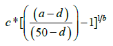
where a is the minimum response, d is the maximum response, c is the middle point (c=(d-2)/2) and b is the slope in the semi-log axis, calculated by finding the location of the maximum difference in response and identifying the change in concentration at the maximum difference, with the below equation:
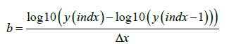
By fitting experimental data to the 4PL model, we were able to determine the IC50 values, which represent the concentration of the compound required to inhibit the response by 50%. This approach allows for the quantitative assessment of the potency of compounds and facilitates comparisons across different experimental conditions.
Statistics
Each experiment included 30 dilutions, in addition to the control samples. Each experiment was repeated in two plates at two locations (top/bottom) at two separate time intervals (Monday/Tuesday). Data from four plates was obtained, with 24 samples. The variation among and within the plates on various days and locations was examined using summary statistics and all samples were included in the analysis.
The raw data was summarized using descriptive statistics (mean, min, max, std, cv). The percentage of control was derived and summarized using descriptive statistics.
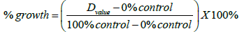

This was calculated for each individual assessment. A value of 100 would indicate the growth was similar to the control, less than 100 would indicate the growth is less than the control and greater than 100 would indicate the growth is greater than control. The mean and the 95% confidence interval were computed and plotted. The mean percent growth and the 95% confidence interval (calculated as mean ±1.96 SE) is presented is graphically plotted below for various viscum species (Figure 2-7). The asterisks (*) denotes significantly different (p<0.05) from the control at the respective dilution (D) value.


Results
IC50 Results (Table 2)
72-hour Incubation of K562 cells
This study was done in order to demonstrate that identical procedures done across columns in the plate are similar enough that no stat. significance was noted. Thus, when statistically significant results are obtained in the main experiment they can be relied upon not to be outliers (Table 3).
Inhibitory and Stimulating Dilutions of Six Different Mistletoe Varieties
Below, in Figure 2-7, are graphical representations of the mean and corresponding 95% confidence intervals of percent of growth in comparison to the control. *indicates a stat. significant dilution (Table 4).


Discussion
We present evidence that the proprietary extract of the antitumor mistletoe (Viscum album L.) can have both inhibiting as well as stimulating effects on the proliferation of the K562 cell line at low as well as high and ultra-high dilution levels. All six mistletoe varieties tested demonstrated significant inhibitory activity but in three of the six varieties also stimulating effects could be observed. Thus, in three of the six varieties enantiodromic effects were observed i.e. effects in opposing directions depending on what concentration of mistletoe the cells were exposed to. These results are significant from multiple points of view. They align themselves with the increasingly well-established finding that contrary to the still wide spread pharmacological dose response model which predicts no effectiveness beyond a minimal low dose [8], a demonstrable activity will appear nevertheless after a certain “low dose” point [9].
Mechanistically, biological phenomena at low doses, such as low mother tincture concentrations, or going up to D23, are accepted, but when the dilutions go beyond the Avogadro number i.e. from a practical point of view no more physical mass of the original substance remain, meaning from D24 up, the existence of any biological activity is still being met with scepticism. This should not be the case since activity in the so called high homeopathic dilutions has been amply demonstrated [10-13].
Consequently, seeing both stimulating and inhibiting activity in range above the D24 dilution of the viscum acer and viscum Crataegus is highly notable. We are not the first to report the existence of such curves that contain both inhibiting and stimulating activity of dilutions of substances. First work in this direction was done in the 1920’s when Rudolf Steiner and Lili Kolisko [14,15] demonstrated that serial dilutions of a multitude of substances resulted in semi-sinusoidal curve patterns containing these double switch effects of stimulating or inhibiting the mother tincture effect by various dilutions of the same substance. In later years this phenomenon was shown again and again f. ex. in an elegant study by Carmine [16].
In her pioneering work Kolisko demonstrated that each substance would have its own curve pattern. We have gathered the results of all 97 studies that she had done and compared the activity of the D30 dilution of each. Fig 8 below shows how different the activity level of each substance can be at the same D30 dilution- going from stimulating to inhibitory or to activity no different than control. These findings are especially significant in the work with the mistletoe extracts which are used clinically in anti-cancer therapy. We have found here that stimulation can occur both at low dilutions (D4, D5, D6, D7) as well as at high and ultra-high (D26-D30). This stimulatory occurrence makes the necessity for future extensive in vitro and pre-clinical testing with each mistletoe variety on each tumor cell line (here on leukemic cells) a requirement.
L. Kolisko [15] had envisioned that in the future doctors would have a detailed knowledge of the concrete dilution curve of each substance before being used clinically to ensure its appropriate and most effective use. This has not happened yet to today, not in the laboratory in vitro, or ex vivo, and not in clinical studies. Much more work is awaiting to be done.
Authors Participation
i. R Rentea MD - designed or wrote the article; also participated in the extraction of the mistletoe material.
ii. M Mueller M. Sc - executed the experimental data and wrote the Materials & Methods section.
iii. M Kamsler MD - participated in the extraction of mistletoe material.
Acknowledgement
i. Our thanks to Ashley Pajak for help with the statistical calculations and Nourhan Shalaby PhD for help with the calculations of the IC50 values and the mechanisms of inhibition chart.
ii. Our thanks to Peter Goedings PhD for invaluable ideas toward the making of the mother tinctures of mistletoe.
iii. Our thanks to Cosmin Borcea for the harvesting of the mistletoe.
Conflict of Interest
The authors report no conflict of interest.
References
- Szurpnicka A, Kowalczuk A Szterk A (2020) Biological activity of mistletoe: in vitro and in vivo studies and mechanisms of action. Arch Pharma Res 43(6): 593-629.
- Burger A, Mengs U, Schueler JB, Fiebig HH (2001) Anti proliferative activity of an aqueous mistletoe extraction on human tumor cell lines and xenografts in vitro. Arzneimittelforschung/Drug Res 51(9): 748-757.
- Park YY, Do YR, Jang BC (2012) Apoptosis of K562 leukemia cells by Abnobaviscum F, a European mistletoe extract. Oncology Rep 28(6): 2227-2232.
- Kienle GS, Kiene H (2024) Influence of Viscum album L. (European Mistletoe) Extracts on Quality of Life in cancer patients: A Systematic Review of controlled Clinical Studies. Rev Articles Integr Cancer Ther 9(2): 142-157.
- Twardziok M, Kleinsimon S, Rolff J, Jaeger S, Eggert A, et al. (2016) Multiple Active Compounds from Viscum album L. Synergistically Converge to Promote Apoptosis in Ewing Sarcoma. Plos One 11(9): e0159749.
- Gabius H, Darro F, Remmelink M, Andre S, Kopitz J, et al. (2001) Evidence for Stimulation of Tumor Proliferation in Cell Lines and histotypic Cultures by clinically relevant low doses of the Galctoside-Binding mistletoe Lectin, A Component of proprietary Extracts. Cancer Investigation 19(2): 114-126.
- Kelter G, Flebig HH (2006) Absence of Tumor Growth Stimulation in a panel of 26 tumor Cell Lines by Mistletoe (viscum album L.) Iscador Extrracts in vitro," Arzneim.-Forschung/Drug Res 56(6a): 435-440.
- Salahudeen MS, Nishtala P (2017) An overview of pharmacodynamic modelling, ligand-binding approach and its application in clinical practice. Saudi Pharma J 25(2): 165-175.
- Calabrese EJ (2006) The Failure of Dose-response Models to Predict low Dose Effects: A Major Challenge to Biomedical, toxicological and Medical Research. Biogerontology 7(2): 119-122.
- Dei A, Bernardini S (2015) Hormetic Effects of Extremely Diluted Solutions on Gene Expression. Homeopathy 104(2): 116-122.
- Vallance A (1998) Can Biological Activity be maintained at Ultr-High Dilution? An Overview of Homeopathy, Evidence and Bayesia Philosophy. J Alternative Complementary Med 4(1): 49-76.
- Witt CM, Bluth M, Albrecht H, Weisshuhn T, Baumgartner S (2007) The invitro evidence for an effect of high homeopathic potencies-A systematic review of the literature. Complementary Therapiesin Med 15(2): 128-138.
- Sunila ES, Kuttan R, Preethi K, Kuttan G (2009) Dynamized Preparations in Cell Cultures. Complementary and Alternative Med 6(2): 257-263.
- Steiner R (1999) Introducing anthroposophical Medicine, hudson, NY, USA: Anthroposophic Press 170-72.
- Kolisko L (1959) Physiologischer und physikalischer Nachweis der Wirksamkeit kleinster Entitaeten, Stuttgart: Arbeitsgemeinschaft anthroposophischer Aerzte.
- Carmine TC (1997) Effects of High Potencies of Tumor Necrosing Factor alpha on H2O2 production in cultured neuroblastoma Cells by enhanced luminol-dependent chemifluorscence (ECL) A positive system for investigating the biological significance of homeopathic high potencies. British Homeopathic J 86: 67-72.


