Pleomorphic Cardiac Sarcoma with Bone Metastases – A Rare Case Report
Deep Shankar Pruthi1*, Puneet Nagpal1, Avnika Jain2, Anurag Jain3 and J B Sharma4
1Department of Radiation Oncology, Action Cancer Hospital, New Delhi, India
2Final Year MBBS, Maulana Azad Medical College, New Delhi, India
3Department of Radiodiagnosis, Action Cancer Hospit1al, New Delhi, India
4Department of Medical Oncology, Action Cancer Hospital, New Delhi, India
Submission: September 11, 2022; Published: September 22, 2022
*Corresponding Address: Deep Shankar Pruthi, Department of Radiation Oncology, Action Cancer Hospital, New Delhi, India
How to cite this article: Deep S P, Puneet N, Avnika J, Anurag J, J B Sharma. Pleomorphic Cardiac Sarcoma with Bone Metastases – A Rare Case Report. Canc Therapy & Oncol Int J. 2022; 22(3): 556087. DOI:10.19080/CTOIJ.2022.22.556087
Abstract
Cardiac tumours are rare and are mostly benign. Amongst malignant cardiac tumors, sarcomas are the most common subtype. Pleomorphic cardiac sarcomas present as an extremely rare clinical entity with challenging diagnosis and treatment. The typical presentation of cardiac sarcomas results from anatomic obstruction of blood flow and patient usually presents with cardio-pulmonary compromise. Presentation of these tumours with multiple bony metastases is highly unusual and only sporadic cases have been reported in literature. In this case report, we describe a case of a young female who presented with dyspnea on exertion along with pain in lower back and pelvis. Cardiac MRI showed an enhancing mass in left atrium and PET CT revealed the same with multiple bony metastases. Biopsy and immunohistochemistry from bony lesion revealed pleomorphic sarcoma. The patient presented with severe cardio-pulmonary compromise and expired before any oncological intervention could be done.
Keyword: Cardiac Sarcoma; Cardiac Magnetic Resonance; Bone Metastases; Cardio-pulmonary compromise
Background
Cardiac tumours are uncommon and can be primary or metastatic. A series of autopsy studies revealed a 0.02% prevalence of cardiac tumours, of which 75% were benign and 25% malignant [1]. Myxoma is the most frequently occurring primary cardiac neoplasm which is benign [2]. Amongst malignant primary cardiac neoplasms, cardiac sarcoma represents the most common type and the 2nd most common of all primary cardiac tumours [3]. These tumours are usually high grade, clinically aggressive and are locally advanced by the time patient presents to the hospital [4]. Clinical presentation depends on the exact location of tumour and patients can present with a variety of cardio-pulmonary symptoms [5]. The workup of cardiac sarcomas involves a variety of imaging investigations like Cardiac Magnetic Resonance Imaging (CMR), Positron Emission Tomography (PET) or Transthoracic Echocardiogram (TTE) [6]. Little is known about the metastatic dissemination patterns of primary cardiac malignancies. In this case report we present a patient of pleomorphic undifferentiated cardiac sarcoma who presented with multiple bony metastases.
Case Presentation
A 33-year-old female with no comorbidities presented to our hospital with dyspnea, along with pain in lower back and pelvis. Blood investigations revealed normal complete blood count, kidney function tests and liver function tests. A transthoracic echocardiography finding was consistent with a mass inside the left atrium with protruding into mitral valve region.
Tumor markers revealed AFP: 1.55 ng/mL, Beta HCG: 0.251 mIU/mL, CA 125: 104.7 U/mL, CEA: 1.54 ng/mL, CA19.9: 20.61 U/mL and LDH: 415 U/L.
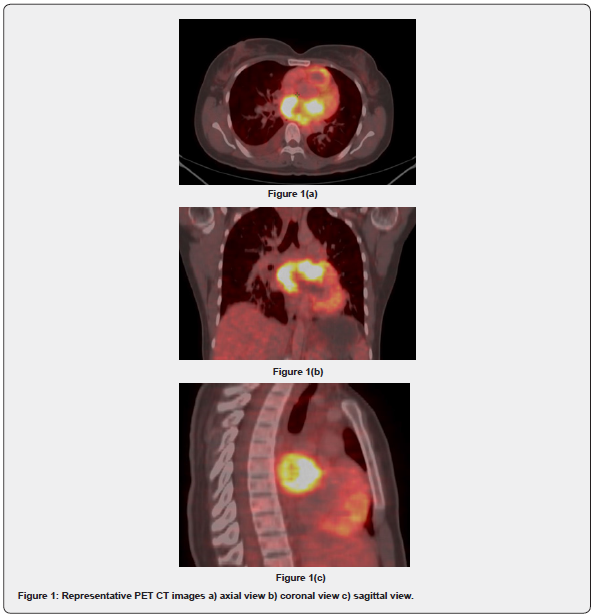
PET CT Scan showed a heterogeneously enhancing mass with increased FDG uptake in the pulmonary venous trunk extending to left atrium. The mass measured 7.0 x 4.1cm with SUVmax of 11.5). It also showed lytic destructive lesion with associated soft tissue component and increased FDG uptake involving L4 vertebrae involving pre vertebral region. There were additional multiple lytic lesions in left humeral shaft, D8-D10 vertebrae, bilateral iliac bones, left acetabulum and left pubic bone (Figure 1).

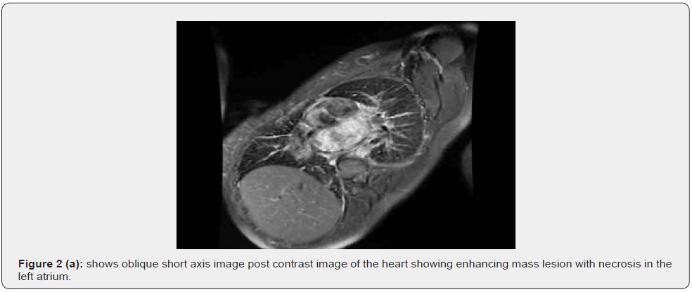
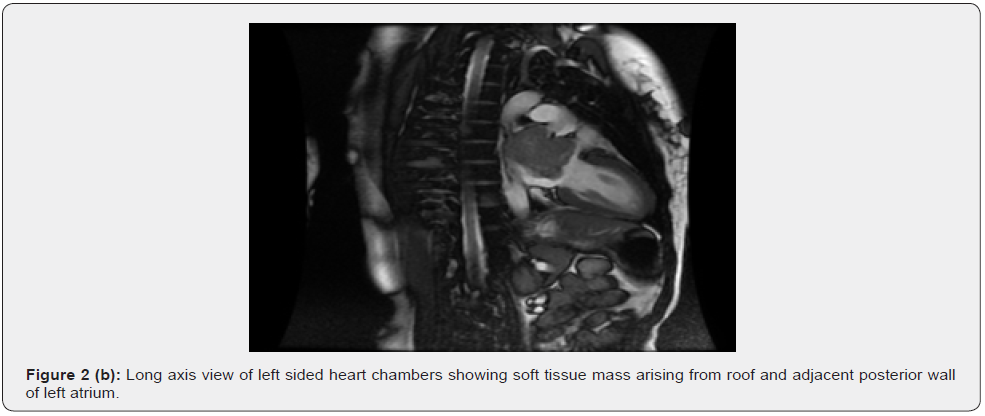
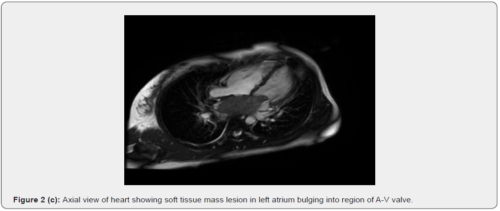
A cardiac MRI was done to better characterize the cardiac mass (Figure 2). It revealed a soft tissue mass lesion of size 6.8 x 3.5 x 4.5cm in left atrium of heart showing a broad-based attachment along its roof and adjacent interatrial septum. The mass was seen to extend into left atrial appendage and protrudes into mitral valve region, however with no further extension across the valvular opening into left ventricle. The mass showed post contrast enhancement with internal non enhancing necrotic areas. There was no obvious gross mural involvement of atrial walls with no obvious epicardial component. Rest cardiac chambers, pulmonary trunk and ascending aorta were unremarkable. CT guided Biopsy was done from right iliac bone lesion which showed high grade sarcoma. Immunohistochemistry revealed immunereactive for vimentin and weakly for SATB2. It was negative for CK, LCA, Desmin, S-100, CD34, HMB-45, Myogenin and EMA with a final impression of pleomorphic sarcoma. Patient was planned for chemotherapy under medical oncology department however patient developed acute cardio-pulmonary compromise and could not be revived.


Discussion
Cardiac sarcomas are distinct and extremely rare subset of soft tissue sarcomas yet represent the majority of malignant cardiac tumours [7]. WHO has established a universal system for classification of cardiac tumours [8]. Angiosarcomas are the most common histological subtype (40%) and typically present in the right atrioventricular groove, with frequent involvement of the pericardium and right atrial wall. Amongst sarcomas that present in the left atrium, undifferentiated pleomorphic sarcomas are the most common subtype [8]. In cardiac sarcomas, histological grading and subtype seem to correlate with prognosis [9]. Cardiac pleomorphic sarcoma is a high-grade malignancy which has fibroblastic and myoblastic differentiation amidst areas of cellular pleomorphism [10].
The presentation of these tumours is usually with life threatening cardio-pulmonary compromise due to obstructed intra cardiac flow wherein there is an interference with valve function, rhythm and tamponade. Symptoms of cardiac tumours are due to the site of the mass rather than their pathology. Many cardiac tumours remain asymptomatic for a long time, however the diagnosis is usually made at advanced stages of the disease [11]. However, presentation of a cardiac sarcoma with distant symptomatic metastases is very rare unlike our patient. There have been a few case reports that describe cardiac sarcomas with bony metastases. In one of these case reports, a 33-year-old female was diagnosed with leiomyosarcoma in the left atrium and underwent surgery followed by adjuvant chemotherapy and radiotherapy. The patient presented after 2 years with bony metastases in right femur and iliac bone [12]. Our patient presented with multiple bony metastases at the time of presentation itself.
The evaluation of cardiac tumours requires a multidisciplinary approach. A good clinical history followed by echocardiography to identify the mass, evaluate its motility and its hemodynamic impact is the first step. By cross sectional cardiac imaging (Contrast Enhanced CT scan or MRI), cardiac sarcomas can be readily distinguished from myxomas or cardiac thrombus by their infiltrative growth into the atrial wall. In this regard, a cardiac MRI is considered to be gold standard in assessing a suspected cardiac tumour [13]. The advantages of MRI over CT scan include higher temporal resolution, ECG gating, additional tissue characterization and non-exposure to ionizing radiation [13]. Large size (>5cm), irregular margins, pericardial effusion, invasion of the wall including adjacent structures and contrast enhancement are features of malignancy. In the absence of gross mural invasive features, it is difficult to distinguish between benign and malignant cardiac neoplasms. The presence of necrotic areas in the mass strongly favours malignant etiology with a very high specificity as per study done by Rahbar et al. [14]. In the same study it was shown that on PET CT scan, malignant cardiac tumours have significantly higher SUV uptake as compared to benign cardiac tumours [14].
The overall prognosis of cardiac sarcomas is very poor with a median survival of less than one year [15]. Metastases in cardiac sarcoma can gravely affect the prognosis. In a series of 34 patients of cardiac sarcomas, the median survival of patients with metastases was a mere 5 months [16]. Cardiac sarcomas do have a propensity for dissemination to lungs, bones, soft tissues and brain. Surgical resection followed by adjuvant chemotherapy with or without radiation therapy is the current standard of care [17]. Complete resection increases the median survival by 7 months compared to non-resected patients [18]. However, the tumours are highly invasive and are often locally advanced which make complete resection difficult. In terms of chemotherapy, there has been no randomized trial to identify an optimal chemotherapy regimen and the drugs commonly used include doxorubicin with ifosfamide and gemcitabine with or without docetaxel [19].
At the molecular level, MDM2 regulates cell cycle by inhibiting p53, so there is a role of MDM2 inhibitors like Nutlin 3a [20]. MDM2 oncogene amplification has been reported in a case of cardiac intimal sarcoma which presented with multiple skeletal metastases [21].
Conclusion
The assessment of cardiac masses requires a multi-parametric approach to locate, characterize tissue, evaluate its etiology and hemodynamic impact. The prognosis of pleomorphic cardiac sarcoma is generally poor and worse than sarcomas at other sites in the body.
Acknowledgement
None.
Conflict of Interest
None.
References
- Lam KY, Dickens P, Chan AC (1993) Tumors of the heart. A 20-year experience with a review of 12,485 consecutive autopsies. Arch Pathol Lab Med 117(10): 1027-1031.
- Hoffmeier A, Sindermann JR, Scheld HH, Martens S (2014) Cardiac tumors--diagnosis and surgical treatment. Dtsch Arztebl Int 111(12): 205-211.
- Hamidi M, Moody JS, Weigel TL, Kozak KR (2010) Primary cardiac sarcoma. Ann Thorac Surg 90(1): 176-181.
- Siontis BL, Zhao L, Leja M, Jonathan B McHugh, Maryann M Shango, et al. (2006) Primary Cardiac Sarcoma: A Rare, Aggressive Malignancy with a High Propensity for Brain Metastases. Sarcoma 2019: 1960593.
- Shanmugam S (2006) Primary cardiac sarcoma. Euro J Cardio-Thoracic Surg 29(6): 925–932.
- Orlandi A, Ferlosio A, Roselli M, Chiariello L, Spagnoli LG (2010) Cardiac sarcomas: an update. J Thorac Oncol 5(9): 1483-1489.
- Butany J, Nair V, Naseemuddin A, Nair GM, Catton C, et al. (2005) Cardiac tumours: diagnosis and management. Lancet Oncol 6(4): 219-228.
- Burke A, Tavora F (2016) The 2015 WHO Classification of Tumors of the Heart and Pericardium. J Thorac Oncol 11(4): 441-452.
- Zhang PJ, Brooks JS, Goldblum JR, Yoder B, Seethala R, et al. (2008) Primary cardiac sarcomas: a clinicopathologic analysis of a series with follow-up information in 17 patients and emphasis on long-term survival. Hum Pathol 39(9): 1385-1395.
- Suh SH, Park TH, Yoo JN, Cha KS, Kim MH, et al. (2007) Primary undifferentiated pleomorphic sarcoma of the left atrium that presented as acute pulmonary edema. Yonsei Med J 48(1): 131-134.
- Siontis BL, Leja M, Chugh R (2020) Current clinical management of primary cardiac sarcoma. Expert Rev Anticancer Ther 20(1): 45-51.
- Strina C, Zannoni M, Parolin V, Cetto GL, Zuliani S (2009) Bone metastases from primary cardiac sarcoma: case report. Tumori 95(2): 251-253.
- Motwani M, Kidambi A, Herzog BA, Uddin A, Greenwood JP, et al. (2013) MR imaging of cardiac tumors and masses: a review of methods and clinical applications. Radiology 268(1): 26-43.
- Rahbar K, Seifarth H, Schäfers M, Lars Stegger, Andreas Hoffmeier, et al. (2012) Differentiation of malignant and benign cardiac tumors using 18F-FDG PET/CT. J Nucl Med 53(6): 856–863.
- Truong PT, Jones SO, Martens B, Cheryl Alexander, Matthew Paquette, et al. (2009) Treatment and outcomes in adult patients with primary cardiac sarcoma: the british columbia cancer agency experience. Ann Surg Oncol 16(12): 3358–3365.
- Simpson L, Kumar SK, Okuno SH, Hartzell V Schaff, Luis F Porrata, et al. (2008) Malignant primary cardiac tumors: review of a single institution experience. Cancer 112(11): 2440-2446.
- Moeri-Schimmel R, Pras E, Desar I, Krol S, Braam P (2020) Primary sarcoma of the heart: case report and literature review. J Cardiothorac Surg 15(1): 104.
- Isambert N, Ray-Coquard I, Italiano A, Maria Rios, Pierre Kerbrat, et al. (2014) Primary cardiac sarcomas: a retrospective study of the French Sarcoma Group. Eur J Cancer 50(1): 128–136.
- Woll PJ, Reichardt P, Le Cesne A, Sylvie Bonvalot, Alberto Azzarelli, et al. (2012) Adjuvant chemotherapy with doxorubicin, ifosfamide, and lenograstim for resected soft-tissue sarcoma (EORTC 62931): a multicentre randomised controlled trial. Lancet Oncol 13(10): 1045–1054.
- Merkel O, Taylor N, Prutsch N, Philipp B Staber, Richard Moriggl, et al. (2017) When the guardian sleeps: reactivation of the p53 pathway in cancer. Mutat Res 773: 1–13.
- Crombe A, Lintingre PF, Le Loarer F, Lachatre D, Dallaudiere B (2018) Multiple skeletal muscle metastases revealing a cardiac intimal sarcoma. Skeletal Radiol 47(1): 125-130.


