




 CTOIJ Home



Atypical Fibroxanthoma of the Eyelid - Surgical Resolution and Unexpected Complication, a Case Report
Martin Gonzalez*
Department of Biochemistry at the school of Medicine, National Autonomous University of Mexico, Mexico
Submission: November 02, 2021; Published: November 12, 2021
*Corresponding Address: Martin Gonzalez, Department of Biochemistry at the school of Medicine, National, Autonomous University of Mexico, Mexico
How to cite this article: Martin G. Atypical Fibroxanthoma of the Eyelid - Surgical Resolution and Unexpected Complication, a Case Report. Canc Therapy & Oncol Int J. 2021; 20(2): 556032. DOI: 10.19080/CTOIJ.2021.20.556032
Abstract
Introduction: A 75-year-old woman with a tumor lesion in the right periorbital region is presented. No signs of orbital invasion according to nuclear magnetic resonance. Underwet surgery with subsequent necrosis of the myocutaneous flap used for reconstruction.
Discussion: It is a mesenchymal tumor that appears in the head and neck of elderly men, being a benign and superficial variant of malignant fibrohistiocytoma. It is related to actinic damage to the skin. It manifests as a solitary, asymptomatic, small pink nodule. Histological diagnosis is a diagnosis of exclusion with immunohistochemistry. The treatment of choice is surgical removal, and they have a good prognosis.
Clinical Case
A 75-year-old female patient with no significant pathological history and a history of facial burn with hot water in adolescence is presented, who consulted for a lesion of progressive growth in the last 9 months in the lateral canthus of his right eyelid (Figure 1). A nodular lesion measuring 5 cm in diameter was evidenced that involved the upper and lower eyelids of the lateral canthus, well defined, not adhered to deep planes, not painful and with a hard elastic consistency on palpation. Visual acuity of 20/40 in both eyes that improved with its correction to 20/20 and the rest of the ophthalmological examination was within normal limits. A nuclear magnetic resonance was requested, which revealed a delimited hypointense lesion in T1 that enhances with contrast and hyperintense in T2 that involved soft tissues and in close contact with the lacrimal gland without compromising the eyeball or invading intraconal structures.
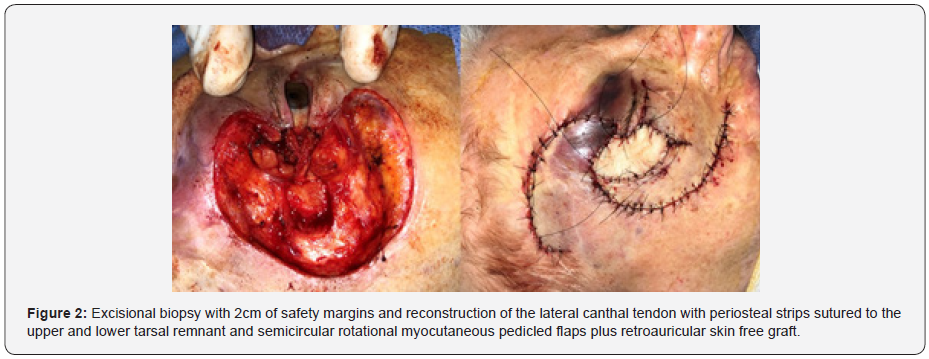
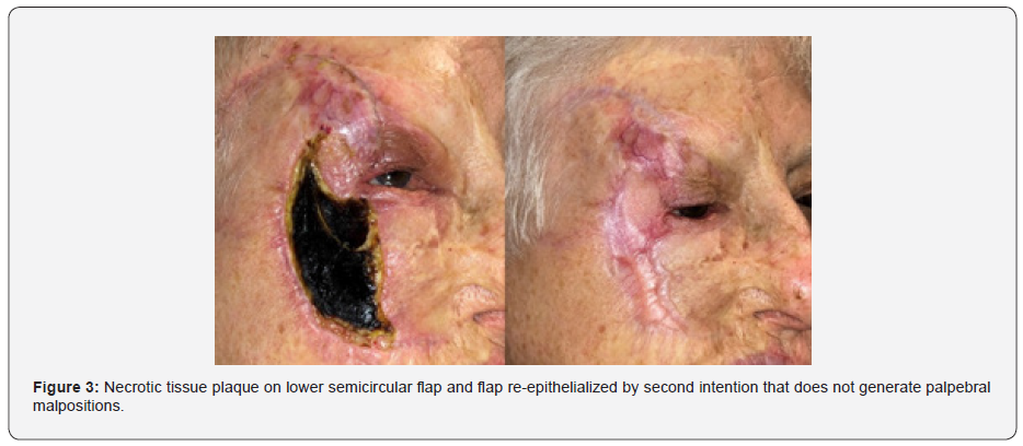
An incisional biopsy was performed for histopathological and immunohistochemical study that was compatible with Atypical Fibroxanthoma. As treatment, excisional biopsy with safety margins was chosen. A periosteal strip technique was used for reconstruction of the lateral canthal tendon and semicircular rotational pedicle flaps and retro auricular free skin graft to replace myocutaneous tissue to the surgical bed. The day after surgery, hematoma was evidenced below the lower flap, which in the following days evolved with paleness and granulation tissue below it. He was treated with antibiotic ointments and corticosteroids associated with oral antibiotics. 15 days after the procedure, necrosis of the lower flap was found, which evolved favorably without additional treatments and healing by second intention. Four months postoperatively, he found a normal ophthalmological examination with no signs of scar retraction or recurrence and under surveillance together with oncology to monitor local and systemic evolution.
Discussion
Atypical Fibroxanthoma (AF) is a fibrous and histiocytic tumor of doubtful origin, rare and of benign course described by Helwig in 1963 [1]. As it has marked pleomorphism, it is considered a potentially aggressive pseudosarcoma with a low probability of metastasis [2,3]. Some authors establish that it is a benign and superficial variant of Malignant Fibrohistiocytoma (FHM). It is a tumor that usually appears in the head and neck in 85% of cases and in the extremities and trunk in 15%, being in this location more frequent in young people4. It is estimated that it affects up to 0.2% of skin tumors [4]. The mean age of onset is 70 years, with a 3: 1 male / female distribution (in other articles the ratio is 7: 1) [5]. Its pathogenesis is not clear, but there are risk factors for developing this type of tumor such as age, exposure to ultraviolet rays and X-rays, Xeroderma Pigmentosum and immunosuppressed [6]. It has been seen that 99% of patients present actinic damage [7].
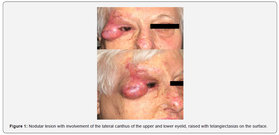
The tumor presents as a solitary, asymptomatic, red or purplish nodule. Most are smaller than 2 cm, but they can grow. They usually bleed to the touch and the center of the tumor may ulcerate. It is of rapid development, with an average of 6 months (2 weeks to 20 years) [8]. The differential diagnosis is with Basal, Squamous and Epidermoid Carcinoma, Desmoplastic or Spindle Cell Melanoma, Merkel’s Carcinoma, Malignant Fibrohistiocytoma, Undifferentiated Pleomorphic Sarcoma, Pyogenic Leiomyosarcoma Granuloma and Angiosarcoma [9]. Immunohistochemical markers are important for diagnosis. There is no marker that confirms the diagnosis of AF, being a diagnosis of exclusion. It has been seen that CD10, CD68 and Vimentin are positive in more than 85% and the Cytokeratin, S-100, Desmin. HMB-45, Melan-A / MART1, P63, Nerve Growth Factor Receptor, CD15, EMA and CD31 negative by more than 85% [10] (Figures 2 & 3).
The prognosis of this tumor is good with a risk of recurrence that is between the 0-20%, being the most widely accepted between 5-10% [10]. Almost 90% of recurrences occur within the first year after removal. The risk of metastasis is rare (0.5 to 10%) affecting subcutaneous cellular tissue, lymph nodes, liver and lung [10]. The histological factors associated with greater aggressiveness are vascular invasion, subcutaneous fat invasion and necrosis, immunosuppressed, history of ionizing radiation and positive margins after excision. These factors condition a worse prognosis since the tumor would be more aggressive and more like malignant fibrohistiocytoma [5]. The treatment of choice is surgical excision. With Mohs surgery or removal with a 2 cm margin, they usually have a good prognosis [2-4].
References
- Helwig EB, May D (1963) Atypical fibroxanthoma. Proceedings of the 18th Annual Tumor Seminar of San Antonio Society of Pathologists. Texas State J Med 59: 664-667.
- Ang GC, Roenigk RK, Otley CC, Kim Phillips P, Amy L Weaver et al. (2009) More than 2 decades of treating atypical fibroxanthoma at Mayo Clínic: what have we learned from 91 patients? Dermatol Surg 35(5): 765-772.
- Helwig EB, May D (1986) Atypical fibroxanthoma of the skin with metastasis. Cancer 57(2): 368-376.
- Koch M, Freundl AJ, Agaimy A, Kiesewetter F, Kunzel J, et al. (2015) Atypical fibroxanthoma - Histological diagnosis, immunohistochemical markers and concepts of therapy. Anticancer Res 5736: 5717-5735.
- Love WE, Schmitt AR, Bordeaux JS (2011) Management of unusual cutaneous malignancies: Atypical fibroxanthoma, malignant fibrous histiocytoma, sebaceous carcinoma, extramammary Paget disease. Dermatol Clin 29: 201–216.
- Iorizzo LJ, Brown MD (2011) Atypical fibroxanthoma: A review of the literature. Dermatol Surg. 37(2): 146-157.
- Beer TW, Drury P, Heenan PJ (2010) Atypical fibroxanthoma: A histological and immunohistochemical review of 171 cases. Am J Dermatopathol 32(6): 533–540.
- Sanchez Herreros C, Diez Recio E, Belmar Flores P, Vergara Sanchez E, Eusebio ME et al. (2008) Fibroxantoma atipico. Estudio clinico-patologico e inmunohistoquímico de diez casos. Med Cutan Iber Lat Am 36(2): 66-71
- Withers AHJ, Brougham NDL, Barber RM, Tan ST (2011) Atypical fibroxanthoma and malignant fibrous histiocytoma. J Plast Reconstr Aesthet Surg 64: e273–278.
- Ziemer M (2012) Atypical fibroxanthoma. JDDG 10(8): 537-548.

























