




 CTOIJ Home



Strychnine, a Natural Alkaloid, induces Liver Cancer cells Apoptosis and G0/G1-Phase Arrest through IL-6/JAK2/Stat3 Signaling Pathway
Yong Wang1, Arong Li2, Bo Hong2, Ruoxuan Liu2, Jiewen Guo3*, Yan Song1, Liming Li2 and Zhijun Deng2
1Department of Gynaecology, Guangzhou Hospital of Traditional Chinese Medicine, Guangzhou, China
2Department of Pharmacy, Guangzhou Hospital of Traditional Chinese Medicine, Guangzhou, China
3Science and Education Section, Guangzhou Hospital of Traditional Chinese Medicine, Guangzhou, China
Submission: October 17, 2021; Published: October 27, 2021
*Corresponding Address: Jiewen Guo, Science and Education Section, Guangzhou Hospital of Traditional Chinese Medicine, Guangzhou, China
How to cite this article: Yong W, Arong L, Bo H, Ruoxuan L, Jiewen G, et al. Strychnine, a Natural Alkaloid, induces Liver Cancer cells Apoptosis and G0/ G1-Phase Arrest through IL-6/JAK2/Stat3 Signaling Pathway. Canc Therapy & Oncol Int J. 2021; 20(1): 556029. DOI: 10.19080/CTOIJ.2021.20.556029
Abstract
Strychnine, an indole alkaloid, mainly obtained from nux vomica, has many pharmacological functions such as anticancer, analgesic and anti-inflammatory. IL-6/JAK2/Stat3 is key signal pathway in the occurrence, development and treatment of liver cancer. This study investigated the effects of Strychnine on hepatocellular carcinoma (HCC). Human hepatoma cell lines (HepG2) and normal liver cell lines(L-02) were cultured and treated with Strychnine. Changes in cell apoptosis and cell cycle distribution were evaluated by flow cytometry. HepG2 cells were examined using western blotting for evaluation of the expression of Proteins involved in apoptosis and cell cycle and IL-6/JAK2/Stat3 signal pathway. We found that Strychnine showed dose- and time- dependent selective cytotoxicity toward HepG2 cells but exhibited no apparent cytotoxicity in L-02 cells. In addition, Strychnine also significantly induced HepG2 cell cycle arrest at G0/G1 phase and apoptosis. In HepG2 cells treated by Strychnine, the protein expressions of Bcl2 and cyclin D1 were downregulated, but upregulated Caspase-3, bax and p21. Strychnine treatment also downregulated the expression of IL-6 and phosphorylated JAK2 and stat3 protein. These results indicate that Strychnine could significantly inhibit the development of hepatocellular carcinoma by targeting IL-6/JAK2/Stat3 signaling network and might be a viable drug candidate in anti-hepatocellular carcinoma.
Introduction
Liver cancer is predicted to be the sixth most frequent cancer and the fourth leading cause of cancer-related mortality. By the end of 2018, there are more than 841,000 confirmed cases and 782,000 deaths worldwide [1]. China has observed an increasing incidence of liver cancer, and there were 466,100 new cases and 422,100 deaths of liver cancer in 2015 [2]. The most common form of liver cancer is hepatocellular carcinoma (HCC), in part because the rise of metabolic disorders, including alcoholic and non-alcoholic fatty liver diseases, and viral hepatitis [3]. Unfortunately, the rapid increase in this malignant disease has not been effectively treated. Liver cancer is particularly insensitive to chemotherapy, and surgical resection and liver transplantation remain the primary options for liver cancer patients [4]. Thus, novel approaches for HCC patients are urgently needed to provide both preventive and curative strategies.
IL-6/JAK/Stat3 signal transduction pathway plays an important role in the occurrence, development and treatment of liver cancer [5]. The IL-6 family of pro-inflammatory cytokine produced primarily by the cells comprising the tumor microenvironment, such as cancer cells, fibroblasts, lymphoid cells and myeloid cells, and coupled with the IL-6 receptor (IL-6R) and gp130 receptor, activates a Janus kinase (JAK)-dependent signaling cascade, mediating tyrosine phosphorylation of Stat3. Stat3 has been broadly characterized as a regulator of tumorigenesis [6]. It is persistently phosphorylated through increased production of positive effectors, such as specific cytokines and cytokine receptors, and decreased expression of negative regulators, such as the SOCS proteins and tyrosine phosphatases [7].
Nux vomica, the dried seed of Strychnos nux-vomica L. (Loganiaceae), has been used as a traditional Chinese medicine for a long history. Nux vomica is native to India, Burma, Thailand, China and Australia. In addition to its anti-tumor effects, nux vomica is also reported have a remarkable effect in improving circulatory system and relieving pains associated with rheumatic diseases [8]. Alkaloids are the main bioactive chemicals in nux vomica, responsible for both the pharmacological and toxic properties possessed by the nux vomica. A total of 16 alkaloids have been separated and identified from the crude nux vomica, among which strychnine and brucine accounted for more than 50% [9].
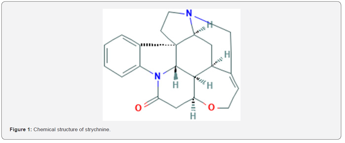
Strychnine, an indole alkaloid, has been shown to possess a variety of biological activities, and its chemical structure was shown in Figure 1. Preliminary studies indicate that strychnine has significant cytotoxic [10], antitumor [11], antiangiogenic [12], analgesic and anti-inflammatory [13]. However, the molecular mechanisms that inhibit the growth of cancer cells is largely unknown. In the present study, we found that Strychnine inhibits the proliferation and induce apoptosis and G0/G1-phase arrest through targeting IL-6/JAK2/Stat3 signaling pathways of liver cancer cells. This research suggested that the traditional Chinese medicine ingredient strychnine could be used as a potential antitumor drug that targets IL-6/JAK2/Stat3 and inhibits liver cancer.
Materials and Methods
Reagents and Antibodies
Strychnine, cisplatin (DDP) and 3-(4,5-dimethylthiazol-2-yl)-2,5-Diphenylterazolium Bromide (MTT) were purchased from Sigma Chemical Co. (St. Louis, MO, USA). Dulbecco’s modified Eagle medium (DMEM), fetal bovine serum (FBS), penicillin/streptomycin and trypsin were obtained from Life Technologies, Gibco BRLProducts (Rockville, MD,USA). Cell Cycle and Apoptosis Analysis Kit (PI) were purchased from KeyGEN BioTECH (Nanjing, China). Antibodies against IL-6, JAK2, p-JAK2, Stat3, p-Stat3, Bcl2, Bax, Caspase-3, Cyclin D1, p21 and β-actin were all obtained from Abcam (Cambridge, MA, USA). RNAiso plus, prime scriptTM RT reagent Kit with gDNAEraser and SYBR Premix EX Taq TM were obtained from Takara (Tokyo,Japan).
Cell lines
Human hepatoma cell lines (HepG2) and normal liver cell lines(L-02) were purchased from the cell bank at chinese Academy of sciences (Shanghai, China). According to the manufacturer’s instructions, the cells were cultured in DMEM supplemented with 10% fetal bovine serum, penicillin (100 U/mL), and streptomycin (100 μg/mL) at 37°C, in a humidified 5% CO2 atmosphere. And cells at logarithmic growth phase were used in the cytological experiments.
Plasmid preparation and cell transfection
The coding sequence of IL-6 cDNA was successfully cloned and the eukaryotic expression vector pcDNA3.1-IL-6 was constructed and confirmed by sequencing. Cells were seeded in 6-well plates (1.5 × 10⁵ or 3 × 10⁵ cells/well) and maintained in complete medium for 12h. The HepG2 cells with IL-6 over-expression was performed by pcDNA3.1-IL-6 transfection using Lipofectamine 2000 (Invitrogen Life Technologies, Carlsbad, CA, USA) according to the manufacturer’s protocol. Cells were harvested after 48h and used for further experiments.
Cell viability assay
The cell viability was measured using MTT assay. Cells (5×10³cells/well) were seeded onto 96-well plates overnight. Then the medium was replaced by the different concentrations of Strychnine medium solution (100, 200, 400, 600 and 800μM). After being cultured for 24h,48h and 72h, 20 μL of MTT solution was added to each well and the plates were incubated for 4h at 37 °C. The medium was removed and the formazan blue, which formed in the cells, was dissolved in 100 μL of DMSO. Cell proliferation was measured 570nm using a microplate reader. Cell viability was calculated as percentage of untreated cells. Three independent experiments were performed from six replicates of each experiment.
Annexin V/PI staining assay for apoptosis
The HepG2 cells at logarithmic growth phase inoculated in 6-well plate were treated with PBS, DDP (10μg/ml), Strychnine for 48h, then the HepG2 cells were collected and suspended in binding buffer at a density of 1× 10⁶ cells/ml. After staining the cells with Annexin V-FITC/propidium iodide (PI; 10μL of Annexin V-FITC+10 μL of PI) in the dark for 15 min, the apoptotic cell death rate was examined using the flow cytometry. The assay was repeated three times independently.
Cell Cycle Analysis
The HepG2 cells at logarithmic growth phase inoculated in 6-well plate were incubated with PBS, DDP (10μg/ml), Strychnine for 48h. The cells were collected, washed twice with ice-cold PBS buffer, fixed with 70% alcohol at 4°C. Then, the HepG2 cells were stained with propidium iodide (PI) in the presence of 1% RNAase A at least for 30 min before the distribution of the cells in cell cycle phases was assessed at 488 nm excitation wavelength by the flow cytometry. The assay was repeated three times independently.
Quantitative reverse transcription PCR
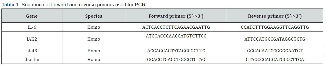
Trizol reagent (Takara, Japan) was used to extract RNA from the cells and total RNA was then reverse transcribed according to the manufacturer’s protocol, and the obtained cDNA was subjected to PCR amplification using the primers detailed in Table 1. PCR cycling conditions were 95 °C for 1 min to denature the cDNA template, followed by 40 cycles at 95 °C for 5s and 60 °C for 20s. The specificity of amplification products was confirmed by melting curve analysis. Data were analyzed using the 2−ΔΔCt method. Independent experiments were performed in triplicate.
Western blot analysis
Total protein was extracted according to the protein extraction kit instructions. Protein concentration was determined using the BCA assay (Beyotime, China). The equal amount of protein was separated by 10% sodium dodecyl sulfate polyacrylamide gel electrophoresis and then the protein was transferred to PVDF membranes (Millipore, Germany), and blocked with 5% skimmed milk for 2h at room temperature. The membranes were incubated with primary antibody overnight at 4 °C. The next day, the membranes were then washed three times and incubated with HRP conjugated secondary antibodies at room temperature for 2h. After extensive washes with TBST, the labeled protein spots were detected by the gel automatic imaging system. Three independent experiments were performed in triplicates. Western blotting was performed using Image-pro plus software for grey value analysis.
Statistical Analysis
The statistical analysis was assessed using SPSS 17.0 software. All the values presented are expressed as mean ± SD. Comparison of different groups was carried out using the One-Way ANOVA Test. P<0.05 was regarded as statistically significant.
Results
Strychnine inhibits the proliferation of HepG2 cells
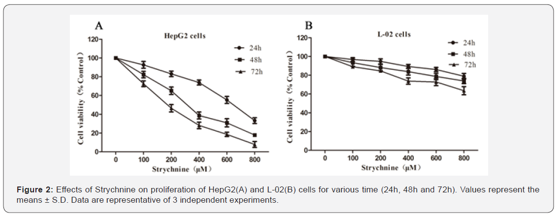
MTT assay was conducted to determine the viability of HepG2 and -02 cells exposed to the trychnine (100, 200, 400, 600, 800 M). As shown in Figure 2, Strychnine could significantly inhibit HepG2 cell growth in a concentration, time-dependent manner. The 50% inhibitory concentration (IC50) of Strychnine on HepG2 cell was about 624.5M (24h), 302.19μM (48h) and 192.4μM (72h). However, the effect of Strychnine on L-02 cells was also evaluated, the IC50 of Strychnine exceeded 800μM at any given time, which was far below that on HepG2 cell lines, indicating that it had weak inhibitory effect on L-02 cells.
Strychnine promotes apoptosis of HepG2 cells
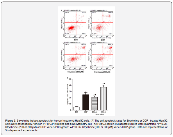
The logarithmic growth of HepG2 cells were treated with PBS, DDP (10μg/ml) and Strychnine (200 or 300μM) for 48h and then the effects of Strychnine on the apoptosis of HepG2 cells were determined using Annexin V/PI staining assay. As shown in Figure 3, HepG2 cells apoptosis rates were .74%, 25.18%, 20.14%, 37.16% in PBS, DDP, Strychnine(200μM) and Strychnine(300μM)-treated groups. Compared with PBS- treated group, HepG2 apoptosis rates were significantly increased in DDP- and Strychnine-treated group(P<0.05), suggesting that Strychnine and DDP could significantly promote HepG2 cells apoptosis.
Strychnine induce G0/G1-phase cell cycle arrest in HepG2 cells
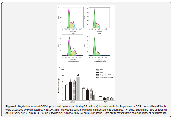
The logarithmic growth of HepG2 cells were treated with PBS, DDP (10μg/ml) and Strychnine (200 or 300μM) for 48h. The PI staining-flowcytometry was applied to analyze the effects of Strychnine onHepG2 cells cycle phase. The results presented in Figure 4 showed that the cell percentages of G0/G1, S and G2/M phases in PBS-treated groups were 47.17%, 40.1% and 12.73%, respectively. whereas the cell percentages of G0/G1, S and G2/M phases in Strychnine(200μM)-treated groups were 64.78%, 28.78% and 6.44%, and whereas the cell percentages of G0/G1, S and G2/M phases in Strychnine(300μM)-treated groups were 77.29%, 16.36% and 6.35%. The analysis showed that Strychnine dose-dependently increased the G0/G1 phase. compared with Strychnine treated group, DDP treated group showed a weaker ability in reducing G0/G1 phase arrest. Also, from the results of cell cycle distribution, HepG2 cells were arrested at G0/G1 phase, so could not enter S phase and the number of G2/M or S phase cells decreased, and then prohibits the propagation of HepG2 cells.
Effect of Strychnine on the expressions of IL-6, JAK and Stat3 mRNA in HepG2 cells
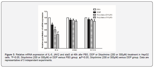
To further explore the apoptotic mechanism of HepG2 cells induced by Strychnine, the effect of Strychnine on IL-6, JAK2 and stat3 mRNA expressions in HepG2 cells was examined by RT-PCR. The experiment results showed that compared with the PBS treated group, the mRNA expression of IL-6 was decreased in the DDP (10μg/ml), Strychnine(200μM) and Strychnine(300μM) -treated group (P<0.05) (Figure 5). However, there were no significant difference in JAK2 and Stat3 mRNA expressions among the groups. In contrast, the expression of IL-6 mRNA was lowest in Strychnine(300μM) -treated group.
Effect of Strychnine on the expressions of IL-6, JAK2 and Stat3 protein in HepG2 cells
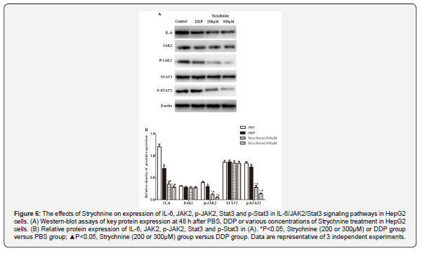
To explore the potential molecular mechanism of Strychnine inhibiting the growth of HepG2 cells, western blot was performed to examine the protein expression level of IL-6/JAK2/Stat3 signaling pathway. As shown in Figure 6, Compared with PBS treated group, IL-6, the phosphorylation of JAK2 and Stat3 protein expression was significantly decreased in DDP-treated group and different concentrations of Strychnine treated groups, and Strychnine had a dose-dependent effect. There was no significant difference in JAK2 and Stat3 proteins expression between groups. The experimental results indicated that Strychnine inhibit the activation of the IL-6/JAK2/Stat3 signaling pathway.
The IL-6/JAK2/Stat3 signaling pathway is involved in the Strychnine-induced inhibition of HepG2 cells
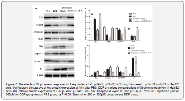
The effects of Strychnine on expression of IL-6/JAK2/Stat3 signaling pathway key proteins were detected using western-blot analysis. As shown in Figure 7, Compared with PBS treated group, the expressions of IL-6, p- JAK2, p-Stat3, Bcl2 and cyclin D1 were decreased respectively, meanwhile Bax, Caspase-3 and p21 protein expression was increased in the DDP (10μg/ml), trychnine(300M) -treated group (P<0.05). However, the protein expressions of IL-6, p- JAK2, p-Stat3, Bax, aspase-3, Bcl2, cyclin D1and p21 of PBS-treated group showed no significant difference compared with Strychnine -treated HepG2 cells with IL-6 over-expression group(P>0.05). The experiment results showed that Strychnine induced G0/G1-phase arrest and promoted HepG2 cells apoptosis might attribute to the activation of IL-6/JAK2/Stat3 signaling pathway.
Discussion
Liver cancer is a malignant tumor derived from the liver cells or intrahepatic bile duct epithelial cells, which is characterized by high degree of malignancy, rapid progression, recurrence and worse prognosis [14]. Despite advances in surgical and radiotherapy techniques, the prognosis of Liver cancer remains poor due to the drug resistance. Therefore, it is necessary to search for the effective therapeutic regimens to combat Liver cancer. Most research suggests that Alkaloids with indole structure, such as vincristine [15], koumine [16] and cephalotaxus alkaloids [17], could inhibit tumor growth and induce apoptosis of tumor cells. Nux vomica has been widely used in anti-tumor therapy for a long time due to its characteristics of activating blood circulation and activating collaterals and combating poison with poison. Strychnine is an indoles active ingredient extracted from the seed of Strychnos which inhibits the growth of various cancer cells in vitro [18]. The experiment results indicate that Strychnine could significantly inhibit the growth of HepG2 cells in a concentration and time dependent manner. But there was almost no significant inhibitory effect of L-02 cells (normal liver cell line).
IL-6 is a pro-inflammatory cytokine produced primarily by cells in the tumor microenvironment and belongs to the interleukin family [19]. Elevated levels of IL-6 are observed in patients with chronic inflammation, such as rheumatoid arthritis and inflammatory bowel disease, and in many hematopoietic malignancies or solid tumours [20]. IL-6 signaling pathways, via either the classical or cross-signaling pathways, involves the engagement of gp130, which leads to activation of gp130-associated JAKs. Four mammalian JAKs (JAK1, JAK2, JAK3, and TYK2) have been identified and all of which are expressed in human cells. The genes encoding JAK enzymes, particularly JAK2, are frequently mutated in malignant tumors, leading to constitutive activation of JAK/ Stat3 signaling [21]. The effects of Stat3 activation on the growth of tumour cells are due to the Stat3-mediated induction of key target genes that regulate cell proliferation and metabolism, inhibit apoptosis, and response to hypoxia [22]. In vitro and in vivo studies have demonstrated that targeting individual nodes in the IL-6/JAK/ Stat3 pathway can have anti-tumour effects [23]. In this study, we demonstrated that Strychnine could reduce IL-6 protein and mRNA levels, thereby inhibiting JAK2/Stat3 activation. The results showed that the inhibitory effect of Strychnine on HepG2 cells may be related to its regulation of IL-6 level and mediated JAK2/Stat3 signaling pathway.
The regulation of cell proliferation is dependent on a balance between cell division and death. Apoptosis is a kind of independent -like programmed cell death, and caspase-3, Bax and Bcl-2 proteins play an important regulatory role in the process of apoptosis. The Caspase family is a class of hydrolytic enzymes which plays an important role in programmed cell death and inflammation [24]. Caspase-3 protein is the "activator" of cell apoptosis. Anti-apoptotic molecule Bcl2 and pro-apoptotic molecule Bax constitute the apoptotic switch in tumorigenesis and therapy [25]. The over-expression of Bcl2 not only prevents the occurrence of apoptosis, but inhibits the release of cytochrome C. However, Bax may increase the permeability of cell membrane, which leads to the release of cytochrome C from mitochondria and activates the caspase activation pathway for apoptosis [26]. In normal cell cycle progression, the transition from G1 to S phase is dependent on the regulation of specific cyclins and cyclin-dependent kinase (CDK), including cyclin D1, cyclin E, p21and p27 [27]. The overexpression of cyclin D1 and cyclin E promotes the development of cancer [28]. The IL-6/JAK/STAT3 pathway is aberrantly hyperactivated in many types of cancer. In the tumour microenvironment, IL-6 acts directly on tumour cells to induce the expression of STAT3 target genes, which encode proteins that then drive tumour proliferation (such as cyclin D1) and/or survival (such as BCL2-like protein 1) [29].
The flow cytometry assay showed Strychnine could significantly promote cells apoptosis and induce G0/G1-phase cell cycle arrest in HepG2 cells. Western-blot assay showed Strychnine significantly increased the protein expressions of Bax and Caspase-3 and decreased the Bcl2 expression in HepG2 cells. Therefore, it may be one of the important cell pro-apoptotic mechanisms of the effective regulation apoptosis-related factors expressions by Strychnine. Meanwhile, the expression of cyclin D1 was significantly decreased and and p21 was significantly increased in HepG2 cells after Strychnine treatment, which indicated Strychnine could also inhibit the growth of HepG2 cells by influencing G0/G1-phase cell cycle arrest.
Conclusion
In brief, this study showed that Strychnine might effectively alleviate or inhibit hepatocellular carcinoma through IL-6/JAK/Stat3 pathway-induced cell apoptosis and GO/G1-phase cell cycle arrest, indicating Strychnine may be a promising drug to treat liver cancer.
Data Availability
All data generated or analyzed during this study are included in this published article.
Conflict of Interest
The authors declare no conflict of interest.
Acknowledgement
This research was supported by Natural Science Foundation of Guangdong Province (2018A030313998), and Administration of Traditional Chinese Medicine of Guangdong Province, China (20191245).
References
- Freddie B, Jacques F, Isabelle S, L SR, A TL et al. (2018) Global Cancer Statistics 2018: GLOBOCAN Estimates of Incidence and Mortality Worldwide for 36 Cancers in 185 Countries. CA: A Cancer Journal for Clinicians 68(6): 394-424.
- Gingold JA, Zhu D, Lee DF, Kaseb A, Chen J (2018) Genomic Profiling and Metabolic Homeostasis in Primary Liver Cancers. Trends in Molecular Medicine 24(4): 395-411.
- Michelotti GA, Machado MV, Anna Mae D (2013) NAFLD, NASH and liver cancer. Nat Rev Gastroenterol Hepatol 10(11): 656-665.
- Poon RTP (2011) Prevention of recurrence after resection of hepatocellular carcinoma: a daunting challenge. Hepatology 54(3): 757-759.
- S Sandra R, Mohamed A, Gabrielle C, Karine P, Sandrine I, et al. (2009) Frequent in-frame somatic deletions activate gp130 in inflammatory hepatocellular tumours. Nature 457(7226): 200-204.
- Marjan B, Sizhi Paul G, Simi A, Kenneth L, Hikmat AA, et al. (2007) Stat3 is tyrosine-phosphorylated through the interleukin-6/glycoprotein 130/Janus kinase pathway in breast cancer. Breast Cancer Research 9(3): R32.
- Hua Y, Drew P, Richard J (2009) STATs in cancer inflammation and immunity: a leading role for STAT3. Nat Rev Cancer 9(11): 798-809.
- Yanhui F, Yu Z, Hongping H, Li H, Yingtong D, et al. (2012) Strynuxlines A and B, alkaloids with an unprecedented carbon skeleton from Strychnos nux-vomica. Journal of Natural Products 75(11): 1987-1990.
- Cai BC, Wang TS, Kurokawa M, Shiraki K, Hattori M (1998) Cytotoxicities of alkaloids from processed and unprocessed seeds of Strychnos nux-vomica. Zhongguo Yao LI Xue Bao 19(5): 425-428.
- Philippe G, Angenot L, Tits M, Monique Tits, Michel Frederich (2004) About the toxicity of some Strychnos species and their alkaloids. Toxicon Official Journal of the International Society on Toxinology 44(4): 405-416.
- Deng XK, Yin W, Li WD, Yin FZ, Lu XY, et al. (2006) The anti-tumor effects of alkaloids from the seeds of Strychnos nux - vomica on HepG2 cells and its possible mechanism. Journal of Ethnopharmacology 106(2): 179-186.
- Saraswati S, Agarwal SS (2013) Strychnine inhibits inflammatory angiogenesis in mice via down regulation of VEGF, TNF-α and TGF-β. Microvascular Research 87: 7-13.
- Chen J, Wang X, Qu YG, Chen ZP, Cai H, et al. (2012) Analgesic and anti-inflammatory activity and pharmacokinetics of alkaloids from seeds of Strychnos nux-vomica after transdermal administration: Effect of changes in alkaloid composition. Journal of Ethnopharmacology 139(1): 181-188.
- Bosch FX, Ribes J, Diaz M, Cleries R (2004) Primary liver cancer: worldwide incidence and trends. Gastroenterology 127(5 Suppl 1): S5-S16.
- HX S, HW H, SH Z, TG L, KH R, et al. (2009) Suppression of N-Ras by shRNA-expressing plasmid increases sensitivity of HepG2 cells to vincristine-induced growth inhibition. Cancer Gene Therapy 16(9): 693-702.
- Zhang X, Chen Y, Gao B, Luo D, Wen Y et al. (2015) Apoptotic Effect of Koumine on Human Breast Cancer Cells and the Mechanism Involved. Cell Biochem Biophys 72(2): 411-416.
- Habli Z, Toumieh G, Fatfat M, Rahal ON, Gali-Muhtasib H (2017) Emerging Cytotoxic Alkaloids in the Battle against Cancer: Overview of Molecular Mechanisms. Molecules 22(2): 250.
- Ren H, Zhao JP, Fan DS, Wang Z, Zhao TJ, et al. (2019) Alkaloids from nux vomica suppresses colon cancer cell growth through Wnt/β-catenin signaling pathway. Phytother Res 33(5): 1570-1577.
- Parvin AK, Pourgholami MH, Richardson DR, Morris DL (2014) Gene of the month: Interleukin 6 (IL-6). J Clin Pathol 67(11): 932-937.
- Kumari N, Dwarakanath BS, Das A, Bhatt AN (2016) Role of interleukin-6 in cancer progression and therapeutic resistance. Tumour Biol 37(9): 1-20.
- Buchert M, Burns CJ, Ernst M (2015) Targeting JAK kinase in solid tumors: emerging opportunities and challenges. Oncogene 35: 939-951.
- Kenny PA, Lee GY, Bissell MJ (2007) Targeting the tumor microenvironment. Front Biosci 12(9): 3468-3474.
- Johnson DE, O'Keefe RA, Grandis JR (2018) Targeting the IL-6/JAK/STAT3 signalling axis in cancer. Nat Rev Clin Oncol 15(4): 234-248.
- Goodsell DS (2010) The molecular perspective: caspases. Oncologist 5(5): 435-436.
- Sadaf F, Zohre G, Sanaz ES, Ali Mohammad A (2015) Mitochondrial targeted peptides for cancer therapy. Tumour Biol 36(8): 5715-5725.
- Narita M, Shimizu S, Ito T, Chittenden T, Lutz RJ, et al. (1998) Bax interacts with the permeability transition pore to induce permeability transition and cytochrome c release in isolated mitochondria. Proc Natl Acad Sci U S A 95(25): 14681-14686.
- Patil MA, Lee SA, Macias E, Lam ET, Xu C, et al. (2009) Role of cyclin D1 as a mediator of c-Met- and beta-catenin-induced hepatocarcinogenesis. Cancer Research 69(1): 253-261.
- Hector B, Yong W, Sanjeev B, Haiming X, Henry H, et al. (2005) Overexpression of cyclin D1 promotes tumor cell growth and confers resistance to cisplatin-mediated apoptosis in an elastase-myc transgene-expressing pancreatic tumor cell line. Clin Cancer Res 11(16): 6075-6086.
- Qing C, Eirini B, Pasquale S, Marjan B, Sizhi Paul G, et al. (2013) The IL-6/JAK/Stat3 feed-forward loop drives tumorigenesis and metastasis. Neoplasia 15(7): 848-862.

























