




 CTOIJ Home



Rare Case of Synchronous Malignancy in Oral Cavity and Esophagus- Case Report
Prachi Kalra*, Diptajit Paul, Abhishek Soni, Rakesh Dhankhar and Vivek Kaushal
Department of Radiation Oncology, PGIMS, Rohtak, India
Submission: July 09, 2021; Published: July 23, 2021
*Corresponding Address: Prachi Kalra, Department of Radiation Oncology, PGIMS, Rohtak, India
How to cite this article: Prachi K, Diptajit P, Abhishek S, Rakesh D, Vivek K. Rare Case of Synchronous Malignancy in Oral Cavity and Esophagus- Case Report. Canc Therapy & Oncol Int J. 2021; 19(3): 556011. DOI: 10.19080/CTOIJ.2021.19.556011
Abstract
Synchronous malignancy in oral cavity and esophagus is a rare entity. Very few case reports have been described on this rarity in literature. Second malignancy is associated with poor prognosis. Herein we report a case of synchronous malignancy of buccal mucosa and esophagus highlighting treatment given and survival outcome.
Introduction
The incidence of head and neck cancer combined with esophageal cancer is approximately around 2%-24%. Esophageal carcinoma must be systematically looked for before the treatment of head & neck carcinomas, after and during follow-up [1]. The definition for classifying a tumor as a second primary malignancy remains consistent since it was first proposed by Warren et al. [2] histological confirmation of malignancy in both the index and secondary tumors, tumors should be separated by at least two centimetres of normal mucosa, if the tumors are recurring at the same site, then they should be separated by at least 5 years in time and metastatic tumor should be ruled out. Synchronous second primary cancers are identified within 6 months of the index tumor. Studies shows that survival of synchronous cancer is lower than the metachronous cancers [3]. The treatment and survival outcomes for Synchronous Head and Neck and Esophageal Carcinoma are not well-known due to its low incidence of this disease. Hence this case report holds value in making treatment decisions and patient counselling for survival outcomes in synchronous malignancies of buccal mucosa and esophagus for future purpose.
Case Report
A 50-year-old man without any comorbidities or illness presented with difficulty in swallowing mainly to solid food for last 2-months, which was insidious in onset and gradually progressive in nature. He also developed hoarseness of voice, which was insidious in onset, gradually progressive and associated with mild throat pain that was relieved partially by oral analgesics. Patient was a chronic smoker and alcoholic for past 25-years. General physical and systemic examination was normal. On local examination, ulcero-proliferative growth of size 3.0×3.0 cm was seen involving right buccal mucosa, extending up to retro-molar trigone, which was firm in consistency and mildly tender on touch. There was no significant lymphadenopathy palpable in cervical region. Complete hemogram and biochemical profile of the patient were within normal limits. The histopathological appearance of the above-mentioned lesion revealed well differentiated squamous cell carcinoma. Upper gastrointestinal endoscopy showed ulcero-proliferative growth causing luminal narrowing at cervical oesophagus, 16.0 cm from the incisor teeth. Biopsy from this lesion revealed moderately differentiated squamous cell carcinoma.
Positron emission tomography-computed tomography (PET-CT) revealed metabolically active lesion involving cervical and proximal thoracic oesophagus extending from C5-D3 vertebra approximately 8.0 cm in length along with metabolically active bilateral multiple cervical, right supra-clavicular & para-esophageal lymph nodes, largest measuring 1.8 × 1.3 cm. It also revealed metabolically active well defined lesion of size 1.6 × 1.9 × 1.2 cm in apical segment of right lung upper lobe suggestive of metastasis with infiltration of overlying pleura and fat along with metastatic gastro-hepatic, supra-pancreatic and mesenteric lymphadenopathy. As patient initially not willing for radiation therapy, 6-cycles of 3-weekly intravenous palliative chemotherapy with docetaxel 120 mg and carboplatin 450 mg were given. Post treatment PET-CT revealed metabolically active lesions in buccal mucosal lesion, cervical and upper thoracic esophagus with near total luminal narrowing extending from C6-D3 vertebrae (Figure 1).
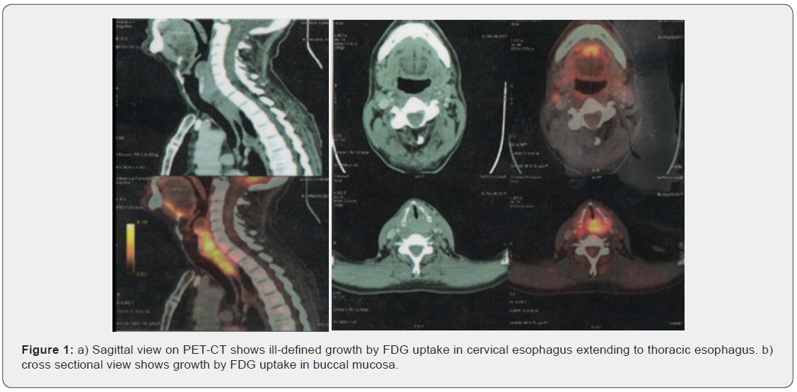
Patient later was also given radiation 20 Gy/ 5 fractions/ 5 days to extended neck and chest. Post treatment patient had partial symptomatic relief and was put on metronomic chemotherapeutic trial with tablet gefitinib 250 mg once daily. Presently patient is on regular monthly follow up with gefitinib therapy for last 6-months and doing well without any progression of disease.
Discussion
In this case report we evaluated the treatment, outcome and survival of a 50-year-old male patient with synchronous malignancy of oral cavity and esophagus. Head and neck squamous carcinoma commonly associated with the carcinoma of the esophagus as synchronous primary. Synchronous or second primary malignancy is a leading long-term cause of mortality in patients with head and neck malignancies and is associated with poor outcome. The prevalence rate remains almost the same in most studies, 5-year OS of 44% has improved compared with historical studies where 5-year OS was <30% [4]. It is suggestive of increased physicians’ awareness to screen for SPMs, improved treatment options and efficacy, and success of lifestyle modifications programs, such as tobacco cessation and cancer-screening programs. Detecting SPMs and cancer screening during follow-up is particularly important for those with HPV-related oropharyngeal cancer, as this subgroup of patients show good prognosis and long-term survival [4]. Genetic event causing loss of heterozygosity is a common in cancer development, LOH analysis is one of the molecular techniques for studying the clonal relationship between two tumors. A recent review on the evolutionary process on field cancerization by Curtius et al. [5] showed role of clonal diversity in the prognoses of many cancers. Thus, primary tumors originating from multiple different fields might be associated with poor prognosis. The ability to accurately distinguish SPM should be distinguished from metastatic tumor for a better treatment plan and follow-up care of HNSCC and esophageal cancer patients.
In our case report patient first underwent upper GI endoscopy guided biopsy as it showed ulcero-proliferative growth in upper esophagus followed by PET scan to rule out multiple sites of metastases. In the absence of PET scan facilities, Triple endoscopy (direct laryngoscopy, esophagoscopy and bronchoscopy) remains an important tool for the identification of synchronous malignancy of the upper aerodigestive tract (UADT) and as a routine procedure in the pre-therapeutic work up or as a screening procedure for the detection of synchronous primary.
In our case report biopsies were taken from the lesion in oral cavity (buccal mucosa) as well as from the lesion in esophagus both showed well to moderately differentiated squamous cell carcinoma in histopathology. After receiving 6-cycles of 3-weekly intravenous palliative chemotherapy with docetaxel 120 mg and carboplatin 450 mg post treatment PET-CT revealed decreased in the size of lesion in esophagus. Patient was then given radiation 20 Gy/ 5 fractions/ 5 days to extended neck and chest. Post treatment patient had partial symptomatic relief and was put on metronomic chemotherapeutic trial with tablet gefitinib 250 mg once daily. Patient is regular follow up since past 6 months with no progression of symptoms or disease.
Conclusion
Synchronous primary cancers of the head and neck region majority of patients can present simultaneously with the index tumor. According to clinical data available the most likely site for the occurrence of synchronous primary cancer along with index primary of the head and neck region is the esophagus. With keeping an insight about the prevalence and risk factors of SPMs in cancer of head and neck region or UADT, critical usage of various diagnostic modalities like PET Scan, triple endoscopies, can help in diagnosis and deciding right treatment for SPMs. Our aim through this case report was to highlight the occurrence of synchronous malignancy in cancer head and neck region with cancer esophagus, its treatment, outcome and survival of the patient of SPMs.
References
- Papazian A, Descombes P, Capron JP, Lorriaux A (1985) Frequency of cancer of the synchronous esophagus of cancers of the upper aerodigestive tract (100 cases): benefit of vital stains with lugol and toluidine blue [Incidence of esophageal cancer synchronous with upper aerodigestive tract cancers (100 cases): value of vital staining with lugol and toluidine blue]. Gastroenterol Clin Biol 9(1): 16-22.
- Warren S, Gates O (1932) Multiple primary malignant tumors: A survey of the literature and a statistical study. Am J Cancer 16: 1358-1414.
- Di Martino E, Sellhaus B, Hausmann R, Minkenberg R, Lohmann M, et al. (200) Survival in second primary malignancies of patients with head and neck cancer. J Laryngol Otol 116(10): 831-838.
- Ng S P, Pollard C, Kamal M, Zeina Ayoub, Adam S Garden, et al. (2019) Risk of second primary malignancies in head and neck cancer patients treated with definitive radiotherapy. npj Precis Onc 3: 22.
- Sunpaweravong S, Bunbanjerdsuk S, Pongrujikorn T, Chaiwat Naktang, Patrapim Sunpaweravong, et al. (2019) Clonal relationship of synchronous head and neck cancer and esophageal cancer assessed by single nucleotide polymorphism-based loss of heterozygosity analysis. BMC Cancer 19: 1174.

























