


Integrating Two Optical Methods to Suppress Diffusive Photons in Ballistic Photons Imaging
Er’el Granot*
Department of Electrical and Electronics Engineering, Ariel Photonics Center, Ariel University, Ariel, Israel
Submission:December 12, 2023; Published:January 05, 2024
*Corresponding author:Er’el Granot, Department of Electrical and Electronics Engineering, Ariel Photonics Center, Ariel University, Ariel, Israel
How to cite this article:Er’el Granot. Integrating Two Optical Methods to Suppress Diffusive Photons in Ballistic Photons Imaging. Curr Trends Biomedical Eng & Biosci. 2024; 22(2): 556082. DOI:10.19080/CTBEB.2024.22.556082
Abstract
A new method to image diffusive media in general and biological tissues in particular, is suggested. The merhod is an integration of two well-known methods: 1) decreasing the collecting detection angle, and 2) using modulated laser light instead of a stationary beam. The two methods have the same purpose – to suppress the diffusive photons with respect to the ballistic ones. The synergy between the two methods has the potential to improve the imaging considerably. Preliminary analysis show that the system’s properties are still challenging – sensitive detectors and high modulation frequencies (>100GHz).
Keywords:Photons; Wavenumber; Ballistic wave; Critical frequency; Stationary; Stationary beam; Scattering; Collimators; Geometrical issue; Detector’s solid angle; Dynamic
Introduction
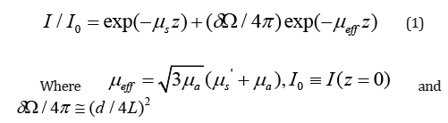
Since biological tissues absorb light in the visible range, light can be a valuable information source for medical imaging [1-4]. The main challenge in using light for tissue imaging is the high scattering coefficient . In the red part of the spectrum, the absorption coefficient (e.g., μa−≅ 0.03cm-1 ) can be negligible compared to the scattering coefficient (e.g., μs−≅ 100cm-1). However, beyond the mean free path (MFP) the image becomes blurry. However, the original image can still be recognized until a distance, which is approximately the reciprocal of the reduced scattering coefficient μs=(1-gμ)μs where  is the mean cosine scattering angle. In this regime, only diffusive methods can be applied, albeit they suffer from low resolution [5-7].
When the tissue layer is relatively thin, the light can then be separated into ballistic component (i.e., unscattered light), and diffusive component ( i.e., scattered light) [8-9]. The diffusive part is scattered in all directions, and therefore the amount detected is proportional to the detector’s solid angle
is the detector’s solid angle. Therefore, there is a critical width,
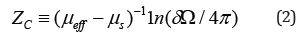
beyond which the medium can be regarded as a diffusive medium, which is governed by the di ffusion equation. When z < zc the image can be reconstructed using optical method, and relatively high resolution can be achieved. Therefore, there is a clear interest in lengthening zc. Several methods were suggested to extend this distance[10]:
decreasing the solid angle of the detector δΩ. This is a geometrical issue, which can be addressed by moving the detector away from the source or using collimators [10,11].
decreasing the scattering coefficient μs. This is a spectral issue, which can be addressed by changing the optical wavelength.
There is a third option, which was not addressed: increasing μeff while decreasing μs. This is a challenging task because the region in which μeff ≅ μs occurs is around 1400nm where the absorption coefficient is extremely high.
In what follows, we suggest a method to increase the attenuation of diffusive photons with minimum effect on the ballistic ones, i.e., to reach the μeff ≅ μs region without increasing the ballistic photons attenuations.
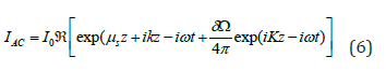
The idea is to use, what was termed in the literature as “diffusive photons waves” [5,6]. But unlike Refs.[5,6] here we suggest using this technology to suppress diffusive light. Instead of a continuous light beam, a modulated laser beam is launched into the medium. Therefore, the stationary diffusion equation should be modified to the temporal one
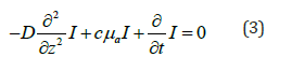
where is the diffusion coefficient. In this case, the light can be separated into a dc (stationary) and an ac (modulated) components

where ℜ represents the real part. After substituting (4) in the dynamic diffusion equation (3) one finds the dispersion relation
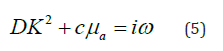
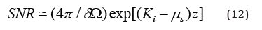
The result is a complex wavenumber. Consequently, the diffusive wave is more sensitive to modulation than the ballistic wave. Therefore, one can use this property to suppress diffusive photons in comparison to ballistic ones. Going back to the experiment presented in Figure1 but with a modulated laser source, the AC part of the detected light looks like
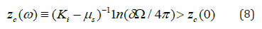
where Ki = ℑK represents the imaginary part of K. Note the difference between the ballistic wavenumber k and the diffusive one K. Since the imaginary part of Kis
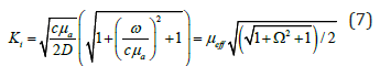
where Ω ≡ω/ωa =ω/cμa . Therefore, the critical distance (2) can be rewritten as
When ω<<ωa ≡ cμa then Ki ≅ μeff , however, ω<<ωa = cμa for


The critical distance (normalized to MFP) as a function of the modulation (normalized to ωa ≡ cμa ) frequency is plotted in Figure 2 for different ratios μeff/μs.
For every given ratio μeff/μs there is a critical frequency, around which the critical distance can increase substantially.
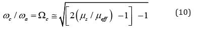
In the important regime, where the ratio is high, the critical frequency is absorption coefficient independent
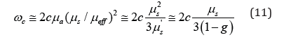
This is a very high critical frequency for wavelengths in the optical domain, around ωc≅11 = 100GHz. This modulation technology is expensive but it is doable.
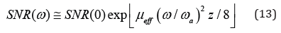
Moreover, these technologies can be used to improve Signal to Noise Ratio (SNR) when the system is within the ballistic regime. Since the SNR is approximately
then even low frequency modulation improves the SNR by a factor of
Conclusion
To conclude, it is shown that modulated light can be used to suppress diffusive photons with respect to ballistic ones. Consequently, modulated light imaging can be. The SNR can be increased substantially, however, to image think layers of diffusive medium, the modulation frequencies should be close to the THz range.
References
- Tuchin V (2000) Tissue Optics, SPIE Press, Bellingham, Washington, US.
- Vaupel P, Thews O, Kelleher D, Hoeckel M (1998) Current status of knowledge and critical issues in tumor oxygenation. Results from 25 years of research in tumor pathophysiology. Adv Exp Med Biol 454: 591-602.
- Gupta PK (1999) Tissue optics. Current Sci 76(10): 1341-1347.
- Bashkatov AN, Genina EA, Tuchin VV (2011) Optical properties of skin, subcutaneous, and muscle tissues: a review. J Innov Opt Health Sci 4(1): 9-38.
- Yodh A, Chance B (1995) Spectroscopy and imaging with diffusing light. Phys Today 48(3): 34-40.
- Jiang H, Keith D, Ulf Osterberg L, Brian W Pogue, Michael Patterson S (1996) Optical image reconstruction using frequency-domain data: simulations and experiments. J Opt Soc Am A 13(2): 253-266.
- McBride TO, Pogue BW, Jiang S, Osterberg UL, Paulsen KD, et al. (2001) Initial studies of in vivo absorption and scattering heterogeneity in near-infrared tomographic breast imaging. Opt Lett 26(11): 822-824.
- Yaroshevsky A, Glasser Z, Granot E, and Sternklar S (2011) Transition from the ballistic to the diffusive regime in a turbid medium. Opt Lett 36(8): 1395-1397.
- Ben I, Layosh Y, Granot E (2016) A study of a simple model for the transition between the ballistic and the diffusive regimes in diffusive media. J Biomed Opt 21(6): 66004.
- Glasser Z, Yaroshevsky A, Barak B, Granot E, Sternklar S (2013) Effect of measurement on the ballistic–diffusive transition in turbid media. J Biomed Opt 18(10): 106006.
- Brezner B, Cahen S, Glasser Z, Sternklar S, Granot E (2015) Ballistic imaging of biological media with collimated illumination and focal plane detection. J Biomed Opt 20(7): 076006.


