Modulatory Role of Aqueous Leave Extracts of Mangifera indica (Linn) on Liver Function Test in Plasmodium - Infected Albino Mice
Olayode Ahmed A*1, Ofusori David A1, Ogunniyi Titus AB2, Saka Olusola S1, Bejide Ronald A3 and Adelodun Stephen T1
1Department of Anatomy and Cell Biology, Obafemi Awolowo University, Nigeria
2Department of Medical Microbiology and Parasitology, Obafemi Awolowo University, Nigeria
3Department of Morbid Anatomy & Forensic Medicine, Obafemi Awolowo University, Nigeria
Submission: March 15, 2017; Published: April 06, 2017
*Corresponding author: Olayode Ahmed A, Department of Anatomy and Cell Biology, Faculty of Basic Medical Sciences, Obafemi Awolowo University, Ile Ife, Osun State, Nigeria, Tel: +2348057077587; Email: olayodeahmed01@gmail.com
How to cite this article: Olayode A A, Ofusori D A, Ogunniyi T A, Saka O S, Bejide R A,et al. Modulatory Role of Aqueous Leave Extracts of Mangifera indica (Linn) on Liver Function Test in Plasmodium - Infected Albino Mice. Anatomy Physiol Biochem Int J. 2017; 2(2): 555584. DIO: 10.19080/APBIJ.2017.01.555584
Abstract
This study was carried out to assess the modulatory role of Mangifera indica on the liver function test in Plasmodium-infected albino mice. Forty-two male albino mice of 20 - 25 g were used for this study. The mice were randomly selected into six groups A, B, C, D, E and F of seven mice each. Mice in groups B, C, D, E and F were infected with Plasmodium berghei while those in group A were administered equivalent volume of normal saline. Mangifera indica extract (50mg/kg, 100mg/kg and 150mg/kg b.w.) dissolved in 0.2ml of distilled water were administered into mice in groups C, D and E respectively for 5days, and those in group F were orally administered with Artesunate for 5days (3mg/kg and 1.5mg/kg b.w. on the first day and on subsequent 4 days respectively) while mice in groups A and B were administered equivalent volume of distilled water, all administration starting 72hours post-infection. The animals were left for another 14days after treatment. Blood samples were collected via cardiac puncture and centrifuged to obtain sera. Activities of alanine aminotransferase (ALT), aspartate aminotransferase (AST) and alkaline phosphatase (ALP) as well as bilirubin concentration were measured from the sera using enzyme colorimetric methods. One-way ANOVA was used to analyze the data, followed by Student Newman-Keuls (SNK) test for multiple comparisons.
The result showed that Mangifera indica extract has an ameliorative effect on the serum level of total bilirubin concentration but appeared to be less effective in reducing the activities of the liver function enzymes. In conclusion, Mangifera indica has an ameliorative effect against hepatocellular injury but appeared to be less effective in the cholestatic injury to the liver.
Keywords: Mangifera indica; Aspartate aminotransferase; Alanine aminotransferase; Alkaline phosphatase; Total bilirubin; Plasmodium berghei
Abbreviations: ALT: Alanine Amino-Transferase; AST: Aspartate Amino-Transferase; ALP: Alkaline Phosphatase; SNK: Student Newman- Keuls; MI: Mangifera Indica; PRBCs: Parasitized Red Blood Cells
Introduction
Genus Plasmodium comprises of various single cell protozoans, transmitted by female Anopheles mosquitoes [1], which causes the world's most devastating human parasitic infection called malaria [2]. Fevers caused by Plasmodium falciparum occur sometimes at 48-hour intervals which lead to most serious illnesses and deaths. Few of the persistent control of malaria are prompt and effective treatment with Artemisinin- based combination therapies, usage of mosquito nets or insecticidal nets by people at risk and indoor residual spraying of insecticide [3].
P. berghei, P. chabaudi, P. vinckei and P. yoelli. P. berghei are some of the Plasmodium species that have been described in African murine rodents to infect the liver after being injected into the bloodstream by a bite of an infected female mosquito. These Plasmodium species are used in research programs for development and screening of anti-malarial drugs and for the development of an effective vaccine against malaria, since they have the same effects on rodents as the P. falciparum has on humans [4,5]. From liver, the parasites spread to several other organs to symptoms like high fever, head ache, drowsiness, confusion and later leads to symptoms like diarrhea, kidney failure and jaundice. Resistance against a number of anti- malarial medicines has developed in malarial parasites in many parts of the world [1].
Limited success had only been achieved during the course of controlling the vectors with the view of disrupting the life cycle of the parasite; the use of anti-malarial drugs has always been hampered by their lack of availability to those most in need and also to the rapid evolution of drug resistant parasites [6]. An adequate and effective vaccination against these parasites would provide a promising approach to controlling the disease but this mechanism has not been available for practice. The infection of liver cells by the sporozoites form of the malaria parasite can cause organ congestion, sinusoidal blockage and cellular inflammation. These changes in hepatocytes can lead to the leakage of parenchyma and membranes of the liver cells thus releasing enzymes (transaminases and phosphatase) of the liver into the circulation. Alanine aminotransferase (ALT) and aspartate aminotransferase (AST) have been established as markers of hepatocellular injury while alkaline phosphatase (ALP) is a marker of cholestasis [7,8].
Genus Mangifera consists of about 30 species of tropical fruiting trees in the flowering plant family Anacardiaceae, which mango belongs to the genus. Some of the chemical constituents of Mangifera indica (MI), a species of genus Mangifera, are polyphenolics, flavonoids, triterpenoids and mangiferin; all being antioxidant and glucosyl xanthone, have strong antioxidant, anti lipid peroxidation, immunomodulation, cardiotonic, hypotensive, wound healing, antidegenerative and antidiabetic activities [9]. Mangiferin (an active component of MI) treatment increased serum iron-binding capacity and decreased serum levels of aspartate aminotransferase (AST) and alanine aminotransferase (ALT) [10]. It is therefore necessary to investigate modulatory role of MI on AST, ALT, ALP and bilirubin concentration in Plasmodium-infected albino mice with a view to improving on the management of malarial infection and complications.
Materials and Methods
Animal Care and Management
Forty-two male albino mice, weighing between 20 and 25 g, obtained from the Animal Holding of College of Health Sciences, Obafemi Awolowo University Ile-Ife, were used for this study. The mice were accommodated in plastic cages in the Animal Holding of the Department of Anatomy and Cell Biology, Obafemi Awolowo University Ile-Ife. They were fed with standard laboratory animal pellets throughout the period of the experiment and water was provided ad libitum. The mice were randomly assigned into six groups A, B, C, D, E and F of seven mice each.
Plant Material and Preparation of Extract
Fresh leaves of mango tree were plucked from plantations in Ile-Ife and authenticated by a plant taxonomist in the Department of Botany, Obafemi Awolowo University, Ile-Ife. A voucher specimen was deposited at the department's Herbarium for future reference (Voucher number: IFE-17465). The leaves were washed with clean water and air-dried. The dried leaves were pulverized with squeezing and crushing machine (Daiki Rika Kogyo Co-ltd, Japan) into fine powder. The aqueous extract was prepared by extracting the powdered leaves, weighing 2.0 kg, with 6 L of distilled water, macerated in a Polytron Homogenizer for 48 hours and the solution was filtered using Buchner funnel and Whatmann No. 1 filter paper (Whatmann International Ltd, Maidstone, UK). The filtrate was evaporated under 56 atm reduced pressure using a rotary evaporator (BuchiRotavapor R110, Schweiz) and later freeze-dried in a lyophilizer (Ilshin Lab. Co. Ltd, Seoul, Republic of Korea). The extract obtained was kept in a desiccator until use. The percentage yield was 10.52%.
Infection with Plasmodium Parasites
The infection was done in the Department of Medical Microbiology and Parasitology, College of Health Sciences, Obafemi Awolowo University, Ile-Ife. Mice were infected with P. berghei intraperitoneally with 0.2 ml of mixture of normal saline and infected blood of the donor mouse whose parasitemia level was approximately 36 % and normal saline at ratio 1:31 respectively. The infection was carried out on the first day of the study and left for two more days to allow for the spread of Plasmodium parasites Basir et al. 2012. The percentage parasitemia was determined on the 3rd day to confirm the Plasmodium status of each mouse.
Experimental Design
- Group A was the negative control group served with equivalent volume of normal saline;
- Group B was infected with Plasmodium parasite (PP) + equivalent volume of normal saline;
- Group C was infected with PP and treated with MI (50 mg/kg body weight) after 72 hours post - infection for 5 days;
- Group D was infected with PP and treated with MI (100 mg/kg body weight) after 72 hours post - infection for 5 days;
- Group E was infected with PP and treated with MI (150 mg/kg body weight) after 72 hours post - infection for 5 days; and
- Group F was infected with PP and treated with Artesunate after 72 hours post - induction for 5 days (3 mg/kg body weight on first day and 1.5 mg/kg body weight for the next 4 days).
The animals were left for another 14 days after treatment before the blood samples were collected for biochemical analysis.
Blood collection
At the end of withdrawal period, blood was collected by cardiac puncture with the aid of non-heparized capillary tubes. About 1 ml was dispensed into clean sample bottles without anticoagulant and left to clot, to be used for biochemical studies.
Estimation of Biochemical Parameters
Blood samples collected were centrifuged and frozen at 4oC before the biochemical procedure was performed. Total bilirubin concentration was measured and the liver serum enzymes such as Alanine aminotransferase (ALT), Aspartate aminotransferase (AST) and Alkaline phosphatase (ALP) were also assayed using commercially available kit (Randox, Northern Ireland).
Statistical Analysis
One-way ANOVA was used to analyze data, followed by Student Newman-Keuls (SNK) test for multiple comparisons. Graph Pad Prism 5 (Version 5.03, Graphpad Inc.) was the statistical package used for data analysis. Data obtained were analyzed using descriptive and inferential statistics. Significant difference was set at p<0.05.
Result
Activity of Aspartate Aminotransferase (AST) in Control and Experimental Groups

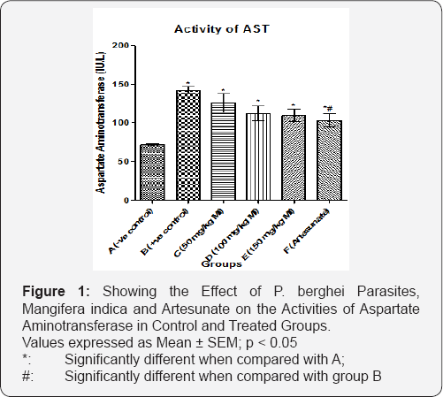
The activities of AST were significantly higher (p = 0.0004; f = 8.03) in groups B (146.00 ± 4.65 IU/L), C (126.00 ± 12.20 IU/L), D (112.00 ± 9.82 IU/L), E (110.00 ± 8.22 IU/L) and F (103.00 ± 8.73 IU/L) when compared with group A (72.00 ± 1.47 IU/L). There was no significant difference (p = 0.4601; f = 0.921) in the activity of AST across groups C (126.00 ± 12.20 IU/L), D (112.00 ± 9.82 IU/L), E (110.00 ± 8.22 IU/L) and F (103.00 ± 8.73 IU/L). Also, there was no significant difference (p = 0.0951; f = 2.67) in the activities of AST across group B (142.00 ± 4.65 IU/L) compared with groups C (126.00 ±12.20 IU/L), D (112.00 ± 9.82 IU/L) and E (110.00 ± 8.22 IU/L), but there was significant increase (p = 0.0076; f = 3.52) in the activities of AST in group B (142.00 ± 4.65 IU/L) compared with group F (103.00 ± 8.73 IU/L) (Figure 1).
Activity of Alanine Aminotransferase (ALT) in Control and Experimental Groups

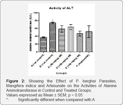
There was significant increase (p < 0.0001; f = 1.45) in the activities of ALT in group B (74.00 ± 2.20 IU/L) compared with the group A (37.00 ± 1.83 IU/L). However, there was an increase but no significant difference (p = 0.2663; f = 1.45) in the activity of ALT as group B (74.00 ± 2.20 IU/L) was compared with groups C (58.50 ± 8.80 IU/L), D (63.30 ± 6.56 IU/L), E (46.80 ± 6.36 IU/L) and F (51.50 ± 14.9 IU/L). There was no significant difference (p = 0.2943; f = 1.36) in the activity of ALT across groups A (37.00 ± 1.83 IU/L), C (58.50 ± 8.80 IU/L), D (63.30 ± 6.56 IU/L), E (46.80 ± 6.36 IU/L) and F (51.50 ± 14.9 IU/L) (Figure 2).
Activity of Alkaline Phosphatase (ALP) in Control and Experimental Groups
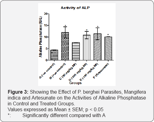
There was significant increase (p = 0.0457; f = 3.15) in the activities of ALP in groups B (12.00 ± 2.40 IU/L), D (10.80 ± 1.59 IU/L), E (11.40 ± 2.56 IU/L) and F (10.10 ± 0.07 IU/L) compared with the group A (4.40 ± 0.11 IU/L). There was no significant difference (p < 0.0001; f = 1.40) in the activity of ALP between group C (7.60 ± 0.02 IU/L) compared with group A (4.40 ± 0.11 IU/L). There was no significant difference (p = 0.4531; f = 0.969) in the activities of ALP across groups B (12.00 ± 2.40 IU/L), C (7.60 ± 0.02 IU/L), D (10.80 ± 1.59 IU/L), E (11.40 ± 2.56 IU/L) and F (10.10 ± 0.07 IU/L) (Figure 3).

Activity of Total Bilirubin Level in Control and Experimental Groups
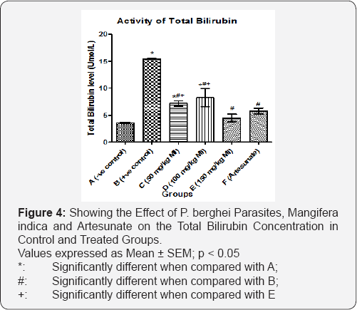
There was significant increase (p < 0.0001; f = 32.3) in total bilirubin level when groups B (15.40 ± 0.13 Umol/L), C (7.20 ± 0.50 Umol/L) and D (8.28 ± 1.65 Umol/L) were compared with the group A (3.60 ± 0.16 Umol/L). However, there was a significant difference (p = 0.044; f = 4.51) when group A was compared with groups E (4.50 ± 0.68 Umol/L) and F (5.75 ± 0.54 Umol/L). There was significant increase (p < 0.0001; f = 28.7) in total bilirubin level in group B (15.40 ± 0.13 Umol/L) when compared to all other groups. There was significant increase (p = 0.0395; f = 6.01) in total bilirubin level in group D (8.28 ± 1.65 Umol/L) when compared with group E (4.50 ± 0.68 Umol/L) (Figure 4).

Discussion
Increase in liver enzymes AST, ALT and ALP observed in malaria infected patients also demonstrated that the serum activities of these liver enzymes increased with the increase in malaria parasite density and the destruction of parasitized red blood cells (PRBCs) by the Kupffer cells. This study showed that serum level of AST was significantly increased in positive control group when compared with the negative control group; this is an indication of impaired liver function due to hepatocellular injury. The serum levels of ALT and ALP also increased in all groups. ALT was significantly reduced in group that received 150 mg/kg MI this supports earlier documentation [11].
There was dose dependent significant change (p < 0.05) in the activities of AST in the extract treated animals with the highest dose (150 mg/kg MI b.w.) treated mice reduced the damage done by PP better than the lower doses but the reduction was not as much as that of artesunate treated group. This was in conformity with the outcome of the study of Ogbe et al. [11]. Artesunate has also been reported to lower the level of AST after Plasmodium infection with lesser dose but couldn't do the same in animals administered with higher doses [12]. There was reduction in the activities of ALT in extract treated animals, although the reduction did not follow a regular pattern. Ogbe et al. [11] also reported reduction in the activity of ALT after the administration of ethanolic extract of MI which conforms with the outcome of this result.
There was no significant change (p > 0.05) in the activities of ALP in the extract treated groups but the lower dose showed more reduction from the damage from PP. This showed there was no significant change between the activities of ALP in positive control and the extract treated mice; it can be deduced that there may be probable no effect of MI on cholestatic injury of the liver. The reduction in the levels of transaminases and phosphatase may be due to the presence of mangiferin which has been reported to increase serum iron-binding capacity and decrease serum levels of AST and ALT [10], but the reduction was not significantly different from the positive control.
Increase in breakdown of PRBCs and centribular damage cause increase in level of bilirubin concentration in acute falciparum malarial infection, leading to hyperbilirubinemia, which is a direct consequence of the impaired drainage capacity of the liver. This also led to the increase in the levels of total bilirubin concentration in the serum of the mice of the positive control groups. Serum bilirubin levels are also well associated with the status and function of the hepatic cells, the serum bilirubin level is elevated normally in hepatotoxicity [13] and it is the probable marker of liver diseases [14]. The level of total bilirubin concentration increased significantly in the positive control. The treated groups were capable of decreasing the bilirubin concentration to near normal probably due to the intense reduction in PRBCs in circulation. Raised serum bilirubin, hepatomegaly along with increase in liver enzymes is an important denominator of liver injury in these patients [15]. Mangiferin possess the ability to increase serum iron binding capacity which eventually reduced the high serum bilirubin [16]. The antioxidant activities possessed by flavonoids, present in MI, has ameliorative effect on oxidative damage in the liver [17].
Conclusion
Mangifera indica has an ameliorative effect on the serum level of total bilirubin concentration and hepatocellular injury but appeared to be less effective in reducing the effects due to the cholestatic injury to the liver.
Conflict of Interest
There was no conflict of interest amongst the authors during the preparation of the work.
Acknowledgment
We acknowledge the technical inputs of Mr. Ajayi O.C. during the infection of the mice with Plasmodium berghei parasites.
References
- WHO (1998) Malarial Chemotherapy, Tech. Reg Ser WHO, Geneva, Switzerland.
- Nicholas JW, Breman JG (2005) Malaria and Babeiosis; Diseases Caused by Red Blood Cell Parasites in Harrisons Principle of Medicine, McGraw Hill, New York, pp. 1218.
- Palmer J (2006) WHO gives indoor use of DDT a clean bill of health for controlling malaria, WHO.
- Hall N, Karras M, Raine JD, Carlton JM, Kooij TW, et al. (2005) A comprehensive survey of the Plasmodium life cycle by genomic, transcriptomic, and proteomic analyses. Science 307(5706): 82-86.
- Kooij TW, Janse CJ, Waters AP (2006) Plasmodium post-genomics: better the bug you know? Nat Rev Microbiol 4: 344-357.
- Good M (2009) The hope but challenge for developing a vaccine that might control malaria. Eur J Immunol 39(4): 939-943.
- Abu AH, Uchendu CN (2010) Assessment of Aqueous Ethanolic Extract of Hymenocardia acida Stem Bark in Wistar Rats. Archives of Applied Science Research 2(5): 56-68.
- Vasudevan DM, Sreekumari S (2007) Textbook of Biochemistry for Medical Students; Jaypee Brothers Medical Publishers Ltd, (5th Edn), New Delhi, India, pp. 266
- Shah KA, Patel MB, Patel RJ, Parmar PK (2010) Mangifera Indica (Mango). Pharmacognosy Reviews 4(7): 42-48.
- Pardo-Andreu GL, Barrios MF, Curti C, Hernandez I, Merino N, et al. (2008) Protective Effects of Magnifera indica L extract (Vimang) and its major component mangiferin, on iron-induced oxidative damage to rat serum and liver. Pharmacol Res 57(1): 79-86.
- Ogbe RJ, Adenkola AY, Anefu E (2012) Aqueous Ethanolic Extract of Mangifera indica Stem Bark Effect on the Biochemical and Haematological Parameters of Albino Rats. Arch Appl Sci Res 4(4): 1618-1622.
- Onovo A, Madusoromuo MA, Ekpo Nta I (2016) Effects of Artesunate on Liver Functions of the Wistar Rats. International Journal of Biochemistry, Bioinformatics and Biotechnology Studies 1(1): 19-29.
- Mondal HT, Chakraborty G, Gupta M, Mazumder UK (2005) Hepatoprotective activity of Diospyros malabarica bark in CCl4 intoxicated rats. Eur Bull Drug Res 13: 25-30.
- Achliya GS, Wadodkar SG, Dorle AK (2004) Evaluation of hepatoprotective effect ofAmalkadighrita against carbon tetrachloride- induced hepatic damage in rats. J Ethnopharmacol 90: 229-232.
- Kochar DK, Kaswan K, Kochar SK, Sirohi P, Pal M, et al. (2006) A comparative study of regression of jaundice in patients of malaria and acute viral hepatitis. J Vect Borne Dis 123-129.
- Rasool M, Sabina EP, Mahinda PS, Gnanselvi BC (2012) Mangiferin, a natural polyphenol protects the hepatic damage in mice caused by CCl4 intoxication. Comp Cli Pathol 21(5): 865-872.
- Sudha A, Sumathik K, Manikandeselvi S, Srinivasan P (2013) Antihepatoxic Activity of Crude Flavonoid Fraction of Lippi nodiflora L. on Ethanol Induced Liver Injury in Rats. Asian Journal of Animal Sciences 7(1): 1-13.


