Abstract
Gold is precious material to the human being while gold nanostar (or nano particle) is also an invaluable extreme probe to the scientists. Nanoscience breakthroughs almost in every field of science through the study of structures and molecules on the scales of nanometers ranging between 1 and 100nm. Ancient people had used nanotechnology without knowing the present-day advanced level theory and technology. Discovery of “Lycurgus Cup”, “Mayan Blue”, “inner portion of black coating Keeladi pottery shards” provide realistic evidence of historical nano-materials used by them. This raises questions: “What technology the ancient people used without knowing the advanced level nanoscience theory and practical knowledge?” or “Is the nanotechnology used in ancient same or different as of the present-day technology?”
One of the mysterious areas is cell functioning in human body. Although scientists have acquired a lot of knowledge in cell biology yet human cell biology remains a mystery to the scientists. Among many unsolved issues related to human cells are:
a) How healthy cell functions at molecular level against diseases?
b) How changes occur in shape and size of a cell over time?
c) How the formation of new cells and removal of death cells are maintained and regulated in human body?
d) The root cause of a disease i.e. what changes are happening to everyday cell processing when cells (i.e. human body) are affected in a disease? etc.
Optical properties of Gold Nanostars (or nano-particles) provide us the opportunity of an extreme probes for studying the human cells. In this review the importance of Gold Nanostars has been discussed indicating its possible role towards solving the secrets of the universe.
Keywords:Cell; Nano-Science; Nano-Technology; Gold Nanostars
Root Pressure
Before the well beginning of humanity gold has long been played as one of the most rare and precious metals on earth. Our ancestors considered gold to be the highest form of matter and have incredibly valuable for scientific, physical and chemical properties. Presently, we know that gold is one of the heaviest stable, naturally occurring atom type found on earth. But main question - “where did gold come from?” Remain unsolved.
Origin of Gold
In the early civilizations human used gold to the deities with a belief that it (i.e. Gold) has a connection with the image of the sun, with light and life-giving warmth, growth and power. For this reason, ancient Egyptians believed that gold represented the earthly form of the sun. According to our present knowledge, scientists / astronomers believed that like many common elements, such as carbon, iron, gold are forged in the fiery furnace of the stars (i.e. cosmic furnace) but special conditions are required for gold and other heavy elements. Such special conditions are the powerful, violent cosmic events.
The Cosmic Furnace
The required powerful, violent events through which gold produces in nature are
i)Supernova i.e. the end stage of exploding stars.
ii)Binary merger i.e. when the remnants of the supernovae, so called neutron stars, collide with one another and created gamma ray bursts.
But as evidence Berger et al [1] suggested that all the gold in the universe is nothing but a byproduct of dense neutron stars
colliding process. Their conclusion was based on the recent
observation of gamma ray burst in a galaxy 3.9 billion light years
away. According to them the pressure and temperature are the key
factors for transforming the chemical structure of iron. The least
values are pressure > 50 GPa and temperature > 15000K. Such
high values of pressure and temperature are only found in star’s
core. In fact, when two massive stars both have died in supernova
explosions, then their remnant neutron stars spiral inwards and
merge. When this merger event occurs, then as a result or cause:
a) A gamma ray burst arises,
b) combine of two neutron stars turn into a black hole, or
c) rather, some 96% of their (neutron stars) masses merges
to form a black hole with a fraction of that mass get ejected a
gamma ray burst as well as in the typical form the heaviest
elements of all, including gold.
Not only that, an estimation shows that a single neutron starneutron star merger can create gold about 20 times the mass of the moon by weight [2].
Historical Gold nanostar (or Nanoparticle)
The prefix term “nano” comes from the Greek word “nanos” meaning “dwarf” or “very small” with a depict of one thousand millionth of a meter (i.e. 10-9 m). For easy, layman type understanding, as a comparison, one must realize that a simple human hair is 60,000 nm thickness while the DNA double helix has a radius of 1 nm (Figure 1) [3,4].
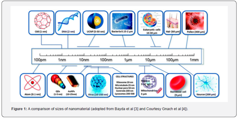
Nanoparticles, therefore, means particles with the sizes ranging in units to hundreds of nanometers. Production of different types of nano-materials began in the early of 1990 [5]. Since then, enormous efforts have led to the interest of knowing the extreme dependence of properties (such as electronic, magnetic, optical, mechanical etc.) on such nanoparticles size and shape in the 1-100 nm regime as well as various applications.
Why gold nanostars
Following quantum-mechanical principles nano-particles, in general, would display electronic structures, reflecting the electronic band structures. This means that the resulting physical properties are the expression of neither those of bulk metals nor those of molecular compounds but strongly depend on the particle shapes, sizes, inter-particle distance, nature of guarding organic shell [6].
As the gold nanostars contain very small sizes several constituent atoms or molecules so they can be considered as an isolated group of atoms or molecules, even they show variation from that of the properties natural in bulk. This implies that the gold nanostars exhibit electronic, magnetic, optical, physical and chemical properties which are completely different from both the bulk and the constituent atoms or molecules. So, gold nanostars (or particles), based on their unique properties (i.e. uniqueness through interpretation in terms of high relativistic contraction of its 6th orbital) and uniqueness offer us its application in almost all fields from micro to macroscopic world covering human biology to cosmology.
Structure of Gold Nanostars
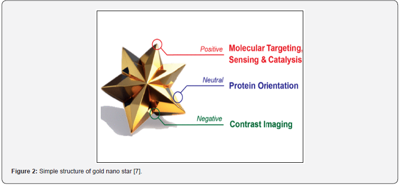
Figure 2 shows the intricate three-dimensional structure of gold nanostars. It uniquely displays its three distinct surface curvature, namely positive, neutral and negative that provide different environment for observing ligands. Not only that, these curvatures are also used for understanding different chemistry properties in gold nanostars, impact on bio-medical, chemical application (including surface enhanced Raman spectroscopy i.e. SERS), contrast agent performance, catalysts, bio-sensing, imaging, local chemical manipulation, etc. [7].
Ancient Evidences of Gold Nanostars
The Mystery of Lycurgus Cup
Regarding the use of gold, as metallurgical metals, can be found
from different surviving documents / works of Greeks during the
period 372 BCE to (40-90) CE (such as Leyden papyrus available
as antiquities in Netherlands). On the other hand, application of
gold nanoparticles (of course, on that time the concept of nanoparticles
were not invented) was found in Lycurgus Cup (Figure
3a, b) [8] in the late Roman period (i.e. in the 4th century CE).
This magic (originally due to nanoparticles) type cup shows two
different colours:
a) A green jade colour due to diffusion of light when
illuminated from outside (Figure 3a)
b) A deep ruby red colour (due to transmission) when it is
illuminated from inside (Figure 3b).
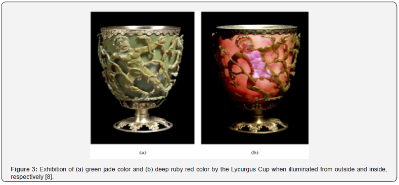
In fact, the secret of this Lycurgus cup came into light in 1965 when Brill [8] first explain the magical behavior of the cup through detailed analysis in the presence of minute amount off gold (i.e. “Au” about 40ppm) and silver (i.e. “Ag” about 300 ppm). Through further analysis by electron microscope Barbar and Freestone [9] showed the presence of silver-gold alloy having nanoparticles of 50-100 nm in diameter and a ratio of silver to gold i.e. Ag/Au = 70:30. This realistic phenomenon was confirmed through a theoretical study by Hornyak et al [10] indicating the emission of deep red colour from the Lycurgus Cup due light absorption~515 nm which is consistent with the presence of Silver-Gold alloy with Ag/Au = 70:30.
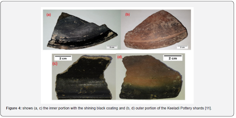
In the 6th Century BC- Recently the inner portion of pottery shards with black coating have been discovered from Keeladi area in Tamil Nadu state, India [11]. Visual observation of this black coating (inside the pottery) shows as a shiny, hard and exhibited endurance (Figure 4). Analysis of this coating with the help of Raman Spectroscopy, transmission electron microscope and x-ray photoelectron spectroscopy revealed the presence of single, multi-walled carbon nanotubes based layered sheets in the coating. The average diameters of this carbon nanotube found ~ 0.6 ± 0.05 nm (theoretical predicted value ~0.4 nm) with the material of Cu and Ag nano-particles. The significance of this discovery is that it shows as evidence of the presence of Cu-Ag nano-particles alloy other than Ag-Au alloy. Another puzzling fact is without knowing the scientific principles of the nano-scale and its unusual properties (in respect to the presently known) what technology the ancient people know and used in glazed Islamic potteries [12] and the Renaissance pottery [13]. Not only that, how these 0-D (i.e. zero dimensional) metal nano-particles they used for improvement of the lustre of these potteries are still unknown.
Another example of use of nano-particle was occurred in 800 AD. In this period ancient people of pre-Columbian Maya city of Chichen Itza produced “Mayan Blue” [14]. This pigment was a complex material consisting of clay with nanopores filled with indigo dye. This “Mayan Blue” was environmentally stable pigment [15].
The Imaginative Pioneers of Nano-technology
As the exact history of nano-science is not yet fully known it
is very difficult to identify who was the first scientist in the nanoworld.
Acknowledgement of literary documents are:
a) 1449-John Utynam made a gold nano-particle based
glass and took patent of it.
b) 16th Century-Swiss doctor Theophrastus von Hohenheim
(known as Paracelsus) used gold nanoparticles for the treatment
of patients suffering from various ailments. This implies that
nano-science field is not a new but presently in the advanced form
over time [16].
c) 1925-Richard Zsigmondy (Nobel Laureate) was the
first to introduce the scientific concept of the nano-meter for
measuring the sizes of the particle like “gold colloidal particles
with the help of microscope.
d) 1940-Silica nano-particles were manufactured as
substitutes to carbon black for rubber reinforcement [17].
e) 1959-Physicist Richard Feynman [18] described the idea
and concept of nano-science and nano-technology through his
lecture entitled “There’s Plenty of Rooms at the bottom” delivered
at American Physical Society meeting held at Caltech on 29th
December 1959. In his lecture he provided clues through hints
how scientists would be able to manipulate and control individual
atoms and molecules. But on that time no ways or techniques were
not invented or developed to see the individual or isolated atoms
until 1981 the scanning tunneling microscope was invented to see
the individual atoms. It can be said that Feynman’s talk, in real
sense, was a source of inspiration for nano-science on that time.
f) 1974-Japanese scientist N. Taniguchi [19] was the first
person, in realistic sense, who coined the term “nanotechnology”.
He described the mechanism and technological processing,
separation, consolidation and deformation of the super thin
nanosized materials with accuracy.
Production of Nano-Material in Modern Science Age
a) 1857-The first scientific report was that Michael Faraday
prepared nanomaterial, so called “Actinated Gold” from the
synthesis of a colloidal solution of gold nanoparticles [20]. In a
lecture delivered at the Royal Society of London he used a purple
color slide containing exceedingly fine particle of gold, diffused in
various preparation of gold for producing a ruby red fluid [21].
b) 1981-Invention of scanning tunneling microscopy
(STM). The impact of this STM was on surface science which
helped the scientists as a new tool to find a new method of direct
measurement and manipulation on nanoscale.
c) 1990-DNA technology a newly born, separate
discipline arose. This field was knocking on the doors of the
physicists, biologists, chemists, engineers and material scientists
(including geologists) for new invention in the development of
nanotechnology. A new material, so called “smart material” [22]
have been discovered which can be influenced by external stimuli
like temperature, moisture, electric and magnetic fields, etc.
d) 2004- Xu et al [23] discovered a new class of carbon
nanomaterial, so called carbon dots (C- dots), with sizes below
10 nm, during the purification of single walled carbon nanotubes.
This C-dots have interesting properties such as benign, abundant
and inexpensive nature [24]. Not only that, it possesses a superior
property such as low toxicity and good bio-compatibility.
Position of Gold nanostar in classification table
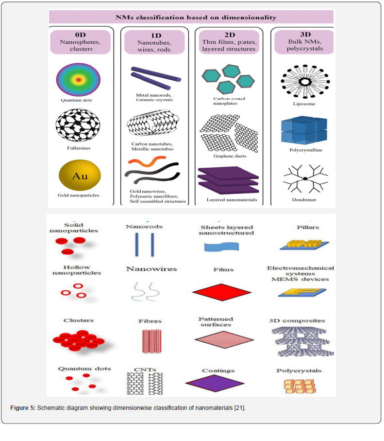
Nanomaterials are classified into four groups on the basis of
the number of dimensions [21] as shown in figure 5.
a) Zero Dimensional (0D)- all dimensions (x, y, z) are at
nanoscale i.e. no dimensions are greater than 100nm. Nanospheres
and nano-clusters are included in this group.
b) One Dimensional (1D)- In this case two dimensions (x,
y) are at nano-scale while the other one is outside the nano-scale.
For this reason, nanomaterials in this group lead to needle shaped.
This includes nanofibers, nanotubes, nanorods and nanowires.
c) Two Dimensional (2D)- In this group only one
dimension (x) is at nano-scale and the other two are outside the
nano-scale. As a result, 2D nanomaterials exhibit plate like shapes.
For example: nanofilms, nanolayers and nanocoating’s with
nanometer thickness.
d) Three Dimensional (3D)- The peculiarity of this group
nanomaterials are that those are not confined to the nanoscale
in any dimension, resulting which they possess three arbitrary
dimensions above 100nm. Thus, 3D nanomaterials have a multiple
arrangement of nano size crystals in different orientations. Nano
materials of this group include: dispersions of nanoparticles,
bundles of nano wires and nano tubes, polycrystals (multi nano
layers), etc.
Optical Probe with Gold Nanostar
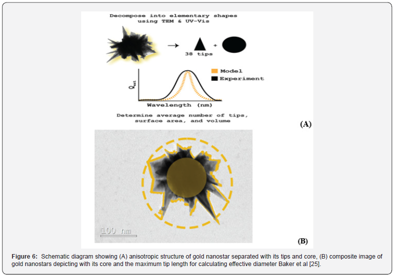
Gold nanostars, basically anisotropic nanostructures with sharp tips (Figure 6A, 6B), are promising candidates suitable for a wide range of applications in various fields (like free electron materials). Like free electron materials such as metals, “Plasmonics” a light-based technology has emerged in employed nano-materials so that in response to incoming light charge coherent oscillations occur (so called Plasmons). If such oscillations are limited (or control) to the metal-dielectric interface, then light intermingles with those particles which are smaller than its own wavelength, resulting which an oscillation of a local charge is produced around the particle. This is known as localized surface plasmon resonance (LSPR). This ability to confine light in nanoscale dimension, thus, makes the plasmonic nanoparticles (in the present case the gold nanostars) to possess various exceptional characteristics such as improved response in the electromagnetic field, rich spectral responses, high photothermal conversion efficiencies, etc. [25].
These special characteristics of gold nanostars are
extremely beneficial for various applications across all fields
like biochemistry, biomedicine, biosensing, nano catalysis,
computational sciences, solar energy technology, etc. Although
there are various existing plasmonic materials, silver (Ag) and
gold (Au) are still at present the most utilized materials. Note that
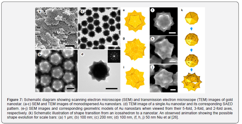
gold nanostars are most effective because (Figure 7) [26]:
a) ranging of the morphology of gold nanostars extends
from a simple 3-branched structure to multiple sharp branches;
b) with the increase of gold nanostar size, the number, the
core size4 and the branch’s aspect ratio increases;
c) with the increase in size, the gold nanostars turn more in
homogenous in shape but the overall size remains homogenous;
d) the properties of gold nanostars are affected by the size,
proximity and a number of the branches;
e) due to the optical properties such as localized surface
plasmon resonance (LSPR), surface enhanced Raman scattering
(SERC), catalystic properties gold nanostars become the extremely
effective probes;
f) hybridization of plasmons both at the cores and the tips
make the gold nanostars to possess intrinsic optical properties;
g) gold nanostar’s core plays an active role in making
electromagnetic field enhancement of the tips to act as an active
antenna. This means that the plasmon frequency and intensity of
the gold nanostars depends on the arrangement and the number
of spikes;
h) the morphology of spikes is controlled by different
fabrication conditions;
i) the gold nanostars exhibit its unique nature of LSPRs
which are related to collective oscillations of conduction electrons
during interaction of the metal with visible or near Infra-Red
(NIR) light. For example-relatively lower values of LSPRs around
600 nm implies the core of gold nanostar while higher LSPRs
values indicates the core-tip interactions.
j) the most abundant asymmetries gold nanostar will
appear when the aspect ratio in synthesis condition reaches to
750-1150 NIR.
k) The most important optical property of gold nanostar is
SERS activity.
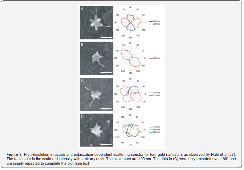
Experimentally Observed Scattering Spectra of Gold Nanostar
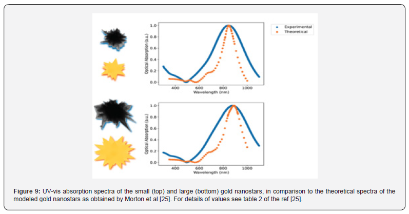
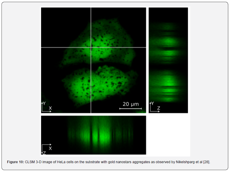
As mentioned above that the localized surface plasmon resonance (LSPR) has a wide application in biological and chemical sensing, biological imaging, nanoscale optical wave guides. On the other hand, shifts of the LSPR resonance to the near-infrared (NIR) enables the gold nanostars as a significant probe for diagnostic and therapaedic. Detailed characterization of the optical properties of complex, heterogeneous gold nanostars as observed by Nehl et al [27] are shown in (Figure 8-10). This treatment can be considered as a single particle spectroscopy. In fact, these images of nanoparticles were placed at the entrance of an imaging spectrometer so that thermoelectrically cooled CCD camera is able to take the shot [28].
Application of Gold Nanostars
Study of Living Cells Through Gold Nanostars
Living cells, in particular human cells, have a special attraction in biochemistry. So, visualizing the structures inside the living cells is essential now-a-days. Gold nanostars with its special characteristic, i.e. surface enhanced Raman Scattering (SERS) can be used as a valuable tool for studying the (i) tracking of chemical composition, reactions, their role in cell growth, internal cellular structures, organelles and the nucleus. Earlier studies [29-31] within a living cell hint the changes in protein conformation during mitosis [32], spatio-temporal changes in cells during different differentiations, etc. But SERS active gold nanostars can be used to investigate the change inside the cells under the influence of magnetic, optical, and other kinds of changes. On the other hand, combined labeled and non-labeled gold nanostars can be used to study the localization and molecular compositions of the plasma membranes and nuclei inside the cell, osmotic shocks, presence of toxic byproducts remaining after chemical synthesis, etc.
Analysis of the Spectra
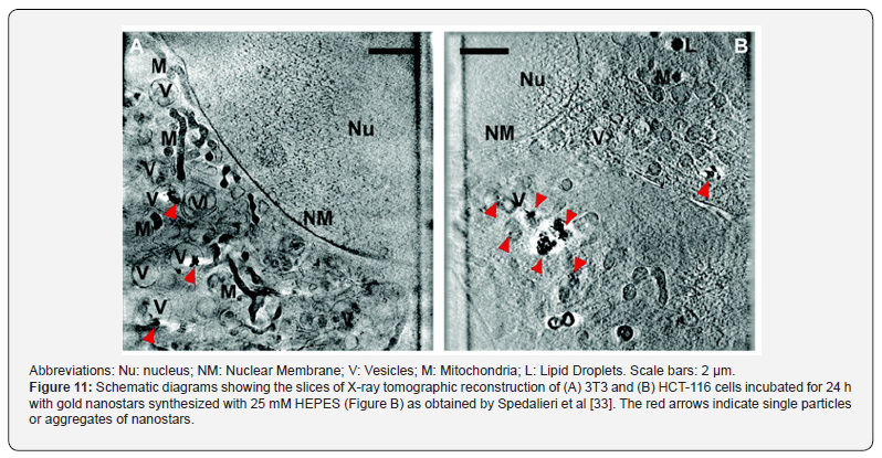
The typical HeLa cell is about 20μm diameter with a 10μm nucleus located in the center of a cell. The observed spectra collected from the center of a cell that can be considered as originated mainly from the nucleus whereas spectra collected from the regions surrounding the nucleus corresponds to the cytoplasm. Due to differences in gold nanostar tip length and sharpness some tips could penetrate a cell wall, some remain in plasma membrane and some do not contact cell. The number of tips with the probability of their penetration into the cell is greater in aggregates of nanostructures than in individual nanoparticles. Thus, the appearance of the peaks from the mitochondria, RNA, DNA in one spectrum may indicate that some spot enhanced gold nanostars signals implies the origin from the nucleus and some enhance signal from surrounding mitochondria which makes it possible to study them simultaneously (Figure 11).
Study of Cell Death by Gold Nanostars
As the cells are crucial for human health, cell death is related to various diseases as well as regulators for disease treatment. Gold nanostars (i.e. nano particles), upon entering the human body, first contact human cells in the blood, targeting organs and then the immune system. Gold nanostars, therefore, could be used in diagnosis and the immune system as well as disturbance of cell function and even cell death purposes.
Regarding cell death the major cell death modalities are (a) apoptosis (b) autophagy (c) necroptosis (d) aponecrosis (e) pyroptosis,and (f) necrosis. Cell death also plays another one important role in maintaining human good health. This cell death is a fundamental biological process which is essential for the development, growth and persistent of human body. Significantly, this contributes to tissue remodeling and the removal of excess cells during the development period. In other words, it can be said that it maintains homostasis by removing death cells. If not, then it poses a danger because they are infected, damaged or autoreactive. Apoptosis process is more significant than the others because it is used by multicellular organism to remove unwanted cells through dedicated, controlled both extrinsic and intrinsic pathways. Not only that apoptosis process occurs during body development, aging, etc. (Figure 11).
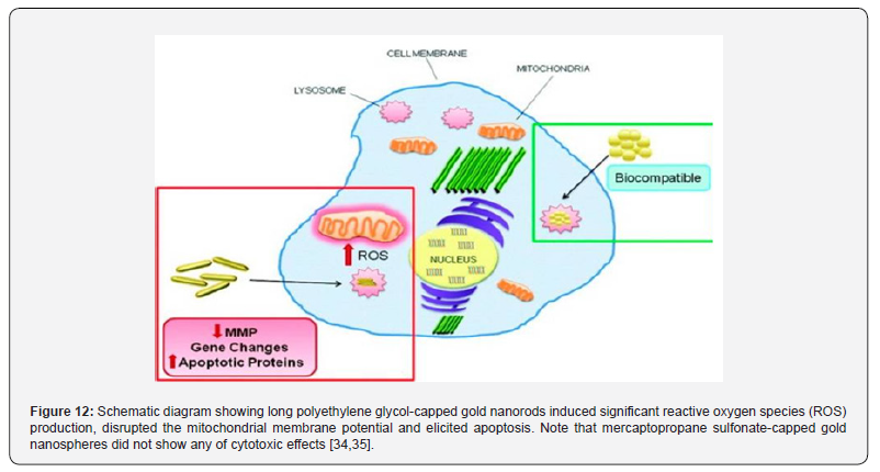
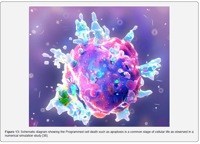
As the gold nanostars have a similar scale to biological nanoscale, thus it may affect them directly. Secondly, due to the stability, size tunable surface plasmon resonance, fluorescence and easy surface functionalization, gold nanostars could be applied to imaging diagnosis. Figure 12 shows how the gold nanostar’s shape affects the apoptosis level i.e. the higher-level reactive oxygen species (ROS). In this figure the apoptosis is induced by gold nanostars in the form of gold nanosphere with 20μm. Figure 13 represents the numerically simulated observed scenario that clearly shows the cell death (white) within a living cell i.e. gold nanostars induced a higher-level apoptosis, ROS generation and mitochondrial membrane depolarization [33-36].
Conclusion and unsolved issues on living cells
Cells, invisible to our eyes, are the fundamental units of human
life. It is known that all the cells of our body turn into tissues,
organs resulting which we are able to take breathe, think, more,
etc. i.e. perform our daily functions. Yet despite of our advanced
research and progress, much of the cell characteristics are remain
a mystery. For example:
a) Can it be possible to know how may i.e. total number of
cells are there in human body?
b) How healthy cells function at molecular level against
diseases?
c) How the shape and size of a cell can change over time? i.e.
how the growth and division of cells are regulated for maintaining
healthy phase?
d) How the new cell formation and removal of death cells
are maintained in human body? i.e. how cells replacement take
place in human body?
e) The root cause of a disease i.e. what changes are
happening to everyday cell processes when cells are affected in
a disease?
Gold nanostars provide itself as an advanced technological probe to the scientists for better understanding the unique characteristics of individual cells and how they function in human body. We are hopeful that scientists will be able to unlock the secret of cells in human body completely in near future.
References
- Berger E, Fong W, Chaornock R (2013) An r-process Kilonova associated with the short-hard GRB 130603B. Astrophys J Lett 774(2): L23.
- Siegel E (2015) The Cosmic origin story of gold.
- Bayda S, Adeel M, Tucciardi T, Cordani M, Rizzolio F (2019) The history of nanoscience and nanotechnology: From chemical-physical application to nanomedicine. Molecules 25(1): 112.
- Gnach A, Lipinski T, Bednorklewicz A, Rybka J, Capobianco JA (2015) Up Converting nano-particles: Assessing the toxicity. Chem Socy Rev 44(6): 1561-1584.
- Alivistos A P (1996) Semiconductor clusters, nanocrystals and quantum dots. Science 271(5251): 933-937.
- Rad AG, Abbasi H, Afzali MH (2011) Gold Nanoparticles: Synthesis, Characterizing and Reviewing novel application in recent years. Physics Procedia 22: 203-208.
- Xi Z, Zhang R, Kiess K, Fabian Z, Twan K, et al. (2024) Role of surface curvature in Gold Nanostar properties and applications. ACS Biomaterial Sci Engg 10(1): 38-50.
- Brill RH (1965) The chemistry of the Lycurgus Cup in Proc. 7th Int Cong Glass Bruxelles, Section B, 223(1).
- Barbar DJ, Freestone IC (1990) An investigation of the origin of the colour of the Lycurgus. Archaeometry 32(1): 33-45.
- Hornyak GL, Patrissi CJ, Oberhauser EB (1997) The Use of Metal Nanoparticles to Produce Yellow, Red and Iridescent Colour, from Bronze Age to Present Times in Lustre Pottery and Glass: Solid State Chemistry, Spectroscopy and Nanostructure. Nano Struc Matter 9: 571.
- Kokarneswaran M, Selvarej P, Ashokan T, Perumal S, Sellappan P, et al (2020) Discovery of Carbon nanotubes in 6th century BC Potteries from Keeladi, India. Scientific Reports Nature 10: 19786.
- Arantegui JP, Molera J, Larrea A, Pradell T, Saz MV, et al. (2004) Luster pottery from the thirteenth century to the sixteenth century: a nanostructured thin metallic film. J Am Ceram Soc 84(2): 442-446.
- Padovani S, Sada C, Mazzoldi P, Brunetti B, Borgia I, et al. (2003) Copper in glazes of Renaissance luster pottery: Nanoparticles, ions, and local environment. J Appl Phys 93: 10058-10063.
- Arnold DE (2005) Mayan Blue and Polygorskite: a second possible pre-Columbian source. Ancient Mesoamerica 16(1): 51-62.
- Heiligtag FJ, Niederberger M (2013) The fascinating world of nanoparticle research. Matter Today 16(7-8): 262-271.
- Sudha P N, Sangeetha K, Vijayalakshmi K, Barhoum A (2018) Nanomaterials history, classification, unique properties, production and model. Chapter 12 in Engineering application of Nanoparticles and Architectural Nanostructures. Elsevier, pp: 341-376.
- Kumar N, Kumbhat S (2010) Essentials in nano science and nano technology. (In: 1st edition), Wiley, USA.
- Feynman R P (1960) There’s plenty of room at the bottom. Engg Sci 23: 22-36.
- Taniguchi N (1974) On the basic concept of Nano-technology. In: Proc Int Conf Prod Engg Part II, Japan Society of Precision Engineering, Tokyo, Japan.
- Rafique M, Tahir MB, Rafique MS, Hamza M (2020) History and Fundamentals of Nanoscience and nano technology, in Nano Technology and Photo-catalysis for Environmental application. Elsevier Inc, pp. 1-25.
- Faraday M (1857) The Bakenian Lecture: Experimental relations of gold (and other materials) to light. Philos Trans Roy Socy Lond 147: 145-181.
- History and Scope, Classification of nanostructured materials. PSCMR college of Engg Tech, Andhra Pradesh, India.
- Xu X, Ray R, Gu Y, et al. (2004) Electrophoretic analysis and purification of fluorescent single walled carbon nanotube fragments. J Am Chem Socy 126: 12736-12737.
- Baker SN, Baker GA (2010) Luminescent carbon nanodots: Emergente nanolights. Angew Chem Socy Inst Ed England 49(38): 6726-6744.
- Morton W, Joyce C, Taylor J, Ryan M, Uberti SA, et al (2023) Modeling Au Nanostar Geometry in Bulk Solutions. J Phys Chem C 127(3): 1680-1686.
- Niu W, Chua YA, Zhang W, Huang H, Lu X (2015) Highly symmetric Gold Nanostars: Crystallographic control and surface enhanced Raman Scattering property. J Am Chem Socy 137(33):10460-10463.
- Nehl C, Liao H, Hafner JH (2006) Optical properties of star-shaped gold nanoparticles. Nano Letters 6(4): 683-688.
- Nikelshparg EI, Prikhozhdenko ES, Verkhovskii RA, Atkin VS, Khanadeev VA, et al. (2021) Live cell Poration by Au Nanostars to probe intracellular molecular composition with SERS. Nanomaterials 11(10): 2588.
- Schlücker S (2009) SERS Microscopy: Nanoparticle Probes and Biomedical Applications. Chem Phys Chem 10(9-10): 1344-1354.
- Kneipp J, Kneipp H, McLaughlin M, Brown D, Kneipp K (2006) In Vivo Molecular Probing of Cellular Compartments with Gold Nanoparticles and Nanoaggregates. Nano Lett 6(10): 2225-2231.
- Yashchenok A, Masic A, Gorin D, Shim BS, Kotov NA, et al. (2013) Nanoengineered Colloidal Probes for Raman-based Detection of Biomolecules inside Living Cells. Small 9(3): 351-356.
- Garcia VC, Strobbia P, Crawford BM, Wang H, Ngo H, et al. (2021) Plasmonic nanoplatforms: From surface-enhanced Raman scattering sensing to biomedical applications. J Raman Spectrosc 52(2): 541-553.
- Panikkanvalappil SR, Hira SM, Mahmoud MA, Sayed MAE (2014) Unraveling the Biomolecular Snapshots of Mitosis in Healthy and Cancer Cells Using Plasmonically-Enhanced Raman Spectroscopy. J Am Chem Soc 136(45): 15961-15968.
- Sun H, Jia J, Jiang C, Zhai S (2018) Gold nanoparticle-induced cell death and potential application in nanomedicine. Int J Mol Sci 19(3): 754.
- Schaeublin NM, Braydich-Stolle LK, Maurer EI, Park K, MacCuspie RI, et al. (2012) Does shape matter? Bioeffects of gold nanoparticles in a human skin cell model. Langmuir 28(6): 3248-3258.
- Magri Z (2023) Cell death is essential to your health-an immunologist explains when cells decide to die with a bang or take their quiet leave.


