




 AJPN Home



Abstract
Background: Anomalous left coronary artery arising from the pulmonary artery (ALCAPA) is a rare coronary artery anomaly that requires surgical repair.
Case Presentation: We report a successful stent implantation for stenosis of the left main coronary artery (LMCA) following ALCAPA surgery in a 15-year-old boy. The patient remained asymptomatic during follow-up.
Conclusions: Serial echocardiography and other cardiac imaging modalities are required after coronary reimplantation to assess outcomes and detect possible postoperative complications. Coronary stent implantation may be an effective long-term treatment for coronary artery stenosis in selected cases, including young patients.
Keywords: Anomalous Coronary Artery; Coronary Stenosis; ALCAPA; Coronary Stent; Case Report
Abbreviations: ALCAPA: Anomalous Left Coronary Artery originating from the Pulmonary Artery; CT Angiography: Computed Tomography Angiography; DOE: Dyspnea on Exertion; ECG: Electrocardiography; LAD: Left Anterior Descending; LMCA: Left Main Coronary Artery; LV: Left Ventricle; MR: Mitral Regurgitation
Introduction
There is a rare cardiac anomaly known as ALCAPA (anomalous origin of the left coronary artery from the pulmonary artery), which affects approximately one in every 300,000 live births [1-3] and accounts for nearly 0.2% of all congenital heart defects [4]. With the decrease in pulmonary vascular resistance, ALCAPA can lead to myocardial ischemia and/or infarction, as well as varying degrees of mitral regurgitation (MR), global left ventricular dysfunction, and annular dilatation. Clinical presentation depends on the extent of coronary collateral development [4]. The early mortality rate may exceed 90% in the absence of timely diagnosis and treatment [5]. Although ALCAPA is typically diagnosed in infancy, it is extremely rare in older children and adolescents [3]. In our case, the patient was diagnosed with ALCAPA at the age of 10 years and remained asymptomatic or minimally symptomatic until diagnosis. After corrective surgery for ALCAPA, stenosis of the left main coronary artery (LMCA) developed.
Case Presentation
We report a 15-year-old boy with a history of ALCAPA who underwent direct reimplantation of the anomalous left coronary artery into the aorta, which later resulted in stenosis at the ostium of the left main coronary artery (LMCA). On clinical evaluation, the electrocardiogram was normal, and the patient reported no chest discomfort during exercise. At 10 years of age, the patient presented with syncope and exertional dyspnea, which prompted cardiac evaluation. Echocardiography revealed reduced left ventricular contractility, enlargement of the left atrium and left ventricle, mitral regurgitation, and a dilated right coronary artery with abnormal flow into the pulmonary artery, findings consistent with ALCAPA. The diagnosis was confirmed by CT angiography. The patient subsequently underwent coronary reimplantation surgery. Approximately five years later, follow-up echocardiography demonstrated stenosis of the left main coronary artery (1.5 mm) as well as ectasia of the left anterior descending artery (LAD) and left circumflex artery (LCx), measuring up to 7 mm. CT angiography confirmed LMCA stenosis (Figure 1).
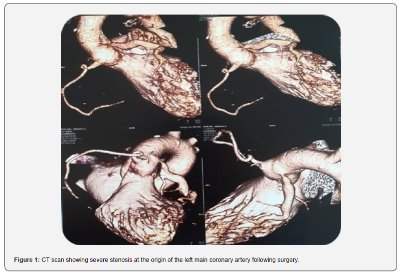
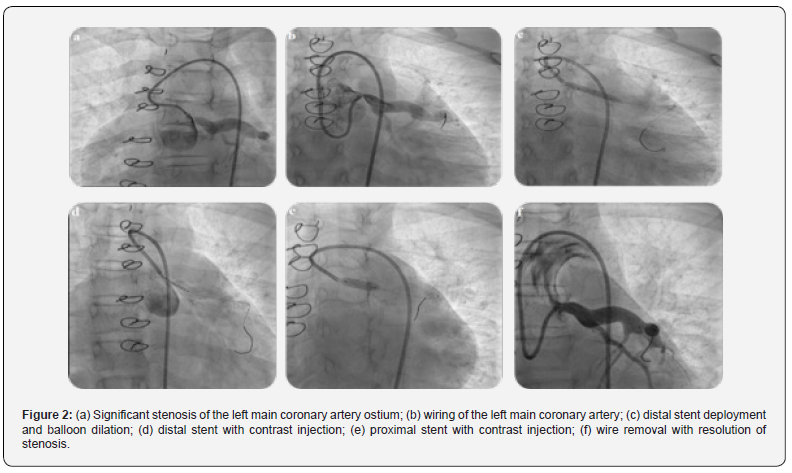
The patient also reported reduced exercise capacity and exertional chest discomfort. Exercise stress testing was not performed due to significant LMCA stenosis. Interventional angiography was performed in the pediatric cardiac catheterization laboratory as part of the treatment plan. The procedure was carried out under general anesthesia via femoral arterial and venous access. An ascending aortic root angiogram in two projections was obtained to assess the ostial LMCA stenosis. A BMW guidewire and a 6F XB 3.5 guiding catheter were used to cross the lesion. Two stents were deployed in the LMCA: a Xience Xpedition 3.5 × 15 mm stent in the distal segment and a Cre8 4.5 × 12 mm stent in the proximal segment, with distal overlap. Post-dilatation was performed using a Sapphire NC 5.0 × 12 mm balloon. The proximal stent slightly protruded (1-2mm) into the aorta. Final angiography confirmed optimal stent position and normal coronary flow. The diagnostic and interventional steps are shown in Figure 2 (A-F). The procedure was completed without complications, and dual antiplatelet therapy with acetylsalicylic acid and clopidogrel was initiated. At one-year follow-up, the patient remained asymptomatic, with no chest pain or exertional dyspnea. Electrocardiography was normal, and no abnormalities were noted on clinical evaluation.
Discussion
Infants with ALCAPA, a rare coronary artery anomaly, may present with myocardial ischemia, myocardial infarction, and heart failure. However, in some cases, the condition may remain asymptomatic during infancy due to antegrade flow of desaturated blood from the pulmonary artery into the left coronary artery and the development of intercoronary collaterals between the right and left coronary arteries. Several surgical techniques have been described for the treatment of ALCAPA, including left subclavian artery–to–coronary artery anastomosis, coronary artery bypass grafting, intrapulmonary baffle reconstruction (the Takeuchi procedure), and direct reimplantation of the anomalous coronary artery into the aorta [2]. Among these, direct coronary reimplantation into the aorta is considered the most physiological and definitive surgical repair for restoring a two-coronary artery system [6]. As demonstrated in our case, some patients may develop left main coronary artery (LMCA) stenosis following surgical repair [7].
Coronary artery stenosis in children is rare. In the pediatric population, etiologies include postoperative coronary stenosis following ALCAPA repair, primary ostial stenosis (isolated or associated with systemic vasculitis such as Takayasu arteritis or Kawasaki disease), and metabolic disorders such as familial hyperlipidemia [8,9]. Although the exact mechanism of postoperative coronary obstruction after ALCAPA repair remains unclear, it has been suggested that mechanical traction on the reimplanted LMCA and disruption of the vasa vasorum during surgery may contribute to its development [10]. Therefore, serial cardiac evaluation is essential following coronary revascularization surgery. In cases of coronary obstruction after reimplantation, percutaneous coronary intervention may offer an alternative to repeat surgery; however, its long-term durability may be limited in lesions resulting from surgical manipulation [11]. Close collaboration between pediatric and adult interventional cardiology teams is crucial for optimal management. In infants and children who develop coronary obstruction after surgical reimplantation for ALCAPA, percutaneous coronary intervention represents a feasible therapeutic option.
Introduction
7. Conclusions
Serial echocardiography and other cardiac imaging modalities are required following coronary reimplantation to assess outcomes and detect potential complications of cardiac surgery. Coronary stent implantation may, in selected cases, be an effective long-term treatment for coronary artery stenosis, even in very young children.
Acknowledgment
The authors are grateful to the patient and his parents for their participation in this study.
Data Availability
The data supporting the findings of this case report are available from the corresponding author upon reasonable request.
References
- Chattranukulchai P, Namchaisiri J, Tumkosit M, Puwanant S, Vorasettakarnkij Y, et al. (2018) Very late presentation of anomalous origin of the left coronary artery from the pulmonary artery: case report. J Cardiothorac Surg 13(1): 70.
- Dodge-Khatami A, Mavroudis C, Backer CL (2002) Anomalous origin of the left coronary artery from the pulmonary artery: collective review of surgical therapy. Ann Thorac Surg 74(3): 946-955.
- Peña E, Nguyen ET, Merchant N, Dennie C (2009) ALCAPA syndrome: not just a pediatric disease. Radiographics 29(2): 553-565.
- Gao Y, Zhang J, Huang GY, Liang XC, Jia B, et al. (2017) Surgical outcomes of anomalous origin of the left coronary artery from the pulmonary artery in children: an echocardiography follow-up. Chin Med J (Engl) 130(19): 2333-2338.
- Wesselhoeft H, Fawcett JS, Johnson AL (1968) Anomalous origin of the left coronary artery from the pulmonary trunk: its clinical spectrum, pathology, and pathophysiology, based on a review of 140 cases with seven further cases. Circulation 38(2): 403-425.
- Dionne PO, Poirier N, Forcillo J, Stevens LM, Chartrand-Lefebvre C, et al. (2013) A rare case of anomalous origin of the left main coronary artery in an adult patient. J Cardiothorac Surg 8: 15.
- Schwerzmann M, Salehian O, Elliott T, Merchant N, Siu SC, et al. (2004) Images in cardiovascular medicine: Anomalous origin of the left coronary artery from the main pulmonary artery in adults: coronary collateralization at its best. Circulation 110(21): e511-e513.
- Bratincsák A, Salkini A, El-Said HG, Moore JW (2012) Percutaneous stent implantation into coronary arteries in infants. Catheter Cardiovasc Interv 79(2): 303-311.
- Viola N, Alghamdi AA, Al-Radi OO, Coles JG, Van Arsdell GS, et al. (2010) Midterm outcomes of myocardial revascularization in children. J Thorac Cardiovasc Surg 139(2): 333-338.
- Huddleston CB, Balzer DT, Mendeloff EN (2001) Repair of anomalous left main coronary artery arising from the pulmonary artery in infants: long-term impact on the mitral valve. Ann Thorac Surg 71(6): 1985-1988.
- Lardhi AA (2010) Anomalous origin of left coronary artery from pulmonary artery: a rare cause of myocardial infarction in children. J Family Community Med 17(3): 113-116.

























