




 AJPN Home



Abstract
Objective: To determine if infants diagnosed with retinopathy of prematurity (ROP) are associated with an increased risk of developing epilepsy and if the severity of ROP is a factor.
Design: Utilizing a large electronic health record database, TriNetX, a retrospective cohort study is performed querying pediatric patients (≤18 years of age) between November 1, 2003, to November 1, 2025.
Subjects: Participants included pediatric patients who had ICD-10 code Z05: encounter for observation and evaluation of newborns for suspected diseases and conditions ruled out. Patients were put into either a ROP positive or ROP naïve cohort. A further sub analysis queried ROP positive patient into low severity (stages 0, 1, 2) ROP or moderate to high severity (stages 3, 4, 5) ROP.
Methods: Propensity score matching was utilized to control covariates before outcomes analysis between cohorts. The main outcome was a diagnosis of epilepsy measured with relative risk and risk difference between cohorts.
Results: The ROP positive cohort displayed a higher risk of developing epilepsy compared to matched controls (3.2% vs. 1.7%; relative risk ratio = 1.85 [95% CI 1.34-2.53], p<0.001). ROP severity did not significantly correlate to an increased risk of epilepsy development.
Conclusions: Our findings indicate that children with a documented history of ROP have a higher risk of developing epilepsy compared to those without ROP. This demonstrates that pediatric patients with diagnosed ROP might warrant more frequent epilepsy screenings during development. Future studies warrant investigating a clearer temporal and causal association between ROP and epilepsy development.
Keywords:ROP; Epilepsy; Convulsions; Pediatric; Real-World Dataset; Database
Abbreviations:ROP: Retinopathy of Prematurity; ICD-10: International Classification of Diseases, 10th Edition; PSM: Propensity Score Matching; UE: Upper Extremity; DD; Diaper Dermatitis
Introduction
Roughly 1 out of 150 children are diagnosed with epilepsy during the first 10 years of life [1]. However, only children considered “high-risk” are recommended to have screening and surveillance for epilepsy [2]. With more than 42-50% of children developing recurrence of seizures after an initial unprovoked seizure within 8 years of life and about 41% of children developing epilepsy by the age of 3 with a history of a neonatal seizure, it is crucial to be able to appropriately stratify those at higher risk for earlier surveillance and management [2-4]. Retinopathy of prematurity (ROP) is a proliferative retinal vascular disorder that affects infants born less than 32 weeks’ gestation. At the time of premature birth (<37 weeks’ gestation), the retina is not yet fully vascularized, and the change of environment causes suspension in the growth of those vessels.
Over time, the hypoxic signals from the halted blood vessels induce angiogenic factors that cause abnormal proliferation of the blood vessels, and ultimately permanent vision loss if not managed properly [5]. Many studies over the past decade have concluded that preterm infants with ROP are at higher risk of neurodevelopmental disabilities [6-10]. Some of this is plausible since neurodevelopmental disabilities are attributed to preterm birth [11]. However, no study thus far has specifically looked at the independent developmental risk of epilepsy within the pediatric population with a history of ROP. This study aims to address this gap and utilize a large electronic health record database to investigate if ROP is independently associated with increased risk of epilepsy development, and whether the severity of ROP further exacerbates that risk.
Methods
A retrospective cohort study was conducted using the TriNetX Research Network, comprised of de-identified patient data from 110 healthcare organizations. Due to the de-identified nature of the electronic health records within TriNetX, local IRB exemption was acquired from the University of New Mexico (IRB# 26-004).
Cohort Construction
The TriNetX database was queried using primary International Classification of Diseases, 10th Edition (ICD-10). Detailed breakdown of the ICD-10 codes used in cohort creation as well as propensity score matching can be found in eTables 6 and 7 in the Supplement. The primary goal of the study was to analyze the risk of developing epilepsy in patients with ROP compared to those without. Pediatric patients (≤18 years of age) were queried between November 1, 2003 to November 1, 2025 that had the ICD-10 code Z05, encounter for observation and evaluation of newborn for suspected diseases and conditions ruled out.
This code was used to make sure that the comparison occurred between infants that were definitively followed from as close to the time of birth as possible. The primary analysis created two cohorts with and without the diagnostic code for ROP (H35.1), for the ROP and No ROP cohorts, respectively. The main outcome analyzed was the risk of epilepsy (G40) development in each cohort. The risk of convulsions (R56) was included to further support the validity of the epilepsy developmental risk outcome. A secondary sub-analysis was performed delineating two cohorts based on severity of ROP. A low-grade ROP cohort was indicated by stages 0, 1, and 2, while the moderate-high grade ROP cohort was defined as ROP stages 3, 4, 5 (eTable 6).
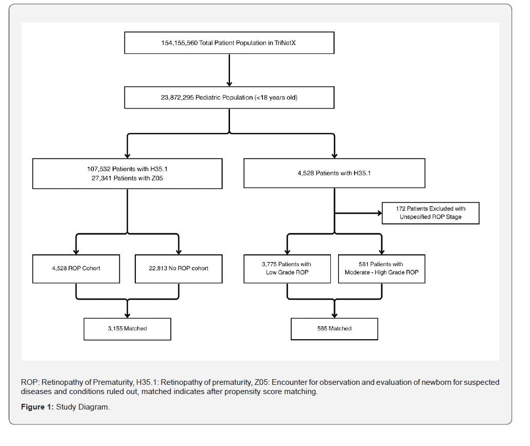
Other parameters remained the same as the primary analysis. Propensity score matching (PSM) was performed for all analyzed cohorts. PSM used baseline demographics and key neonatal covariates including gestational age, birthweight, and major comorbid conditions (i.e intracranial hemorrhage, cerebral palsy, sepsis, hypoxic-ischemic encephalopathy, and neonatal cerebral leukomalacia). Detailed breakdown of all covariates used for propensity matching can be found in eTable 7 in the Supplement. Flowchart indicating creation of study population is shown in Figure 1.
It is important to disclose that TriNetX has built-in restrictions on using specific pediatric ICD-10 diagnostic codes during cohort construction due to patient privacy, such as low birth weight, preterm gestation, and hypoxic-ischemic encephalopathy. Therefore, these codes were unable to be used during cohort construction for purposes of inclusion or exclusion criteria. However, the usage of these terms has no restrictions for the purposes of propensity score matching. eTable 1 (ROP vs No ROP), eTable 4 (Low Grade ROP vs Moderate-High Grade ROP), and eTable 5 (Negative Control Exposure) showcase the before and after PSM breakdown.
Statistical Analysis
All statistical analysis was done within the TriNetX platform utilizing their in-house analysis tools. Measures of association analysis were performed for all cohort comparisons with the selected option of excluding patients who had the outcome prior to the designated time window. PSM analysis was performed with a 1:1 greedy matching algorithm with a caliper of 0.25 times the pooled standard deviations. A standardized mean difference of less than or equal to 0.1 indicated a successful propensity matching process.
Negative Control Analysis
Two forms of negative control analysis were performed to ensure that any results were controlled for any residual bias present. First, negative control exposure was performed by creating a separate cohort comparing pediatric patients with and without diaper dermatitis. Diaper dermatitis was chosen as an exposure control as it is common, well-coded, and biologically unrelated to epilepsy development. All other parameters and propensity score matching was identical to the ones performed for the primary ROP analysis (eTable 5). Secondly, a negative control outcome analysis was performed for the primary ROP vs No ROP cohorts using common pediatric upper extremity injuries as the measured outcome (eTable 7). These were chosen as a common, trauma-based, and non-biologically linked outcome to ROP history. Negative controls were prespecified to evaluate whether the analysis produced false associations.
Results
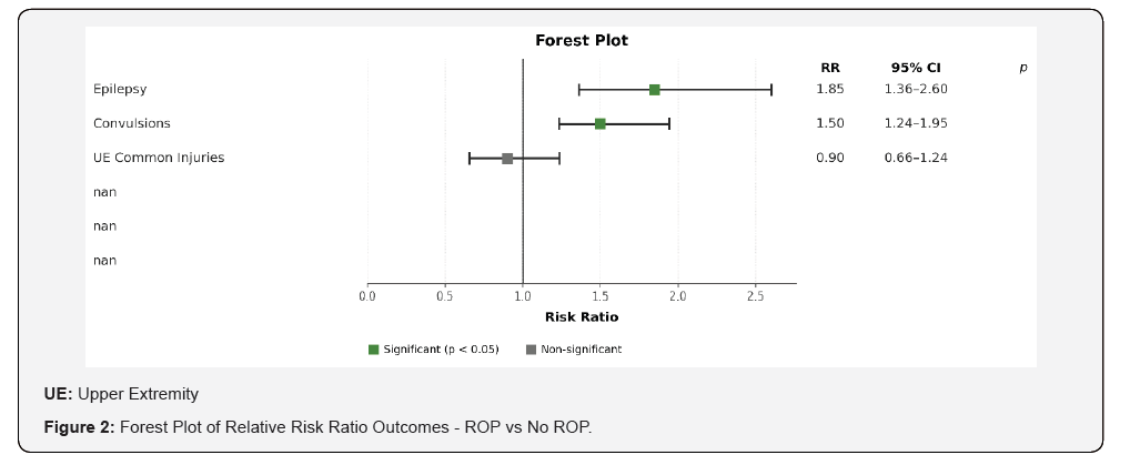
A combined population of 27,341 individuals (ROP: n=4,528; No ROP: n=22,813) were extracted from the TriNetX database. After PSM, 3,155 patients were allocated in both the ROP cohort (46.9% female; mean age 4.5 ± 2.7 years) and No ROP cohort (45.9% female; mean age 4.5 ± 2.8 years). Further baseline characteristics of ROP and No ROP cohorts are displayed in eTable 1. The ROP cohort showcased a nearly two-fold increase of developing epilepsy when compared to the No ROP cohort (Risk Difference 1.6%; 95% CI [0.8%-2.3%]) (Table 1). The ROP cohort had a relative risk of 3.3% of epilepsy development while the No ROP cohort had a relative risk of 1.8%, resulting in a relative risk ratio of 1.85 (95% CI [1.363, 2.601]) (Figure 2). Similarly, the ROP cohort also displayed a significantly higher risk of convulsions compared to the No ROP cohort (Risk Difference 2.1%; 95% CI [1.0%-3.2%) (Table 1). The ROP cohort had a significant relative risk ratio of 1.55 (95% CI [1.235, 1.945]) for the outcome of convulsions (Figure 2).
Secondary analyses revealed no statistically significant difference between PSM low grade ROP cohort compared to a moderate-high grade ROP in epilepsy developmental risk (Risk Difference 1.1%; 95% CI [-1.9% - 4.1%]) (Table 2). Convulsion risk also shared no significant difference between the low grade and moderate to high grade ROP cohorts (Risk Difference 0.7%; 95% CI [-3.4%-4.9%]) (Table 2). Two forms of negative control analysis were performed. Negative control exposure analysis utilizing diaper dermatitis showcased no significant difference in epilepsy developmental risk (Risk Difference -0.2%; 95% CI [-1.2%-0.6%) (eTable 2). Negative control outcome analysis utilizing common upper extremity injuries displayed no significant difference in epilepsy developmental risk (Risk Difference -0.2%; 95% CI [-1.0%-0.5%]) (eTable 3). Similarly, the relative risk ratio for common upper extremity injuries was 0.902 (95% CI [0.657, 1.238]) as shown in Figure 2.
Discussions
This study showed that infants who had a history of ROP had a significantly higher risk of developing both epilepsy and convulsions. ROP and the development of epilepsy share multiple common risk factors such as preterm birth, low birth weight, and intraventricular hemorrhage [9,12-16]. Looking at the risk of ROP in preterm infants, a multicenter study showcased approximately 43.1% of screened preterm infants developed ROP [17]. However, a Finnish national cohort study found that the incidence of epilepsy was 2.53% in very preterm infants (<32 weeks’ gestation), with a lower incidence in moderately preterm infants (1.08%, 32-33 weeks’ gestation) [16]. Given that ROP demonstrates a significantly greater prevalence among preterm infants relative to epilepsy, a diagnosis of ROP may function as a useful proxy marker to identify infants who warrant epilepsy screening.

ROP: Retinopathy of Prematurity; Percent populations were rounded to nearest tenths.

ROP: Retinopathy of Prematurity; Percent populations were rounded to nearest tenths.
Delayed or missed diagnosis of epilepsy in infants can lead to persistent and worsening neurodevelopmental outcomes. Studies have shown that a delay of diagnosis by even one month can lead to a significant drop in both motor and cognitive scores, which can persist and worsen in later childhood [18,19]. Therefore, any stratification of infants at birth into a higher epilepsy risk subgroup may result in an improvement in diagnosis and, consequently, optimize the child’s neurodevelopment later in life. Indeed, this analysis showed a 1.85-fold increase in the risk of developing epilepsy when a child had a historical diagnosis of ROP and thus should be considered when counseling families about future potential medical issues. The subgroup analysis of severity of ROP delineated between lower grade ROP and moderate-high grade ROP. Due to the fact one of the limitations of this study was the oxygen supplementation received by children during infancy, this subgroup analysis was used to both quantify if increased severity had any link to increased epilepsy risk, as well as controlling for possible increased oxygen supplementation.
This was important to include because increased supplementation of oxygen is known to increase the risk of developing more severe stages of ROP [5]. Interestingly, a difference in the low-grade ROP and moderate-high grade ROP was not observed. Most likely the lack of support for increased epilepsy risk correlated to ROP severity is hypothesized to be influenced by the small sample size within each cohort. The moderate-high risk ROP cohort only contained 581 patients before matching which immediately limited the available number of infants that could have obtained an epilepsy diagnosis. However, further studies should be performed to further delineate if severity of ROP is a factor that affects the likelihood of predicting epilepsy development in preterm infants.
Negative control exposure analysis used diaper dermatitis as a diagnostic exposure group with no plausible biological link to epilepsy development to rule out any residual exposure bias within the cohorts. Analysis revealed no statistical difference between the cohorts. Negative control outcome analysis was also completed with the outcome of common pediatric upper extremity injuries, as they are common and should not have a direct causal relationship with epilepsy development. The common pediatric injuries included fractures, dislocations, and superficial injuries of the wrist, hand, fingers, forearm, elbow, and shoulder girdle [20].
Study Limitations
The retrospective nature of the study limits the ability to create a causal relationship between the development of epilepsy and ROP. It is important to acknowledge that the restrictions of TriNetX on the usage of specific ICD-10 codes limit the ability of the study to be able to further delineate and/or exclude patients with definitive and pre-established links to epilepsy.
Since the PSM process was performed through TriNetX’s inhouse statistical software, there were limitations to the depth of confounders that could be controlled for using only the ICD- 10 codes. Oxygen supplementation is a common requirement for preterm infants due to their underdeveloped respiratory systems. However, the level of oxygen supplementation is also a direct risk factor for ROP development and exacerbation [5]. TriNetX has no way of quantifying supplemental oxygen therapy and therefore could not be included as a confounding factor.
Conclusion
Overall, studies have shown that infants with a diagnosis of ROP have an increased risk of neurodevelopmental outcomes, but no studies have yet quantified an individual risk of epilepsy within this population. This study aimed to use a large, electronic health record database to observe infants using an observational ICD-10 code (Z05) and to compare ROP and No ROP cohorts to determine the individual risk of epilepsy development with appropriate propensity score matching. Infants with a diagnosis of ROP had a 1.85-fold increase in the risk of developing epilepsy when compared to the no ROP cohort.
Subgroup analysis showed no statistical significance between the severity of ROP history and the induvial risk of developing epilepsy. This study emphasizes that the history of ROP may be clinically useful in further stratifying infants at high risk for epilepsy to allow for more specific monitoring. Additionally, this information can be used during the newborn period to inform those at higher risk of developing epilepsy later in childhood. Further research is needed to investigate the biological mechanism that could potentially link ROP and epilepsy or seizure disorder development.
Credit:
HUZEFA Y. SARIA, Writing – review and editing, Writing – original draft, Formal analysis, Investigation, Conceptualization; JESSIE R. MAXWELL, Writing – review and editing, Validation, Supervision./p>
References
- Kari Modalsli Aaberg, Nina Gunnes, Inger Johanne Bakken, Camilla Lund Søraas, Aleksander Berntsen, et al. (2017) Incidence and Prevalence of Childhood Epilepsy: A Nationwide Cohort Study. Pediatrics 139(5): e20163908.
- Epilepsies I of M (US) C on the PHD of the, England MJ, Liverman CT, Schultz AM, Strawbridge LM (2025) Next Steps and Recommendations. In: Epilepsy Across the Spectrum: Promoting Health and Understanding. National Academies Press (US); 2012.
- Altınbezer P, Çolak R, Çalkavur Ş, Yılmaz Ü (2023) Epilepsy Frequency and Risk Factors Three Years After Neonatal Seizures. Pediatr Neurol 149: 120-126.
- Guerrini R (2006) Epilepsy in children. Lancet Lond Engl 367(9509): 499-524.
- Retinopathy of Prematurity (2025) A Global Perspective and Recent Developments. Pediatrics 150(3): e2021053924.
- Nagamani Beligere, Vijayalaksmi Perumalswamy, Manish Tandon, Amit Mittal, Jayasheele Floora, et al. (2015) Retinopathy of prematurity and neurodevelopmental disabilities in premature infants. Semin Fetal Neonatal Med ;20(5): 346-353.
- Hellström A, Smith LEH, Dammann O (2013) Retinopathy of prematurity. Lancet Lond Engl 382(9902): 1445-1457.
- Daniela Ricci, Simona Lucibello, Lorenzo Orazi, Francesca Gallini, Susanna Staccioli, et al. (2020) Early visual and neuro-development in preterm infants with and without retinopathy. Early Hum Dev 148: 105134.
- Fang L, Wang C, Yang Y, Feng J, Chen F (2023) A meta-analysis of adverse effects of retinopathy of prematurity on neurodevelopment in preterm infants. Medicine (Baltimore) 102(50): e36557.
- Tan H, Blasco P, Lewis T, Ostmo S, Chiang MF, Campbell JP (2021) Neurodevelopmental outcomes in preterm infants with retinopathy of prematurity. Surv Ophthalmol 66(5): 877-891.
- Inder TE, Volpe JJ, Anderson PJ (2023) Defining the Neurologic Consequences of Preterm Birth. N Engl J Med 389(5): 441-453.
- Chang JW (2019) Risk factor analysis for the development and progression of retinopathy of prematurity. PloS One 14(7): e0219934.
- Bahmani T, Karimi A, Rezaei N, Daliri S (2022) Retinopathy prematurity: a systematic review and meta-analysis study based on neonatal and maternal risk factors. J Matern-Fetal Neonatal Med Off J Eur Assoc Perinat Med Fed Asia Ocean Perinat Soc Int Soc Perinat Obstet 35(25): 8032-8050.
- Chou IC, Sung FC, Hong SY (2020) Incidence of epilepsy in children born prematurely and small for gestational age at term gestation: A population-based cohort study. J Paediatr Child Health 56(2): 324-329.
- Tu YF, Wang ST, Shih HI, Wu PM, Yu WH, et al. (2019) Epilepsy occurrence after neonatal morbidities in very preterm infants. Epilepsia 60(10): 2086-2094.
- Mikko Hirvonen, Riitta Ojala, Päivi Korhonen, Paula Haataja, Kai Eriksson, et al. (2017) The incidence and risk factors of epilepsy in children born preterm: A nationwide register study. Epilepsy Res 138: 32-38.
- Graham E Quinn, Gui-Shuang Ying, Edward F Bell, Pamela K Donohue, David Morrison, et al. (2018) Incidence and Early Course of Retinopathy of Prematurity: Secondary Analysis of the Postnatal Growth and Retinopathy of Prematurity (G-ROP) Study. JAMA Ophthalmol 136(12): 1383-1389.
- Helen O'Reilly, Christin Eltze, Kate Bennett, Kristien Verhaert, Rebecca Webb, et al. (2018) Cognitive outcomes following epilepsy in infancy: A longitudinal community-based study. Epilepsia 59(12): 2240-2248.
- Berg AT, Loddenkemper T, Baca CB (2014) Diagnostic delays in children with early onset epilepsy: impact, reasons, and opportunities to improve care. Epilepsia 55(1): 123-132.
- Watkins RA, De Borja C, Ramirez F (2022) Common Upper Extremity Injuries in Pediatric Athletes. Curr Rev Musculoskelet Med 15(6): 465-473.

























