Intraoral Vesicular Lesions in Neonate
Chelsea M Matthia*, Kendall R Steadmon and Thao N Vu
Department of Pediatrics, University of Florida College of Medicine, Gainesville, USA
Submission: October 10, 2021; Published: December 15 2021
*Corresponding author:Chelsea M. Matthia, Department of Pediatrics, University of Florida, Gainesville, USA
How to cite this article:Chelsea M M, Kendall R S, Thao N. Intraoral Vesicular Lesions in Neonate. Acad J Ped Neonatol 2021; 11(2): 555863. 10.19080/AJPN.2021.11.555863
Abstract
Intraoral sebaceous hyperplasia is an uncommon yet benign finding in neonates. Familiarity with this process can prevent unwarranted procedures and infectious workup.
Case Report
A full-term female infant was born to a 31-year-old G2P2002 mother via spontaneous vaginal delivery. Rupture of membranes showed clear amniotic fluid and occurred two hours prior to delivery. Delivery was otherwise uncomplicated. The mother received adequate prophylaxis with Penicillin prior to delivery in the setting of positive group B Streptococcus testing. Other serologies were negative. The pregnancy was uncomplicated, with adequate prenatal care and normal findings on routine anatomy imaging. Prenatal cell-free DNA testing performed during the first trimester showed female sex and no detected aneuploidy for chromosomes 13, 18, 21, or the sex chromosomes. The mother and father are of Ashkenazi Jewish descent.A three-vessel cord was noted at delivery. The infant required only routine suctioning and stimulation. Apgar scores were 8 and 9 at 1 and 5 minutes of ife, respectively.

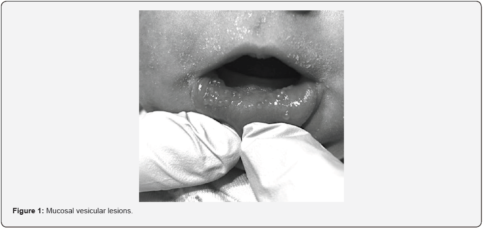
On initial evaluation, the neonate was well-appearing and active. She was noted to have numerous 3-5mm nonerythematous vesicular lesions scattered across the mucosal surface of the lower inner lip (Figure 1). All lesions appeared intact without erosion and were fluctuant to palpation. There was no irritability with manipulation of the lip and palpation of the lesions. No other lesions were noted on dermatologic and mucosal examination. The newborn had been exclusively breastfed since birth. The mother denied a History of Herpes Simplex Virus (HSV). The physical exam was otherwise notable for posterior ankyloglossia and a right preauricular pit. The remainder of the physical exam was unremarkable. Weight, length, and head circumference were appropriate for age.
Diagnosis
Intraoral Sebaceous Gland Hyperplasia.
Hospital Course
He infant received routine prenatal care. HSV testing of the intraoral lesions and blood was obtained due to the vesicular appearance of the lesions. These tests were negative. Further infectious workup, including lumbar puncture and blood culture, was deferred, as the lesions did not appear consistent with herpetic vesicles. The infant remained normothermic with appropriate heart rate, temperature, and respiratory rate for age.The newborn passed the hearing screen bilaterally and the Critical Congenital Heart Defect (CCHD) screen. She received the Hepatitis B immunization, Vitamin K injection, and erythromycin ophthalmic ointment within the first 24 hours of life. The state newborn screen was obtained at 24 hours of life and ultimately resulted as normal. The infant was discharged in stable condition on the second day of life and has had an uneventful infancy to date, with resolution of the lesions by one month of age.
Discussion
Sebaceous gland hyperplasia has a significantly different presentation in adults and should not be confused with the neonatal form. In adults, this presents as flesh-colored to yellowish papules with a central dell. Heterotopic sebaceous glands can also be found on the eyelids (meibomian glands), areolae (Montgomery tubercles), and labia minora and prepuce (Tyson glands) 5, 7 Sebaceous gland hyperplasia affecting the oral mucosa may present as vesicular lesions and could be quite alarming to the neonatal team. Unfamiliarity with this process could lead to unnecessary testing and extensive workup. This condition is selfresolving within weeks and does not warrant treatment [8].
Sebaceous gland hyperplasia is a common benign dermatologic finding in neonates with a recorded incidence of greater than 40% [1-3]. However, the incidence of neonatal intraoral sebaceous gland hyperplasia, otherwise known as Fordyce spots or granules, is approximately one percent [4]. Fordyce granules are heterotopic or anomalous sebaceous glands that tend to localize in the oral mucosa and vermilion area [5]. Fetal and neonatal sebaceous gland function is regulated by maternal androgens, and newborns experience an increase in sebum excretion within the first few hours of life [6, 7]. Premature infants tend to be less affected.
Conflict of Intrest
The authors have no conflicts of interest or financial conflicts to disclose.
References
- Kanada K, Merin M, Munden A, Friedlander S (2012) A prospective study of cutaneous findings in newborns in the United States: correlation with race, ethnicity, and gestational status using updated classification and nomenclature. J Pediatr 161(2): 240-245.
- Moosavi Z, Hosseini T (2006) One-year survey of cutaneous lesions in 1000 consecutive Iranian newborns. Pediatr Dermatol 23(1): 61-63.
- Rivers J, Frederiksen P, Dibdin C (1990) prevalence survey of dermatoses in the Australian neonate. J Am Acad Dermatol 23(1): 77-81.
- Flinck A, Paludan A, L Matsson, A K Holm, I Axelsson (1994) Oral findings in a group of newborn Swedish children. Int J Paediatr Dent 4(2): 67-5.
- Bolognia JL, Schaffer JV, Cerroni L, Allen CM, Camisa C, et al. (2018) Oral Diseases. Dermatol 2(4): 1425- 1446.
- Zouboulis C (2010) Die Talgdrüse/The Sebaceous Gland. Hautarzt 61(6): 467-468.
- Farci F, Rapini R (2021) Sebaceous Hyperplasia. StatPearls.
- Quierós C, Santos M, Rita Pimenta, Cristina Tapadinhas, Paulo Filipe (2021) Transient Cutaneous Alterations of the Newborn. EMJ 6(1): 97-106.


