Case Report: Giant Coronary Artery Aneuyrism, Arteriovenous Malformation of Cerebral Vessels, in a Kawasaki Disease Patient. Cause or Association?
Zeinab Anwar El Kabbany1, Mohamed Alaa El-Din Habib2, Omneya Ibrahim Youssef3, Yosra Mohamed Mohsen Awad4 and Amira Abdelghany Sabra Mahfouz5
1 Professor of pediatrics at Ain Shams University, Egypt
2Professor of neurosurgery Ain Shams University, Egypt
3Assistant professor of pediatrics, Ain Shams university, Egyp
4 Lecturer of pediatrics, Ain Shams University, Egypt
5 Lecturer of pediatrics, Ain Shams University, Egypt
Submission: November 11, 2019; Published: January 24, 2020
*Corresponding author: Amira Abdelghany Sabra Mahfouz, lecturer of pediatrics, Ain Shams University, 56 Ramsis street, Abbasiya, postcode 11566, Egypt
How to cite this article: Zeinab A E K, Mohamed A E-D H, Omneya I Y, Yosra M M A and Amira A S M. Case Report: Giant Coronary Artery Aneuyrism, Arteriovenous Malformation of Cerebral Vessels, in a Kawasaki Disease Patient. Cause or Association?. Acad J Ped Neonatol. 2020; 8(5): 555806. DOI: 10.19080/AJPN.2020.08.555806
Abstract
Kawasaki disease is an acute systemic vasculitis, It represents the most prominent cause of acquired coronary artery disease in childhood, yet coronary aneurysms in children are not only caused by vasculitic syndromes, sometimes it has genetic background and is associated with aneurysms elsewhere, we present a case of a child with both coronary and cerebral aneurysms.
Introduction
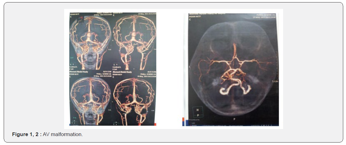
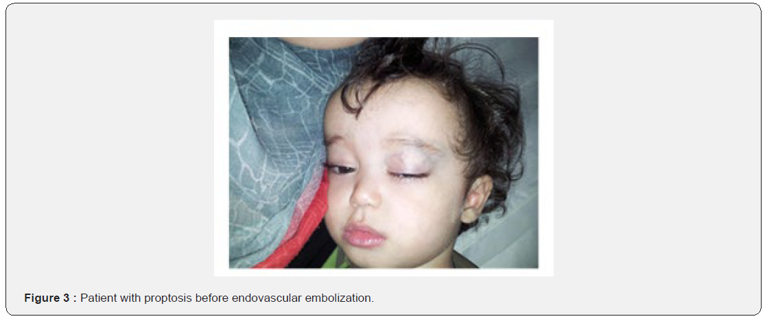
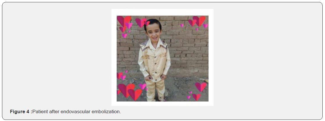
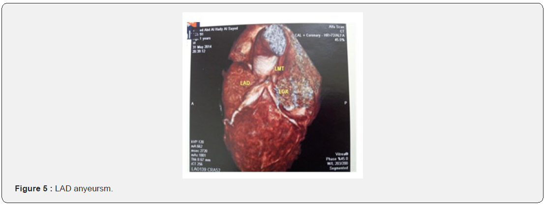
A twenty months old male patient, presented to the Paediatrics Ain Shams university hospital, with the classic clinical picture of Kawasaki disease: High grade fever of 15 days duration, swelling of hands and feet, maculopapular skin rash, strawberry tongue, and right sided cervical lymphadenopathy, where fever plus four out of five criteria are found. On admission it was noticed that the occipito- frontal circumference of the patient exceeded the 95th centile (52cm), and mild proptosis was seen. His weight (12kg) and length (83cm) were between 25th and 50th centile. Patient was also noted to have mild proptosis (Figure3). Laboratory investigations on admission showed elevated ESR (77mm first hour) and positive CRP, while his CBC was normal. Echocardiography done at the time of presentation showed left main coronary aneurysm of 0.31cm (Z score of 2.78mm), while the LAD artery showed giant aneurysm of 0.72cm (Z score of 13.65). Patient received intravenous immunoglobulins and aspirin, with resolution of the fever one day later. Patient was also investigated for his macrocephaly, where MRI brain was done which showed cerebral AV malformation. MRA and MRV followed, confirming type I AV fistular malformation of vein of Galen. Multi-slice CT angiography and CT venography of the cerebral vessels revealed Figures 1&2: abnormal arteriovenous malformations are seen at the cavernous sinus regions, posterior fossa and periventricular regions, dilated intraorbital vessels and superior ophthalmic veins, aneurismal dilatation of the vein of Galen is also seen. Patient underwent four times endovascular embolization for this aneurysm, his proptosis now improved, (Figure 4) yet he still needs further sessions. Follow up echocardiography on weeks 2, 6, 12 and later after 6, 9, 12 and 18 months did not show any change in diameter of aneurysm. Multi-slice CT coronary angiography also showed: left main trunk appears dilated reaching maximal luminal diameter of about 5.2 mm, the proximal left anterior descending shows a fusiform aneurysm measuring about 16.3mm long and 7.6mm wide, the proximal left circumflex appears dilated reaching maximal luminal diameter of about 5.5mm. The patient is currently on aspirin alone on 5mg/kg/day, while warfarin was not given, although it was indicated for fear of bleeding from intracranial aneurysm. Whether this aneurysm resulted from Kawasaki disease, or was it present from the start and was discovered accidentally, remains the question.


Discussion
Our patient was diagnosed as Kawasaki disease according to the American heart Association criteria [1]. The coronary aneurysm in our patient was discovered during the acute febrile illness and it remained unchanged 20 months later. The aneurysm in the LAD was considered giant since it is more than 8mm internal diameter according to the Japanese ministry of health criteria [2]. Lin et al. [3] reported upon their study on 1073 patients with coronary aneurysm in Kawasaki patients stated that coronary aneurysms were present in 40.6% of patients at their time of the acute febrile illness, of whom the giant aneurysms persisted. Yet in another study by Levy et al. [4], showed that giant aneurysms regressed in 32% and 22% among patients receiving warfarin and patients not receiving warfarin respectively, indicating that nearly more than 70% of giant’s aneurysms did nor regress too. Our patient received IVIG, once on 2gm/kg per dose on day 15 of illness, although late, yet still recommended as he was still febrile and ESR and CRP were elevated according to the American Heart Association guidelines [1] Cerebral aneurysms as well, may be congenital or acquired. A case of Kawasaki disease associated with cerebral vasculitis was reported by Gitiaux et al. [5], but in our case the brain aneurysm was congenital. Intracranial aneurysms have been reported in certain genetic variants and many studies are conducted in this field, common variants of 9p21.3 have been reported to be associated with cardiovascular phenotypes as coronary artey disease and intracranial aneurysms [6]. We may recommend from this case report that any child with coronary aneurysm, even in case of kawasaki, if it is big from the start or does not regress in size, should be screened for other aneurysms especially cerebral. Written informed consent was obtained from the patient for publication of this case report and accompanying images. A copy of the written consent is available for review by the Editor-in-Chief of this journal.
References
- Freeman A, Shulman S (2006) Kawasaki disease: Summary of the American Heart Association guidelines. Am Fam physician74(7): 1141-1148.
- Akagi T, Rose V, Benson LN, Newman A, Freedom RN (1992) Outcome of coronary artery aneurysms after Kawasaki disease. J Pediatr121: 689-694.
- Lin MT, Sun LC, Wu ET, Wang JK, Lue HC (2015) Acute and late coronary outcomes in 1073 patients with Kawasaki disease with and without ɣ-immunoglobulin therapy. Arch Dis Child 100(6): 542-547.
- Levy DM, Silverman ED, Massicotte MP, McCrindle BW, Yeung RS (2005) Long term outcomes in patients with giant aneurysm secondary to Kawasaki disease. J Rheumatol 32(5): 928-934.
- Gitiaux C, Kossorotoff M, Berqounioux J, Adjadi E, Lesage F (2012) Cerebral vasculitis in severe Kawasaki disease: early detection by magnetic resonance imaging and good outcome after intensive treatment. Dev Med Child Neurol 54(12): 1160-1163.
- Bendjilali N, Nelson J, Weinsheimwer S, Sidney S, zaroff J (2014) Common variants on 9p21.3 are associated with brain arteriovenous malformations with accompanying arterial aneurysms. J Neurol Neurosurg Psychiatry 25(11): 1280-1283.


