




 AJOP Home



Poly(1-Vinyl-1,2,4-Triazole-Co-N-Vinylpyridone) Matrix Decorated with Iron Nanoparticles: Synthesis and Properties
SH Sargsyana*, AS Sargsyanb, KM Khizantsyana*, TS Sargsyanb, I G Aghajanyana and K S Margaryanb
aNational Polytechnic University of Armenia, Yerevan, Armenia
bYerevan State Medical University named after Mkhitar Heratsi, Yerevan, Armenia
Submission:August 19, 2026August 05, 2026;Published: August 19, 2026
*Corresponding author: SH Sargsyan & KM Khizantsyan, National Polytechnic University of Armenia, Yerevan, Armenia
How to cite this article: SH Sargsyana, AS Sargsyanb, KM Khizantsyana, TS Sargsyanb, I G Aghajanyana, et al. Poly(1-Vinyl-1,2,4-Triazole-Co-NVinylpyridone) Matrix Decorated with Iron Nanoparticles: Synthesis and Properties. Academ J Polym Sci. 2026; 7(1): 555704. DOI: 10.19080/AJOP.2026.07.555704
Abstract
This paper reports the electrochemical synthesis of metal–polymer nanocomposites and iron-containing coatings on pure iron, steel, and magnesium electrodes during the combined process of electrochemical copolymerization of 1-vinyl-1,2,4-triazole with N-vinyl pyridone and cathodic metal deposition.
Eectron Paramagnetic Resonance (EPR) and Transmission Electron Microscopy (TEM) analyses revealed that, in addition to metallic iron nanoparticles, iron oxide particles are also formed, predominantly of spherical morphology. The nanocomposites obtained during electrochemical synthesis contain nanoparticles with diameters ranging from 2 to 4 nm. Thermal decomposition of these nanocomposites occurs in stages. The resulting materials are promising for applications in medicine, construction, and catalysis.
Keywords: Electrochemical synthesis; 1-vinyl-1,2,4-Triazole; Iron nanoparticles; Polymerization; N-vinylpyridone
Introduction
Metal nanocomposites are materials in which metal nanoparticles are embedded within a macroscopic matrix, leading to strong interparticle interactions that often mask the properties of isolated particles [1, 2]. Understanding how different interactions influence material properties is essential. Iron-containing nanocomposites (INCs) are of particular interest due to their magnetic properties. For example, Fe³⁺-gallium acid polyvinylpyrrolidone complexes have been shown to function as contrast agents for photoacoustic and magnetic resonance imaging, with rapid in vivo degradation and natural excretion of products, making them attractive for clinical use [3]. Zero-valent iron nanoparticles also enhance microbial denitrification processes in water purification [4,5].
Electrosynthesis of functional polymers and nanomaterials has become a rapidly growing field in modern chemical science. Such nanomaterials are used in medicine as antibacterial agents, drug delivery systems, biosensors, and contrast agents [1-7]. Previous studies have synthesized iron nanocomposites in polyvinyl-1,2,4-triazole (PVT) matrices by chemical reduction of Fe²⁺ ions withNaBH4 [8], and in polyvinylpyrrolidone (PVP) matrices by thermal decomposition of iron pentacarbonyl or iron acetylacetonates [9-16]. These composites typically contained spherical nanoparticles of Fe₂O₃ or Fe₃O₄, ranging from 3 to 30 nm, and exhibited high biocompatibility.
However, despite numerous reports on chemical synthesis of iron nanocomposites in PVT or PVP matrices, there are no documented examples of electrochemical synthesis in copolymer matrices of 1-vinyl-1,2,4-triazole (VT) with N-vinylpyridone (VP). Such copolymers are highly promising for biomedical use due to their hydrophilicity, biocompatibility, biological activity, and ability to bind various substances, including drugs, via functional groups [17-20]. This study presents the results of electrochemical synthesis of iron-containing nanocomposites and coatings on iron, magnesium, and steel electrodes through simultaneous copolymerization of VT with VP and cathodic metal deposition.
Experimental
Electrochemical initiation of polymerization was carried out in a glass electrolysis cell without a diaphragm. Elemental analysis was performed on a FLASH EA 1112 Series analyzer (Germany). UV spectra were recorded on a "Perkin Elmer Lambda 35 UV/VIS" (UAS) spectrophotometer. IR spectra of polymers were taken on "Specord M-80" (Germany) and "Bruker Vertex 70" (Germany) spectrometers using finely ground powders pressed into KBr tablets. The metal content in composites was determined by elemental and atomic absorption analysis on a Perkin Elmer Analyst 200 spectrometer (UAS). EPR spectra were recorded on a FT Bruker ELEXSYS E-580 (X-97PI) (Germany) spectrometer in continuous mode. The distribution of iron nanoparticles was determined using a Leo 906 E TEM (Germany). Thermogravimetric analysis (TGA) was performed on a MOM derivatograph (Hungary), with a temperature rise rate of 5°C/min-1. Electrical conductivity was measured using a standard teraohmmeter E6-13A (USSR).
VT was obtained and purified according to the method described in [21]. VP (Tboiling=91-92°C/1mmHg) was obtained via vinyl exchange reaction in a pyridone-2-vinylacetate system according to the method in [22].
General Method for Electrosynthesis of Nanocomposites and Coatings
In a 50 mL glass electrolytic cell, electrolysis [E=-0.1…-1.2 V (vs. SCE) or j=1-20 mA/cm2] was carried out in aqueous or aqueous-ethanol solutions containing 0.5-1 mol/L VT, 0.5-1 mol/L VP, 1.5-4 mmol/L FeSO4·H2O, 0.02-0.05% 4-tert-butylperoxy-4-oxobutanoic acid (TBOPA), and in some cases, 0.05-0.07% chitosan, which facilitates the electro-reduction of iron. A pure iron, magnesium, or steel plate (1-2 cm2 area) was used as the working electrode, and a platinum or glass-carbon (SU-12, SU-20) plate of the same area was used as the counter-electrode. According to our data, the nature of the electrode material does not affect the composition and morphology of the electrode material and the distribution of nanoparticles. At low current densities j≤10 mA/cm2, nanocomposite coatings are formed, while at higher current densities j>10 mA/cm2, the nanocomposite is deposited at the bottom of the electrolysis cell. After electro-polymerization, the electrode package was removed, the cathode with the formed coating was separated, washed thoroughly with distilled water, and dried to a constant mass. The synthesized coatings containing iron had an orange color. Previously, we defined mechanical properties, impact strength and thermomechanical properties, and biocompatibility. All chemical reagents from Sigma-Aldrich according to standards.
Results and Discussions
During the electrolysis of aqueous and aqueous-ethanol solutions of VT and VP, or their mixtures in various ratios in the presence of , nanocomposites and nanocomposite coatings (NC and NCC) are formed with iron content ranging from 0.8 to 6%, in the presence of a peroxide-type initiator such as TBOBA, whose electro-reduction potential is close to the cathodic metal separation potentials – 0.6-1.2 (h.s.e.).
By varying the molar ratio of monomers, iron sulfate, and current density, NC and NCCs with iron content from 0.4 to 6% in orange color, soluble in water, were obtained.
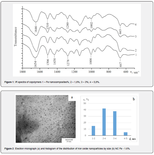
In the IR spectra of the copolymers (Figure 1) of TR-VP, the characteristic frequencies of the valence and deformation vibrations of the triazole ring at 1654, 1506, 1438, 1278, 1008, 667 cm⁻¹ (C-N, C=N), 1278 cm⁻¹ (N-N), 1008 cm⁻¹ (C-H), 667 cm⁻¹ (C-N), 1654 cm⁻¹ (C=O) are present.
Analysis of the IR spectra shows that the formation of iron-containing polymer nanocomposites leads to slight shifts (2-12 ) in the characteristic absorptions. This phenomenon indicates the involvement of heterocycles in the coordination of both iron ions and its metallic nanoparticles. According to Transmission Electron Microscopy (TEM) data, in addition to the metallic iron nanoparticles, iron oxide particles are formed, which predominantly have a spherical shape (Figure 2). According to our data, the ratio of iron oxides to pure iron is approximately 65 to 35%.
The dispersion of nanoparticles by size depends on the iron content. NCs with the lowest iron content are characterized by a uniform distribution of nanoparticles within the polymer matrix, with sizes ranging from 1-6 nm. With increasing iron content, NCs with a wider nanoparticle size distribution (14 nm) are formed (Figure 3). This dependence is a consequence of the fact that with increasing metal content, more nanoparticles are formed, which are less effectively incorporated into the copolymer matrix, inevitably leading to coagulation processes and resulting in the formation of larger nanoparticles (Figure 3).
In addition to isolated, uniformly distributed nanoparticles, aggregated clusters of stabilized nanoparticles appear in the NC, which are stabilized by the copolymer matrix. The size of these aggregates does not exceed 50-80 nm in most cases. As the iron content increases, the number of disordered polymer chains also increases.
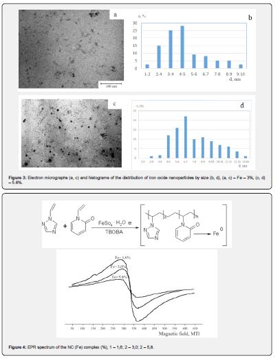
The results of electron paramagnetic resonance (EPR) spectroscopy are presented in (Figure 4). In the EPR spectra of all investigated NCs, a broad asymmetric signal with an effective g-factor in the range of 2.04-2.1 and a width of 70-100 mT is observed. This signal is characteristic of iron-containing substances and corresponds to the predominance of high-spin iron. Additional signals in the g-factor region from 1.876 to 4.3 are also present, indicating the presence of iron in the trivalent state [23,24].
Changes in the double integral of the signal, the g-factor, and the line width with increasing iron content in the NC may be related to changes in the size and shape of the NCs, as well as to changes in the relaxation characteristics and the nature of the formed iron oxides and their ratios in the polymer matrix.
In the synthesized nanocomposites, a signal with a g-factor of 4.3 corresponding to γ-Fe₂O₃ nanoparticles in polymer matrices is observed [25,26]. The broadest asymmetric signal is given by the NC containing 1.6% iron, most likely because the amounts of iron oxides Fe₂O₃ and Fe₃O₄ are comparable.
All EPR spectra of the nanocomposites (NCs) exhibit signals in the range of g = 1.9–2.1, which indicates the presence of zero-valent iron. This suggests the formation of core–shell type nanoparticles [27], where a metallic iron core is surrounded by an oxide shell.
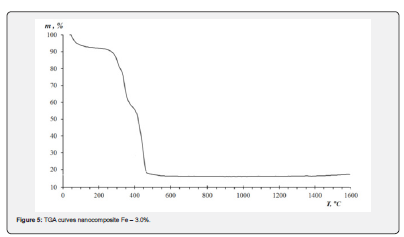
Thermal stability studies of the polymer nanocomposites (Figure 5) showed that the pristine copolymer has higher thermal stability (325°C) compared to the NCs (250-285 °C). Increasing the metal content in the NCs leads to a further decrease in stability of 35-70 °C. In the temperature range of 60-155 °C (mass loss ~8%), the weight reduction is attributed to the release of physically adsorbed water. Between 240-385 °C, the mass loss is associated with the elimination of functional fragments from the macromolecular chains. At higher temperatures (390-500 °C), oxidation of the hydrocarbon skeleton of the polymer matrix occurs, predominantly forming carbon dioxide.
The electrical conductivity of the NCs (10⁻¹¹-10⁻¹⁰ S/cm) is two to three orders of magnitude higher than that of the pristine copolymer (3.8·10⁻¹³ S/cm). This enhancement is most likely due to localized tunneling currents [28], which arise between closely spaced conducting iron nanoparticles embedded in the dielectric polymer matrix. Preliminary tests demonstrated that the composition and structure of the nanocomposites remain stable under ambient conditions for at least one month.
Conclusion
The possibility of synthesizing iron and iron oxide nanocomposites (NCs and NCPs) by electrochemical methods based on VT and VP using pure iron, steel, and magnesium electrodes has been demonstrated.
According to EPR data, in addition to zero-valent iron, iron oxides are also formed. The solubility of the nanocomposites is due to interstructural bonding between VT-VP macromolecules and both nanosized iron particles and iron oxides. The obtained nanocomposite materials can be applied in medicine and electronics.
References
- Gusev Ai (2007) Nanomaterial Nanostructure Nanotekhnologii M Fizmat lit: 416.
- Sergeev G B, Klabunde K J (2013) Nano chemistry Book Second Edition.
- Chen L, Chen J, Qiu Sh (2018) Biodegradable Nano agents with Short Biological Half-Life for SPECT/PAI/MRI Multimodality Imaging and PTT Therapy of Tumors. Nano-micro-Small 14(4).
- Jiang Ch, Liu Y, Chen Z, Megharaj M, Naidu R, et al. (2013) Impact of iron-based nanoparticles on microbial denitrification by Paracoccus sp. strain YF1. Aquat Toxicol 15: 142-143: 329-335.
- Yan Liu, Shibin Li, Zuliang Chen, Mallavarapu Megharaj, Ravi Naidu, et al. (2014) Influence of zero-valent iron nanoparticles on nitrate removal by Paracoccus sp. Chemosphere 108: 426-432.
- Lee N, Yoo D, Ling D, Cho MH, Hyeon T, et al. (2015) Oxide Based Nanoparticles for Multimodal Imaging and Magnetoresponsive Therapy. Chemical Reviews 115(19): 10637-10689.
- Nikiforov VN, Filinova E, Yu (2009) Biomedical Applications of Magnetic Nanoparticles / Book Editor(s): Prof Sergey P. Gubin; First published.
- Prozorova GF, Korzhova SA, Emel'Yanov AI, Pozdnyakov AS, Khutsishvili SS, et al. (2013) Polymer nanocomposites with iron oxide nanorarticles. Russian Journal of Applied Chemistry 86: 1452-1455.
- Ha-Young L, Sang-Hoon L, Chenjie Х, Jin X, Jin-Hyung L, et al. (2008) Synthesis and characterization of PVP-coated large core iron oxide nanoparticles as an MRI contrast agent. Nanotechnology 19(16):165101.
- Young L, Nak Hyun L, Seo Jin A (2006) Preparation and magnetic resonance imaging effect of polyvinylpyrrolidone-coated iron oxide nanoparticles. Journal of Biomedical Materials Research 79(1):142-50.
- Huang H, Xie Q, Kang M (2009) Labeling transplanted mice islet with polyvinylpyrrolidone coated superparamagnetic iron oxide nanoparticles for in vivo detection by magnetic resonance imaging. Nanotechnology 20(36): 365101.
- Lu X, Niu M, Qiao R, Gao M (2008) Superdispersible PVP-coated Fe3O4 nanocrystals prepared by a "one-pot" reaction. J Phys Che 112(46): 14390-14394.
- Zhang B, Tu Z, Zhao F, Wang J (2013) Superparamagnetic iron oxide nanoparticles prepared by using an improved polyol method. Applied Surface Science 266: 375-379.
- Tu Z, Zhang B, Yang G (2013) Synthesis of poly(ethylene glycol) and poly(vinyl pyrrolidone) co-coated superparamagnetic iron oxide nanoparticle as a pH-sensitive release drug carrier. Colloids and Surfaces A: Physicochemical and Engineering Aspects 436: 854-861.
- Jurkin T, Gotić M, Štefanić G, Pucić I (2016) Gamma-irradiation synthesis of iron oxide nanoparticles in the presence of PEO, PVP or CTAB. Radiation Physics and Chemistry 124: 75-83.
- Xu Y, Zhang X, Hsing Y, Fang Y (2011) Ultrasonic-assisted Synthesis of Fe Nanoparticles in the Presence of Poly(N-vinyl-2-pyrrolidone). Chin J Chem 29: 1829.
- Malekigorji M, Curtis ADM, Hoskins C (2014) The Use of Iron Oxide Nanoparticles for Pancreatic Cancer Therapy. Journal of Nanomedicine Research 1(1).
- Prozorova GF, Korzhova SA, Pozdnyakov AS, Emel yanov AI, Ermakova TG, et al. (2015) Immunomodulatory properties of silver-containing nanocomposite on the basis of polyvinyltriazole. Russian Chemical Bulletin 64(6): 1437-1439.
- Kavas H, Durmus Z, Baykal A, Aslan A, Bozkurt A, et al. (2010) Synthesis and conductivity evaluation of PVTri–Fe3O4 nanocomposite. Journal of Non-Crystalline Solids 356(9-10): 484-489.
- Uzun L, Kara А, Osman B, Yılmaz E, Beşirli N, et al. (2009) Removal of heavy-metal ions by magnetic beads containing triazole chelating groups. First published 114(4): 2246-2253.
- Ermakova TG, Kuznetsova NP, Pozdnyakov AS (2016) 1-Vinyl-1,2,4-triazole in copolymerization reaction with 1-vinyl-4,5,6,7-tetrahydroindole: synthesis and properties of copolymers. Russ Chem Bull 65: 485-489.
- Lavlinskaya MS, Shatalov GV, Mokshina NY, Pakhomova OA, Kuznetsov VA (2016) Extraction and reextraction of histidine and riboflavin by N-vinylcaproctame copolymers with 1-vinyl- and 1-metrhacryloyl-3,5-dimethylpyrozole. ChemChemTech 59(7).
- Hevus I, Kohut A, Voronov A (2012) Micellar assemblies from amphiphilic polyurethanes for size-controlled synthesis of silver nanoparticles dispersible both in polar and nonpolar media. J Nanopart Res 14(820)
- Ulitin NV, Deberdeev RYa, Deberdeev TR (2012) Kinetics of styrene radical polymerization proceeding at conditions of reversible chain transfer using trithiocarbonates. ChemChemTech 55: 9.
- Chakrabarti S, Mandal SK, Nath BK (2003) Synthesis of 𝛾-Fe2O3 nanoparticles coated on silica spheres: Structural and magnetic properties. Eur Phys B 34: 163-171.
- Vereshchagina TA, Anshits NN, Maksimov NG (2004) The Nature and Properties of Iron-Containing Nanoparticles Dispersed in an Aluminosilicate Matrix of Cenospheres. Glass Physics and Chemistry 30(3): 247-256.
- Amstad E, Textor M, Reimhult E (2011) Stabilization and functionalization of iron oxide nanoparticles for biomedical applications. Nanoscale 3(7): 2819-2843
- Sargsyan SH, Sargsyan AS, Khizantsyan KM (2024) Electrochemical synthesis of silver nanocomposites based on 1-vinyl-1,2,4-triazole and N-vinyl-caprolactam. Polym Bull.

























