Optical Imaging Methods for Volumetric Additive Manufacturing
Johanna J Schwartz1 and Dongping Terrel-Perez2*
1Materials Science Division, Lawrence Livermore National Laboratory, USA
2Computational Engineering Division, Lawrence Livermore National Laboratory, USA
Submission: April 27, 2023; Published: May 08, 2023
*Corresponding author: Dongping Terrel-Perez, Computational Engineering Division, Lawrence Livermore National Laboratory, Livermore, CA 94550 USA
How to cite this article: Johanna J S, Dongping Terrel-P. Optical Imaging Methods for Volumetric Additive Manufacturing. Academ J Polym Sci. 2023; 6(1): 555679. DOI: 10.19080/AJOP.2023.06.555679
Abstract
Tomographic volumetric additive manufacturing (VAM) is a technique that enables light-induced curing of photoresins into complex 3D end use objects within a single step. This is made possible through projecting tomographically patterned light energy into a photo-curable resin volume within a rotating container. In order to monitor, quantify, and control curing during tomographic VAM, researchers need to visualize the curing parts in real-time. This may enable advancements toward dynamic, controlled and closedloop VAM methods in the future, as some researchers have shown. Herein we briefly review various optical imaging methods used to monitor printing in real-time, as well as provide some perspectives for future needs and considerations for imaging methods in VAM.
Keywords: Optics; Imaging; Volumetric Additive Manufacturing; Metrology; Photoresin
Abbreviations: VAM: Volumetric Additive Manufacturing; RI: Refractive Index; OST – Optical Scattering Tomography
Introduction
Tomographic volumetric additive manufacturing (VAM) [1] has revolutionized light-driven AM by concurrently printing freeform 3D objects all-at-once without layering artifacts. This ability expands the geometric freedom and material scope accessible, achieving fully printed end use objects quickly. To ensure a successful print, the ability to “see” the structures form and correctly control the light exposure is essential. Otherwise, objects may not be completely formed, or have resulting outgrowth, limiting use and VAM success rates. Fundamentally, insitu quantitative monitoring systems that can quantify the print progress are much needed. Herein we briefly review existing methods for imaging during VAM and provide some short perspectives on future needs.
Optical imaging Methods
The simplest imaging method for VAM is direct viewing with a standard camera or by eye [1-4]. This method, however, is difficult as the photoresins themselves are optically transparent. Instead, methods that enable greater contrast, sensitivity, and quantitative monitoring of refractive index (RI) change provide one solution for better monitoring. Optical light rays propagating through a transparent medium with spatially varying RI experience refraction - light rays deflect towards the region with greater refractive index. The angular deflection can be calculated as:

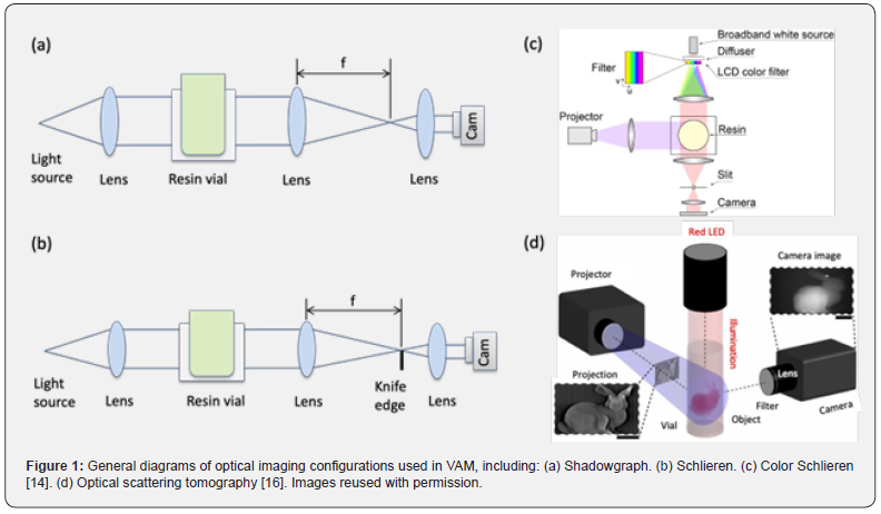
where n0 is the RI of surrounding media (in this case uncured resin), z is imaging light propagation or viewing direction, and x is one direction orthogonal to the viewing direction. The equation above indicates that ray deflection depends not only on the RI of the medium but mainly on its gradient in the orthogonal direction x; it is also dependent on the extent or depth of the region where RI variation occurs along the viewing direction, z, because of the integral or accumulation in the z direction. These fundamentals lay the foundation for shadowgraph and Schlieren imaging techniques [5]. It is important to note that, in general, cylindrical vials or vats of resin are commonly used in VAM. To reduce the impact of the curvature of the vial for imaging techniques, negative lenses or straight-walled containers of RI-matching fluid outside of the vat are often used [6]. These are denoted as square boxes around the resin vial in Figure 1a-c.
Shadowgraph
Shadowgraph is a simple and commonly used optical imaging technique to visualize polymerization processes in VAM [7-11]. A red LED light source is chosen whose wavelength is outside of the absorption band of the photoinitiator in the resin so that it will not interfere with printing process or induce photopolymerization. The collimated red LED light propagating through the resin is refracted by the RI variations within resin during curing. The light is then collected and focused by a lens beyond the VAM resin vat and further imaged by another lens and a camera, as shown in Figure 1(a). This essentially creates a backlit image of the curing object, corresponding to the second derivative of the RI. Regions with non-constant second derivative of RI appear dark, or as a shadow, providing increased contrast relative to direct imaging of these optically transparent resins. While not quantitative, the ease of use of this technique makes it readily amenable for monitoring many resins, including acrylate, thiol-ene, and even glass-filled and cell-laden polymerizations [6-10]. These resins, in general, have large changes in RI, making them easy to visualize with this method.

Schlieren
A Schlieren setup is nearly identical to that of a shadowgraph but with the addition of a knife edge at the focal point of the focusing lens to cut off certain light rays as shown in Figure 1(b). Schlieren images the first derivative of RI. Contrast is created when light propagates through a resin with a non-constant first derivative of RI: if light rays are deflected towards the knife edge, the image of the region where those light rays originate from will appear darker, more than that with constant RI. If light rays deflect away from the knife, they will appear brighter. Schlieren is more sensitive than shadowgraph. However, the sensitivity is only along one direction where the knife edge is placed. Shadowgraphs have an advantage over Schlieren setups for visualizing things such as resin flow because shadowgraphs are uniformly sensitive in all directions. As VAM is often in a viscous, non-flowing regime however, a Schlieren setup’s increased sensitivity may be useful. Regardless, swapping between the two methods is facile, relying on the movement of the razor blade in or out of the focal point region. The original Schlieren technique is a qualitative method but using a calibration object with a known RI variation, the Schlieren system can achieve quantitative measurements [12,13]. The sensitivity of a Schlieren system depends on the light source, focal length of the focusing lens, and the degree to which the knife edge is cutting off the beam. Schlieren can visualize RI change as low as ~10-5 [12].
Color Schlieren
Color schlieren is a variation of the basic Schlieren system described above that also enables quantification of RI change. In a color Schlieren system, a color filter is used instead of a knife edge, resulting in a colored Schlieren image where RI variation is encoded in color instead of intensity. Light rays from a broadband white light source are diffused by a diffusor and then pass through a transparent color LCD color filter. The spectrum separated light rays are collimated by a lens and propagate through the VAM resin container. The light rays after the resin volume are then focused by a focusing lens which is placed one focal length away from the resin vial. A slit is placed at the focal plane of the focusing lens [14]. Depending on the ray deflection by the resin going through gelation, only light of certain color can pass through the slit and get collected by the camera. In this way, ray deflection information is encoded into color changes on Schlieren images (Figure 1(c)). A ray deflection versus hue calibration curve is generated using a known planoconvex lens before VAM experiments. Changes of hue observed in color Schlieren images of resins are quantitatively mapped back to ray deflection and further RI change during print based on the calibration curve. Color Schlieren technique is more robust against changes in scattering and achromatic absorbance. Color Schlieren provides contrast along one direction but holds the potential to encode RI along both vertical and horizontal directions by using a more sophisticated color filter [14,15]. RI changes as low as 10-4 of a urethane dimethacrylate-based resin were detected by color Schlieren system [14,15].
Optical Scattering Tomography
Optical scattering has been exploited to visualize the polymerization process in VAM. Taking advantage of the sharp increase in light scattering by some resins as they undergo gelation, researchers [16] have developed optical scattering tomography (OST) and used it to monitor the side-scattered light of the resin volume to directly measure the geometry of the gelled print. A collimated red LED illuminates the resin volume from the top of the vial and a camera oriented orthogonal to both the vial and LED monitors the side-scattered LED light from resin undergoing gelation (Figure 1(d)). A bandpass filter is used to block the blue projection light and only allows the red LED light to pass through and detected by the camera. For each resin used, a calibration process is performed by printing a standard cylinder and finding its OST intensity value Ip corresponding to the gelation threshold of this resin. During actual prints, the object being printed is visualized by thresholding its scattering volume at Ip and assigning the voxels above this value to 1 and otherwise 0, thus a thresholded volume corresponding to the actual polymerized object is obtained. OST is simpler to implement than color Schlieren system as a broadband light source and color filter are not needed. Additionally, OST provides contrast in 2D direction as opposed to 1D in color Schlieren system. OST relies on scattering contrast whereas shadowgraph and Schlierenbased methods measure ray deflection, thus comparatively more strongly scattering resins are compatible with OST. RI changes of 0.007 to 0.01 from methacrylates resins have been investigated with OST. Other resins which show dramatic increase in scattering at the onset of gelation will also be compatible with this method, however resins with little change in scattering during gelation would be challenging to visualize with this method.
Perspective
The imaging methods highlighted within this review provide a means to accurately monitor curing during VAM printing. Shadowgraph and Schlieren imaging methods are simple, robust and easily translatable across a wide range of material toolsets, however they are generally qualitative measurements unless consistently benchmarked against a known standard. Color Schlieren and OST are exciting as they provide methods to quantify curing with the resins, opening opportunities for feedback and dynamic curing control. These methods, however, still require more study to determine the sensitivity ranges and limitations in terms of RI change and scattering, respectively.
A single method for monitoring and quantifying curing during VAM that completely meets the need of the process and the diverse resins has yet to be identified or standardized. Variations in sensitivity, versatility, RI ranges, scattering, and quantification approaches create an exciting challenge for further study. Robust in-situ quantitative methods that can quantify the polymerization progress will provide valuable insights on material development and characterization, crosslinking kinetics study, and print quality inspection. In addition, the methods described above may still be unsuitable imaging methods for low RI change materials (such as hydrogels). Identifying methods with heightened sensitivity for these material systems will expand the pool of material candidates accessible for controlled VAM and increase print success. Ultimately, imaging methods able to handle composite, scattering, and more absorbing resins will also be needed [17]. Through the generation of these robust quantitative metrological approaches, feedback and feedforward systems of adaptive and dynamic VAM for high-fidelity printing, increased accuracy, and precision will become possible. This could include feeding images from the print directly back into the tomographic slicing algorithms to update the projected light patterns during printing. In essence, if an area of the part is “done”, turning off light dosage within that region would eliminate over-exposure, and improve resolution. Additionally, if a part sinks or rises during curing [7], effective imaging methods could enable object tracking, moving the projection set with the object as it cures in VAM. No two photoresins are the same, providing the need for a monitoring setup with both sensitivity and robustness to push the field of VAM forward. In this way, researchers can “see” the resolution and curing challenges in real time, and hopefully use this information to remove any obstacles to VAM print success.
Acknowledgement
This work was supported by Lawrence Livermore National Laboratory Laboratory-Directed Research and Development funding 23-FS-020 (to L.T.P.). This work was performed under the auspices of the U.S. Department of Energy by Lawrence Livermore National Laboratory under Contract DE-AC52-07NA27344.
Funding provided by the LLNL LDRD Program LLNL-JRNL-848304.
References
- Kelly BE, Bhattacharya I, Heidari H, Shusteff M, Spadaccini CM, et al. (2019) Volumetric additive manufacturing via tomographic reconstruction. Science 363(6431): 1075-1079.
- Xie M, Lian L, Mu X, Luo Z, Garciamendez MCE, et al. (2023) Volumetric additive manufacturing of pristine silk-based (bio) inks. Nature Comm 14(1): 210.
- Boniface A, Maître F, Madrid WJ, Moser C (2023) Volumetric helical additive manufacturing. Light Adv Manuf, p. 18.
- Cook CC, Fong EJ, Schwartz JJ, Porcincula DH, Kaczmarek AC, et al. (2020) Highly tunable thiol‐ene photoresins for volumetric additive manufacturing. Adv Mater 32(47): 2003376.
- Settles GS (2001) Schlieren and shadowgraph techniques- Visualizing phenomena in transparent media(Book). Springer-Verlag GmbH, Berlin, Germany.
- Moran BD, Fong EJ, Cook CC, Shusteff M (2021) Volumetric additive manufacturing system optics. In Emerging Digital Micromirror Device Based Systems and Applications XIII. SPIE, Vol 11698, pp. 8-15.
- Bernal PN, Delrot P, Loterie D, Li Y, Malda J, et al. (2019) Volumetric bioprinting of complex living‐tissue constructs within seconds. Adv Mater 31(42): 1904209.
- Rackson CM, Toombs JT, De Beer MP, Cook CC, Shusteff M, et al. (2022) Latent image volumetric additive manufacturing. Optics Letters 47(5): 1279-1282.
- Toombs JT, Luitz M, Cook CC, Jenne S, Li CC, et al. (2022) Volumetric additive manufacturing of silica glass with microscale computed axial lithography. Science 376(6590): 308-312.
- Schwartz JJ, Porcincula DH, Cook CC, Fong EJ, Shusteff M (2022) Volumetric additive manufacturing of shape memory polymers. Polymer Chemistry 13(13): 1813-1817.
- Loterie D, Delrot P, Moser C (2020) High-resolution tomographic volumetric additive manufacturing. Nature Comm 11(1): 852.
- Settles GS, Hargather MJ (2017) A review of recent developments in schlieren and shadowgraph techniques. Measurement Science and Technology 28(4): 042001.
- Hargather MJ, Settles GS (2012) A comparison of three quantitative schlieren techniques. Optics and Lasers in Engineering 50(1): 8-17.
- Li CC, Toombs J, Luk SM, de Beer M, Schwartz J, et al. (2022) Computational optimization and the role of optical metrology in tomographic additive manufacturing. In: Advanced Fabrication Technologies for Micro/Nano Optics and Photonics XV. SPIE, Vol. 12012, pp. 38-44.
- Chung Li C, Toombs J, Taylor H (2020) Tomographic color Schlieren refractive index mapping for computed axial lithography. In Proceedings of the 5th Annual ACM Symposium on Computational Fabrication. pp. 1-7.
- Orth A, Sampson KL, Zhang Y, Ting K, van Egmond DA, et al. (2022) On-the-fly 3D metrology of volumetric additive manufacturing. Additive Manufacturing 56: 102869.
- Madrid WJ, Boniface A, Loterie D, Delrot P, Moser C (2022) Controlling light in scattering materials for volumetric additive manufacturing. Advanced Science 9(22): 2105144.


